. 2002 Dec 24;99(26):16684-8.
doi: 10.1073/pnas.262655299. Epub 2002 Dec 13.

Polyphosphate kinase (PPK2), a potent, polyphosphate-driven generator of GTP

Affiliations

Polyphosphate kinase (PPK2), a potent, polyphosphate-driven generator of GTP

Kazuya Ishige et al. Proc Natl Acad Sci U S A. .

Abstract

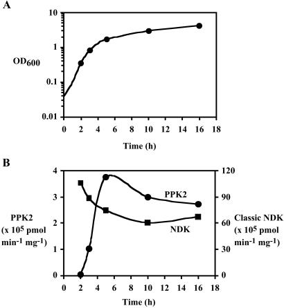
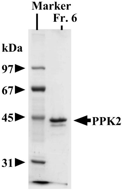
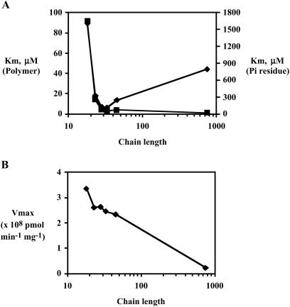
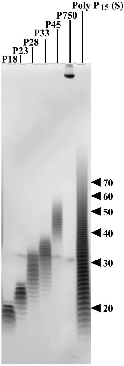
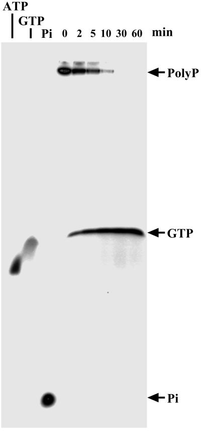
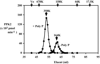
An enzyme that uses inorganic polyphosphate (poly P) as a donor to convert GDP to GTP has been purified 1,300-fold to homogeneity from lysates of Pseudomonas aeruginosa PAOM5. Poly P chains of 30-50 residues are optimal; those of 15-700 residues can also serve. GDP is preferred over ADP among nucleoside diphosphate acceptors. This nucleoside diphosphate kinase (NDK) activity resides in the same protein isolated for its synthesis of poly P from GTP and designated PPK2 in an accompanying report. The reaction that synthesizes poly P and the reaction that utilizes poly P differ in their kinetic features. Especially notable is the catalytic potency of the NDK activity, which is 75-fold greater than that of poly P synthesis. PPK2 appears in the stationary phase of growth and reaches NDK levels of 5-10% that of the classic NDK; both kinase activities may figure in the generation of the guanosine precursors in the synthesis of alginate, an exopolysaccharide essential for the virulence of P. aeruginosa.


Figures

Fig 1.
Fig 1.
Growth-phase dependency of PPK2 and the classic NDK activities. After a 1% inoculation with an overnight preculture, P. aeruginosa PAOM5 was grown in 500 ml of LB media at 37°C with vigorous shaking and its OD600 was monitored. At time points indicated as symbols (corresponding to middle log, late log, early stationary, middle stationary, and late stationary phases, respectively), cells were harvested from 10-ml culture samples. After suspending cells to 1 ml of TED buffer (50 mM Tris⋅HCl, pH 8.0/0.5 mM EDTA/1 mM DTT), cells were sonically disrupted and centrifuged (10,000 × g, 10 min) to give crude lysates, which were assayed for both PPK2 and the classic NDK activities as described in Materials and Methods. (A) Growth curve of P. aeruginosa PAOM5. (B) Both NDK activities in crude lysates of the cells from various growth phases. Circles represent PNDK (PPK2) activity and squares represent the classic ATP-driven NDK (NDK) activity.
Fig 2.
Fig 2.
Thermostabilization of PPK2. Crude lysate (Table 1, Fraction 1) was incubated at stated temperatures and assayed for PPK2 activity: diamonds at 37°C, squares at 40°C, triangles at 50°C, and circles at 50°C with poly P15 (Sigma; expressed as phosphate residues).
Fig 3.
Fig 3.
SDS/PAGE analysis of PPK2. SDS/PAGE (10%) was performed as described (33), and proteins were visualized by Coomassie blue staining: 1 μg of Fraction 6 (Fr. 6) in Table 1; marker proteins were phosphorylase b (97 kDa), BSA (67 kDa), ovalbumin (45 kDa), and carbonic anhydrase (31 kDa).
Fig 4.
Fig 4.
Km and Vmax values with poly P of various chain lengths. The assay mixture contained 50 mM Hepes-KOH (pH 8.0), 80 mM (NH4)2SO4, 10 mM MgCl2, 1 mM [8-3H]GDP (20 cpm/pmol), various concentrations of poly P, and various amounts of PPK2 (Fraction 6 in Table 1). Poly P chain lengths were poly P18 (18 ± 2), poly P23 (23 ± 3), poly P28 (28 ± 5), poly P33 (33 ± 6), poly P45 (45 ± 8), and poly P750 (as described in ref. 13). For more details, see Fig. 5 legend. In each case, the Km and Vmax were determined by a Lineweaver–Burk plot (data not shown). (A) Km values for poly P: squares for molar concentration, diamonds for Pi residue concentration. (B) Vmax values with various chain lengths.
Fig 5.
Fig 5.
PAGE analysis of poly P samples. Commercial poly P15 (Sigma, 250 mg) was solubilized in 100 ml of 25 mM Hepes-KOH (pH 7.6) and applied to HiTrap Q (5 ml, Amersham Pharmacia) equilibrated with 25 mM Hepes-KOH (pH 7.6). Bound poly P was eluted with a 20 column-volume gradient (0–1 M NaCl in the same buffer) and fractionated. PAGE analysis using 20% acrylamide gel containing 7 M urea was carried out as described (34). Poly P was visualized by toluidine blue staining. Applied samples and chain lengths were: P18 (7.5 μg, 18 ± 2); P23 (5 μg, 23 ± 3); P28 (5 μg, 28 ± 5); P33 (5 μg, 33 ± 6); P45 (3.5 μg, 45 ± 8); P750 (1.5 μg; as described in ref. 13); and commercial poly P15 (S) (80 μg, obtained from Sigma). These poly P samples were also used in Fig. 4.
Fig 6.
Fig 6.
PPK2 as a processive enzyme. The assay mixture (10 μl) contained 50 mM Hepes-KOH (pH 8.0), 80 mM (NH4)2SO4, 4 mM MgCl2, 1 mM GDP, 100 μM [32P]poly P (88 cpm/pmol), and 11 ng of PPK2 (Fraction 6). After incubation of the mixture at 37°C, the products were separated by PAGE using 20% acrylamide gel containing 7 M urea (34) and visualized by PhosphorImager (Molecular Dynamics). [32P]ATP, [32P]GTP, and [32P]inorganic phosphate were used as standards.
Fig 7.
Fig 7.
Mass of PPK2 by size-exclusion chromatography. To measure the molecular mass of PPK2 complexed with poly P, the assay mixture (500 μl) contained 50 mM Tris⋅HCl (pH 8.0), 50 mM (NH4)2SO4, 5 mM MgCl2, 6 μg of PPK2 (Fraction 6 in table 1), and 2 mM poly P15 (Sigma). After incubation for 10 min at 37°C, 0.8 M KCl was added. The mixture was applied to a HiLoad 16/60 Superdex 200-pg column (Amersham Pharmacia) equilibrated with buffer PC [50 mM Tris⋅HCl, pH 8.0/0.5 mM EDTA/1 mM DTT/2 mM poly P15 (Sigma)/0.1% CHAPS] containing 0.8 M KCl. PPK2 was eluted with equilibration buffer and monitored by PNDK activity (diamonds). For measuring molecular mass of PPK2 without poly P, poly P was omitted from the incubation mixture and the equilibration (elution) buffer (circles). Positions of molecular mass standards are shown at the top of the panel: blue dextran (Vo), thyroglobulin (670 kDa), bovine gamma globulin (158 kDa), chicken ovalbumin (44 kDa), and equine myoglobin (17.5 kDa).

References

    1. Kulaev I. S., (1979) The Biochemistry of Inorganic Polyphosphate (Wiley, New York).
    1. Kornberg A. (1995) J. Bacteriol. 177, 491-496. - PMC - PubMed
    1. Kornberg A., Rao, N. N. & Ault-Riché, D. (1999) Annu. Rev. Biochem. 68, 89-125. - PubMed
    1. Kuroda A. & Kornberg, A. (1997) Proc. Natl. Acad. Sci. USA 94, 439-442. - PMC - PubMed
    1. Ishige K. & Noguchi, T. (2001) Biochem. Biophys. Res. Commun. 281, 821-826. - PubMed

Publication types

MeSH terms

Substances

LinkOut - more resources