. 2003 Jan;23(1):1-13.
doi: 10.1128/MCB.23.1.1-13.2003.

Transcriptional repression of peri-implantation EMX2 expression in mammalian reproduction by HOXA10

Affiliations

Transcriptional repression of peri-implantation EMX2 expression in mammalian reproduction by HOXA10

Patrick J Troy et al. Mol Cell Biol. 2003 Jan.

Abstract

HOXA10 is necessary for mammalian reproduction; however, its transcriptional targets are not completely defined. EMX2, a divergent homeobox gene, is necessary for urogenital tract development. In these studies we identify and characterize the regulation of EMX2 by HOXA10. By using Northern analysis and in situ hybridization, we found that EMX2 is expressed in the adult urogenital tract in an inverse temporal pattern from HOXA10, suggestive of a negative regulatory relationship. Constitutive expression of HOXA10 diminished EMX2 mRNA, whereas blocking HOXA10 through the use of antisense resulted in high EMX2 mRNA expression. Deletional analysis of the EMX2 5' regulatory region revealed that a 150-bp element mediated transcriptional repression when cotransfected with pcDNA3.1/HOXA10 in transient-transfection assays. Binding of HOXA10 protein to this element was demonstrated by electrophoretic mobility shift assay and further localized to a consensus HOXA10 binding site within this element by DNase I footprinting. Site-directed mutagenesis abolished binding, as well as the negative transcriptional regulation. Transcriptional activation of empty spiracles, the Drosophila ortholog of EMX2, by Abdominal-B (HOXA10 ortholog) has been previously demonstrated. These findings demonstrate conservation of the transcription factor-target gene relationship, although the direction of regulation is reversed with possible evolutionary implications.


Figures
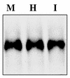
FIG. 1.
FIG. 1.
EMX2 is expressed in the adult reproductive tract. Persistent expression of Emx2/EMX2 is seen in adult mouse and human endometrium and in Ishikawa cells. Total RNA from estrus cycle day 1 adult mouse uteri or adult human proliferative-phase endometrium was analyzed by Northern blotting with a 3′-UTR EMX2 riboprobe. Northern blot results of representative samples are shown (M, mouse; H, human). Ishikawa (I) cells are a well-differentiated endometrial adenocarcinoma cell line (39). Expression of estrogen and progesterone receptors, as well as HOXA10, in this cell line has been previously well characterized (23, 24, 30, 54). Expression of EMX2 mRNA in this cell line is also shown.
FIG. 2.
FIG. 2.
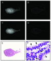
Localization of adult uterine Emx2 and Hoxa10 expression. Both Emx2 and Hoxa10 expression are evident throughout the uterus, with the highest expression in the endometrium. To localize Emx2 and Hoxa10 expression within the adult uterus, in situ hybridization was performed with 33P-labeled EMX2 and HOXA10 riboprobes, respectively. Representative photomicrographs of results are shown (×100 magnification). (A) Dark-field photomicrograph of mouse uterus hybridized to the EMX2 riboprobe. (B) Lack of hybridization to a labeled EMX2 sense probe. (C) Dark-field photomicrograph of mouse uterus hybridized to a HOXA10 riboprobe. (D) Lack of hybridization to a labeled HOXA10 sense probe. (E) Hematoxylin-eosin-stained section revealing the layers of a mouse uterus on a transverse section. The endometrium adjacent to the lumen is comprised of surface epithelium immediately lining the uterine cavity and of glands. The interglandular tissue comprises the stroma. The endometrium is surrounded by a few layers of circular smooth muscle (myometrium) and finally the external layer (serosa), comprised of loose connective tissue. (F) High-power photomicrograph (×400) showing results of in situ hybridization of human endometrium with silver grains over both the glandular epithelium and endometrial stroma. Arrow, endometrial stroma; arrowhead: endometrial glands.
FIG. 3.
FIG. 3.
EMX2 expression varies throughout the human reproductive cycle. EMX2 is expressed in the endometrium throughout the reproductive cycle. Endometrium from 32 subjects representing each developmental phase of the reproductive cycle was analyzed by Northern blotting and RT-PCR. (Top panel) Northern analysis was performed with a riboprobe complementary to the 3′-UTR of EMX2. Hybridization with a G3PDH probe was used as control. EMX2 mRNA expression rises significantly throughout the proliferative phase of the reproductive cycle (days 1 to 14 or 28) and peaks in the early secretory phase. This is rapidly followed by an ca. 50% decrease. Representative results of the Northern blot are shown. P1, P2, and P3 correspond to the early (days 1 to 5), middle (days 6 to 10), and late (days 9 to 14) proliferative phases, respectively. Similarly, S1, S2, and S3 correspond to the early (days 15 to 18), middle (days 19 to 23), and late (days 24 to 28) secretory phases, respectively, of the human reproductive cycle. (Bottom panel) Semiquantitative RT-PCR was performed simultaneously with primers that specifically amplify HOXA10 and EMX2. Amplification of G3PDH was performed as a control. EMX2 expression increases throughout the proliferative phase of the reproductive cycle (days 1 to 14 of 28) to peak in the late proliferative and early secretory phases. Expression declined by >50% (normalized to G3PDH) in the mid-secretory phase (S2), with continued decline in the late secretory phase (S3). Simultaneously, HOXA10 was expressed at low levels throughout the proliferative phase (P1, P2, and P3) and increased by the middle and late secretory phases (S2 and S3).
FIG. 4.
FIG. 4.
Alteration of HOXA10 expression affects EMX2 expression. (A) The sex steroids estrogen and progesterone do not directly regulate EMX2 expression. Ishikawa cells were pretreated with cycloheximide (to block the effects of sex steroids on HOXA10) and then treated with 5 × 10⁻⁸ M 17β-estradiol and/or 10−7 M progesterone. Control cells were treated with cycloheximide only. Analysis by Northern blotting with a 3′-UTR EMX2 riboprobe showed no significant change in EMX2 expression in response to sex steroids. The results were quantified by densitometry and normalized to G3PDH expression. (B) To evaluate the temporal effect of sex steroid treatment on HOXA10 and EMX2 expression, Northern analysis with 32P-labeled 3′-UTR EMX2 and HOXA10 riboprobes was performed in Ishikawa cells treated with 5 × 10⁻⁸ M 17β-estradiol and 10−7 M progesterone. RNA for Northern analysis was extracted at specified time points after sex steroid treatment over a 48-h period. G3PDH was used as a control. Expression of HOXA10 (normalized to G3PDH) is shown. Expression of HOXA10 increased from basal (0 h) levels by 30 min and continued to increase for 4 h. EMX2 mRNA expression declined to 30% of basal levels after 1 h, with a further decline by 2 h. The expression stabilized thereafter at 50% basal expression levels. EMX2 repression occurred subsequent to HOXA10 activationby sex steroids. (C) To further characterize the effect of HOXA10 on EMX2 in endometrial cells, Ishikawa cells were treated with constructs that alter HOXA10 expression and corresponding changes in EMX2 mRNA were determined by Northern blotting. HOXA10 mRNA and protein expression were determined by Northern and Western blotting, respectively, in transfectants treated with either HOXA10 antisense or the HOXA10 expression vector pcDNA3.1/HOXA10. The HOXA10 antisense construct was a 30-bp antisense phosphothiorate-modified oligodeoxynucleotide complementary to the HOXA10 translation start site. Corresponding results from controls treated with either a missense oligonucleotide (of the same length and nucleotide composition, but in random order) or with pcDNA3.1 were used for comparison. G3PDH or actin were used as loading controls in Northern or Western blots, respectively. Representative results of the Northern (panels 1 and 2) and Western (panels 3 and 4) blots are shown. Antisense treatment did not alter HOXA10 mRNA expression but decreased HOXA10 protein levels by ca. 50%. HOXA10 mRNA and protein levels were increased after transfection with pcDNA3.1/HOXA10, which constitutively expresses HOXA10 cDNA. (D) Treatment with HOXA10 antisense results in simultaneously increased EMX2 mRNA expression. In cells treated with pcDNA3.1/HOXA10, EMX2 mRNA levels correspondingly decrease. (E) In transfectants treated with HOXA10 antisense, EMX2 mRNA levels increase significantly 30 min after treatment and remain elevated at 72 h. (F) Results of densitometric analysis of EMX2 mRNA levels normalized to G3PDH expression over a time course of 72 h since treatment with HOXA10 antisense are shown. The asterisk indicates a statistically significant difference from the control of P ≤ 0.001 (as determined by analysis of variance). The results are an average of four experiments ± the standard error of the mean.
FIG. 5.
FIG. 5.
HOXA10 regulates EMX2 expression via two 150-bp negative regulatory elements. The effect of HOXA10 on EMX2 enhancer-luciferase reporter gene constructs (pGL3promoter/EMX2) was tested in BT-20 cells. (A) BT-20 cells are a breast adenocarcinoma cell line that we demonstrated by RT-PCR not to express HOXA10. (B) Schematic representation of the EMX2 gene regulatory region. Transcriptional suppression by HOXA10 was demonstrated in constructs containing the oligonucleotide termed EMXE (450 bp; nt −700 to −250). This region was further subdivided into oligonucleotides termed EMXA (150 bp; nt −250 to −400) and EMXD (300 bp; nt −250 to −550). EMXD was then divided to EMXB and EMXC (both 150 bp; nt −401 to −549 and −550 to −700, respectively). These oligonucleotides were tested in reporter constructs. The portion of the 150-bp EMXC sequence containing the consensus HOXA10 binding site is shown. (C) Overexpression of HOXA10 in BT-20 cells repressed reporter gene expression from artificial promoter constructs containing elements from the EMX2 5′ regulatory region. BT-20 cells were transfected with 4 μg of an artificial promoter construct that contained a single copy of an EMX2 regulatory element (EMXA-D), a minimal promoter and a luciferase reporter. Additionally, the cells were cotransfected with either 4 μg of a HOXA10 expression vector (pcDNA3.1/HOXA10) or control vector (pcDNA3.1) and 4 μg of pcDNA3.1/LacZ as a control for transfection efficiency. Luciferase and β-galactosidase activity were measured in the cellular lysate. Results are reported as luciferase activity normalized to β-galactosidase activity and each experiment was repeated at least three times. pGL3 promoter, cotransfected with either pcDNA3.1 or with pCDNA3.1/HOXA10 is the negative control. As a positive control we used a construct (pGL3/β3INTS) that, when cotransfected with HOXA10, has been previously demonstrated to drive high luciferase expression (15). Overexpression of HOXA10 did not cause transcriptional repression of the artificial reporter construct pGL3/EMXA, which contains the 150-bp EMXA region. Transcriptional repression of reporter gene expression by HOXA10 was seen with constructs pGL3promoter/EMXB, -C, -D, and E. pGL3/EMXB demonstrated a slight but significant reduction in luciferase activity when cotransfected with HOXA10. Overexpression of HOXA10 with pGL3promoter/EMXC reduced luciferase activity expression to ca. 50% of levels in the absence of HOXA10. Cotransfection of HOXA10 with either pGL3promoter/EMXD or pGL3promoter/EMXE resulted in an equivalent (ca. 65%) reduction in luciferase activity compared to cotransfection with pcDNA3.1. The asterisk indicates statistically significant differences fromthe control of P ≤ 0.001 for EMXB, P ≤ 0.004 for EMXC, and P ≤ 0.002 for EMXD and EMXE as determined by using the Mann-Whitney rank sum test. (D) In order to further characterize HOXA10-mediated transcriptional repression, BT-20 cells were cotransfected with pcDNA3.1/HOXA10 and the artificial promoter constructs pGL3control/5XEMXB and pGL3control/5XEMXC, containing five copies of either EMXB and EMXC, respectively, linked to a simian virus 40 promoter and enhancer expressing high basal luciferase activity. The luciferase activity was significantly repressed in transfectants treated with pcDNA3.1/HOXA10 compared to controls (treated with pcDNA3.1). Luciferase repression was significantly greater with pGL3/5XEMXC than with pGL3/5XEMXB (P < 0.0003). Each experiment was done in triplicate and was repeated three times. An asterisk indicates a statistically significant difference from the control of P ≤ 0.001 as determined by the Student t test.
FIG. 6.
FIG. 6.
EMX2 transcriptional repression by HOXA10 is mediated by HOXA10 binding to the 150-bp EMX2 regulatory element. To evaluate HOXA10 protein binding, Ishikawa nuclear extract, primary endometrial epithelial cell nuclear extract, or Flag-HOXA10 protein was used. Ishikawa cells were transfected with a Flag-pcDNA3.1/HOXA10 construct. Flag-HOXA10 protein was isolated and used. (A) EMSA of Flag-HOXA10 protein and 32P-labeled oligonucleotides EMXC encompassing the HOXA10 binding site TTAT or MUTC containing the mutated HOXA10 binding site GCAT. Lane 1, no protein; lane 2, Flag-HOXA10 protein and 32P-labeled EMXC demonstrate a shift; lane 3, Flag-HOXA10 protein and 32P-labeled EMX/MUTC demonstrate absent binding. (B) Flag-HOXA10 protein binding to 32P-labeled EMXC (lanes 1 to 5) is competed for by excess unlabeled competitor and supershifted with antibody directed against HOXA10. Lane 1, no protein; lane 2, Flag-HOXA10 protein; lane 3, a 160-fold molar excess of unlabeled oligonucleotide EMXC; lane 4, HOXA10 antibody, no protein; lane 5, Flag-HOXA10 protein and HOXA10 antibody. The arrow indicates the shift; an asterisk indicates a supershift, and “NS” indicates nonspecific binding. (C) EMSA of Ishikawa cell nuclear extract binding to 32P-labeled EMXC (lanes 1 to 6) is dose responsive. Lane 1, no nuclear extract; lanes 2 to 6, increasing concentrations of nuclear extract (0.1 to 5 μg). The arrow and arrowhead indicate two specific shifts, and “NS” indicates nonspecific binding. (D) EMSA of Ishikawa cell nuclear extract and 32P-labeled EMXC (lanes 1 to 5) further demonstrates that binding is competed for by excess unlabeled competitor and unaffected by unlabeled oligonucleotide EMXMUTC containing the mutated HOXA10 binding site. Lane 1, no nuclear extract; lane 2, Ishikawa cell nuclear extract; lanes 3 and 4, 160- and 320-fold molar excesses of unlabeled EMXC; lane 5, a 160-fold molar excess unlabeled oligonucleotide EMXMUTC. The arrow and arrowhead indicate two specific shifts, and “NS” indicates nonspecific binding. (E) EMSA of Ishikawa cell nuclear extract and 32P-labeled EMXC (lanes 1 to 5). Lane 1, two specific shifts with nuclear extract indicated by the arrow and arrowhead; lane 2, a 160-fold excess of unlabeled EMXC effectively competed away detectable protein-32P-labeled EMXC complexes; lane 3, complex resulting from Flag-HOXA10 binding to EMXC comigrates with the nuclear extract-32P-labeled EMXC complex (lane 1, indicated by arrow). In lane 4, nuclear extracts were supershifted with an antibody directed against HOXA10 (indicated by the asterisk). Lane 5, HOXA10 antibody with no added protein; lane 6, absence of supershift in lane containing nuclear extract and antibody to Pbx1. (F) EMSA of primary endometrial epithelial cell nuclear extract and 32P-labeled EMXC demonstrates binding of a single complex in a dose-responsive manner. Lane 1, no nuclear extract; lanes 2 to 6, increasing concentrations of nuclear extract (0.1 to 5 μg).
FIG. 6.
FIG. 6.
EMX2 transcriptional repression by HOXA10 is mediated by HOXA10 binding to the 150-bp EMX2 regulatory element. To evaluate HOXA10 protein binding, Ishikawa nuclear extract, primary endometrial epithelial cell nuclear extract, or Flag-HOXA10 protein was used. Ishikawa cells were transfected with a Flag-pcDNA3.1/HOXA10 construct. Flag-HOXA10 protein was isolated and used. (A) EMSA of Flag-HOXA10 protein and 32P-labeled oligonucleotides EMXC encompassing the HOXA10 binding site TTAT or MUTC containing the mutated HOXA10 binding site GCAT. Lane 1, no protein; lane 2, Flag-HOXA10 protein and 32P-labeled EMXC demonstrate a shift; lane 3, Flag-HOXA10 protein and 32P-labeled EMX/MUTC demonstrate absent binding. (B) Flag-HOXA10 protein binding to 32P-labeled EMXC (lanes 1 to 5) is competed for by excess unlabeled competitor and supershifted with antibody directed against HOXA10. Lane 1, no protein; lane 2, Flag-HOXA10 protein; lane 3, a 160-fold molar excess of unlabeled oligonucleotide EMXC; lane 4, HOXA10 antibody, no protein; lane 5, Flag-HOXA10 protein and HOXA10 antibody. The arrow indicates the shift; an asterisk indicates a supershift, and “NS” indicates nonspecific binding. (C) EMSA of Ishikawa cell nuclear extract binding to 32P-labeled EMXC (lanes 1 to 6) is dose responsive. Lane 1, no nuclear extract; lanes 2 to 6, increasing concentrations of nuclear extract (0.1 to 5 μg). The arrow and arrowhead indicate two specific shifts, and “NS” indicates nonspecific binding. (D) EMSA of Ishikawa cell nuclear extract and 32P-labeled EMXC (lanes 1 to 5) further demonstrates that binding is competed for by excess unlabeled competitor and unaffected by unlabeled oligonucleotide EMXMUTC containing the mutated HOXA10 binding site. Lane 1, no nuclear extract; lane 2, Ishikawa cell nuclear extract; lanes 3 and 4, 160- and 320-fold molar excesses of unlabeled EMXC; lane 5, a 160-fold molar excess unlabeled oligonucleotide EMXMUTC. The arrow and arrowhead indicate two specific shifts, and “NS” indicates nonspecific binding. (E) EMSA of Ishikawa cell nuclear extract and 32P-labeled EMXC (lanes 1 to 5). Lane 1, two specific shifts with nuclear extract indicated by the arrow and arrowhead; lane 2, a 160-fold excess of unlabeled EMXC effectively competed away detectable protein-32P-labeled EMXC complexes; lane 3, complex resulting from Flag-HOXA10 binding to EMXC comigrates with the nuclear extract-32P-labeled EMXC complex (lane 1, indicated by arrow). In lane 4, nuclear extracts were supershifted with an antibody directed against HOXA10 (indicated by the asterisk). Lane 5, HOXA10 antibody with no added protein; lane 6, absence of supershift in lane containing nuclear extract and antibody to Pbx1. (F) EMSA of primary endometrial epithelial cell nuclear extract and 32P-labeled EMXC demonstrates binding of a single complex in a dose-responsive manner. Lane 1, no nuclear extract; lanes 2 to 6, increasing concentrations of nuclear extract (0.1 to 5 μg).
FIG. 7.
FIG. 7.
HOXA10 binding is localized to two HOXA10 binding sites located within EMXC and EMXB. By using DNase I footprinting analysis, HOXA10 transcription factor binding within the 150-bp regions EMXC (A) and EMXB (B) was localized to a 10-bp region (EMXC) or a 6-bp region (EMXB), respectively. The EMXC footprint contained one HOXA10 consensus site. The EMXB footprint demonstrates weak protection of a nonconsensus site. The solid bar on the right indicates the extent of the footprint. No Flag-HOXA10 protein was added to lanes labeled “0.” The triangle above indicates serially increasing Flag-HOXA10 protein concentrations. The oligonucleotide sequences containing the nucleotides protected by DNase I footprinting are displayed. The consensus HOXA10 binding site in EMXC is underlined.
FIG. 8.
FIG. 8.
Mutation of the HOXA10 binding site results in loss of negative regulation. Mutation of the HOXA10 binding site in the EMX2 regulatory region resulted in loss of HOXA10-mediated transcriptional repression. BT-20 cells were cotransfected as described earlier (Fig. 5) with either 4 μg of pcDNA3.1/HOXA10 or pcDNA3.1 and with 4 μg of reporter constructs containing mutated EMX2 elements (pGL3/MUTC and pGL3/MUTD). In vitro mutagenesis was used to alter the HOXA10 binding site from TTAT to GCAT. Results demonstrating the relative luciferase activity normalized to the β-galactosidase activity are shown. The effect of HOXA10 on the corresponding nonmutated elements pGL3/EMXC and -D described earlier (Fig. 5) are shown here for comparison only. Each experiment was repeated at least three times.

Similar articles

Cited by

References

    1. Akam, M. 1989. Hox and HOM: homologous gene clusters in insects and vertebrates. Cell 57:347-349. - PubMed
    1. Asahara, H., S. Dutta, H. Y. Kao, R. M. Evans, and M. Montminy. 1999. Pbx-Hox heterodimers recruit coactivator-corepressor complexes in an isoform-specific manner. Mol. Cell. Biol. 19:8219-8225. - PMC - PubMed
    1. Awgulewitsch, A., and D. Jacobs. 1992. Deformed autoregulatory element from Drosophila functions in a conserved manner in transgenic mice. Nature 358:341-344. - PubMed
    1. Bagot, C. N., H. J. Kliman, and H. S. Taylor. 2001. Maternal Hoxa10 is required for pinopod formation in the development of mouse uterine receptivity to embryo implantation. Dev. Dyn. 222:538-544. - PubMed
    1. Bagot, C. N., P. J. Troy, and H. S. Taylor. 2000. Alteration of maternal Hoxa10 expression by in vivo gene transfection affects implantation. Gene Ther. 7:1378-1384. - PubMed

Publication types

MeSH terms

LinkOut - more resources