. 2003 Jan;23(1):38-54.
doi: 10.1128/MCB.23.1.38-54.2003.

The mammalian SIR2alpha protein has a role in embryogenesis and gametogenesis

Affiliations

The mammalian SIR2alpha protein has a role in embryogenesis and gametogenesis

Michael W McBurney et al. Mol Cell Biol. 2003 Jan.

Abstract

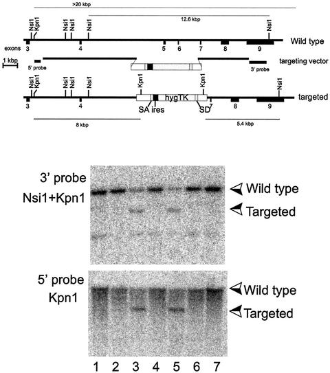
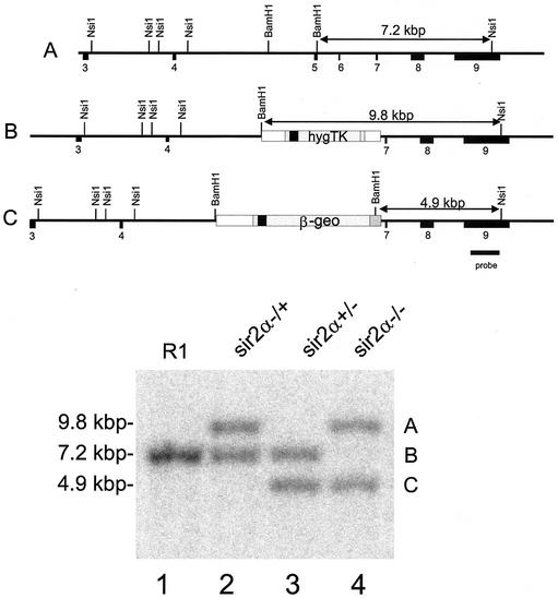
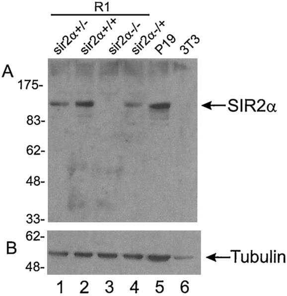
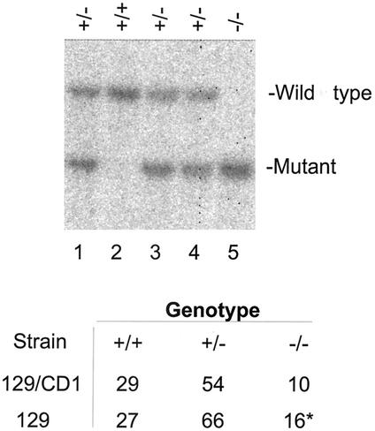
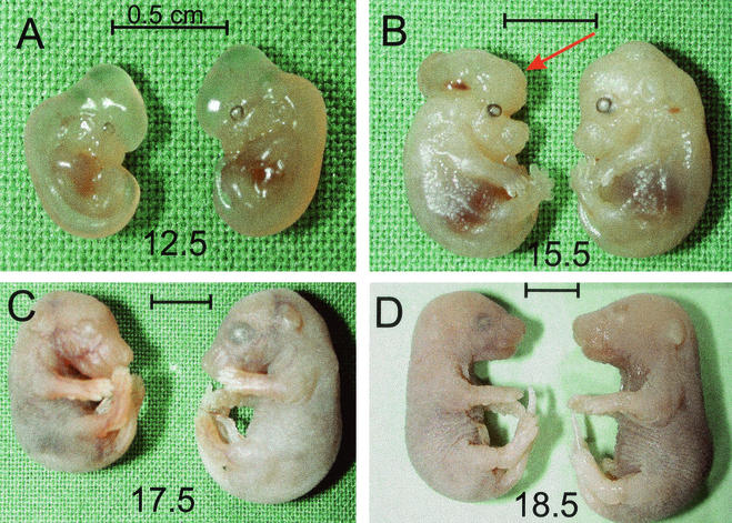
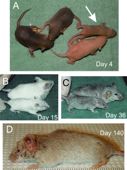
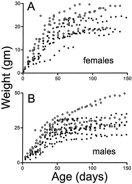
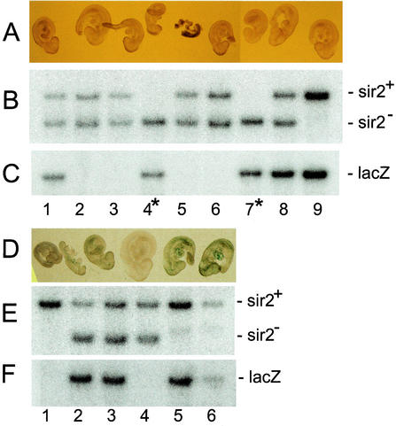
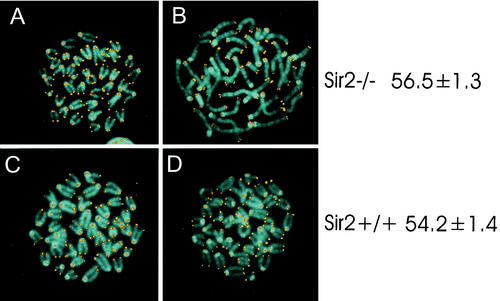
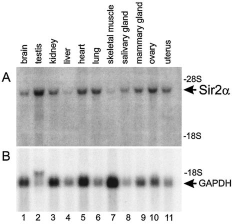
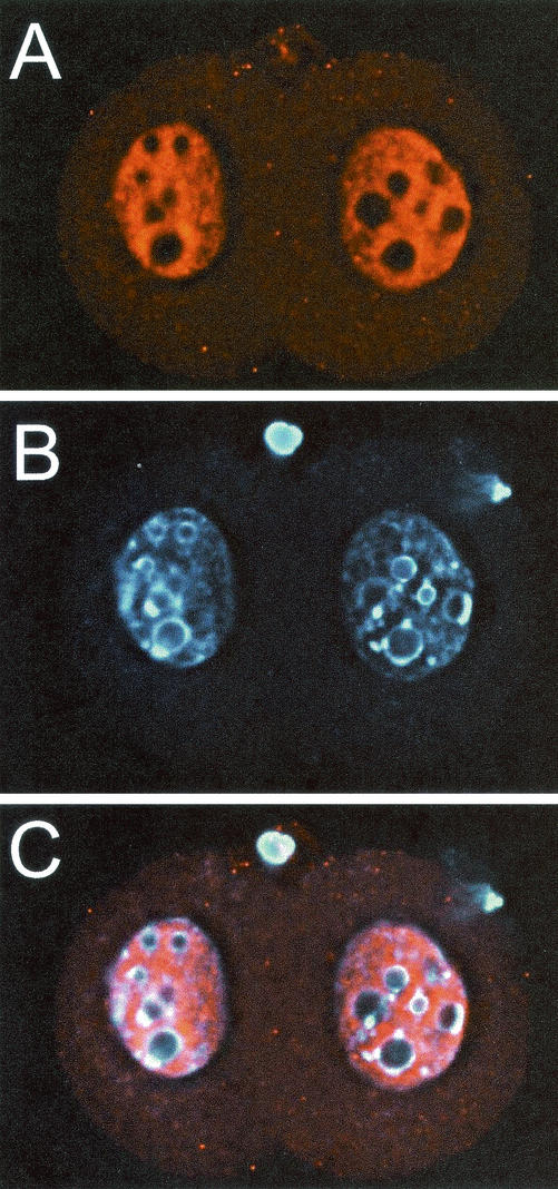
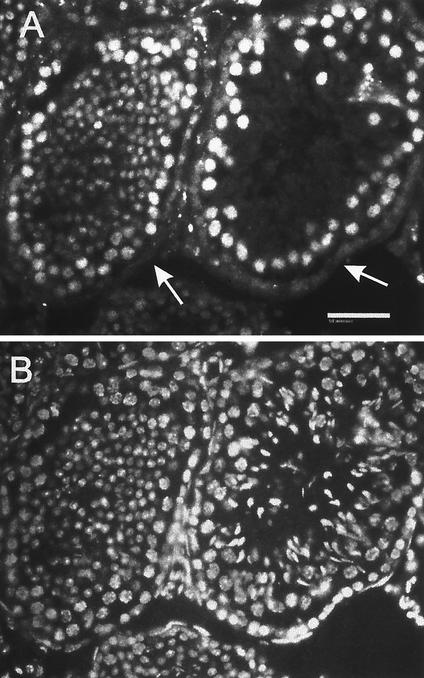
The yeast Sir2p protein has an essential role in maintaining telomeric and mating type genes in their transcriptionally inactive state. Mammalian cells have a very large proportion of their genome inactive and also contain seven genes that have regions of homology with the yeast sir2 gene. One of these mammalian genes, sir2alpha, is the presumptive mammalian homologue of the yeast sir2 gene. We set out to determine if sir2alpha plays a role in mammalian gene silencing by creating a strain of mice carrying a null allele of sir2alpha. Animals carrying two null alleles of sir2alpha were smaller than normal at birth, and most died during the early postnatal period. In an outbred background, the sir2alpha null animals often survived to adulthood, but both sexes were sterile. We found no evidence for failure of gene silencing in sir2alpha null animals, suggesting that either SIR2alpha has a different role in mammals than it does in Saccharomyces cerevisiae or that its role in gene silencing in confined to a small subset of mammalian genes. The phenotype of the sir2alpha null animals suggests that the SIR2alpha protein is essential for normal embryogenesis and for normal reproduction in both sexes.


Figures

FIG. 1.
FIG. 1.
Homologous recombination into the sir2α locus. The sequence for sir2α mRNA was inferred from the expressed sequence tag database, and reverse transcription-PCR was used to amplify and clone a cDNA corresponding to exons 3 to 9. This cDNA sequence was confirmed and used to isolate two overlapping genomic clones derived from strain 129/Sv mice. DNA sequencing was used to locate the exons, as shown in the upper panel. The targeting vector was constructed as shown, where the selectable sequence replaces exons 5 and 6, which encode highly conserved regions of the catalytic domain of the SIR2α protein. The selectable sequence consists of splice acceptor (SA) and splice donor (SD) sequences derived from exons 3 and 5 of the mouse Pgk-1 gene (3) surrounding the poliovirus internal ribosome entry site (ires) (35) and the coding region for a gene fused from the hygromycin and herpes simplex virus thymidine kinase (TK) genes (30). The targeting vector was linearized and electroporated into the R1 line of ES cells (36), which were subsequently selected for hygromycin resistance. Individual clones were isolated and expanded, and their DNA was analyzed by Southern blotting following digestion with the enzymes shown in the two lower panels. The two clones shown in lanes 3 and 5 had patterns of hybridizing bands that are consistent with homologous recombination having occurred at both the 5′ and 3′ ends of the targeting vector.
FIG. 2.
FIG. 2.
Isolation of ES clones deleted for both sir2α genes. R1 ES cells (lane 1) that had homologously integrated the hygromycin resistance vector (lane 2) were electroporated with a second knockout construct carrying the lacZ-neomycin resistance (β-geo) gene (35), and the cells were selected for resistance to G418. Among the clones isolated, some, like that shown in lane 3, had the β-geo sequence in place of the hygromycin resistance-thymidine kinase sequence, while others, like that shown in lane 4, had both the β-geo and the hygromycin resistance-thymidine kinase sequences homologously integrated into the two sir2α genes. The Southern blot shown in the lower panel was derived from DNA digested with NsiI plus BamHI and probed with the sequence shown, derived from DNA outside the region used for the knockout vectors.
FIG. 3.
FIG. 3.
SIR2α protein is absent from sir2α−/− ES cells. Protein was isolated from the cells indicated and run on sodium dodecyl sulfate-polyacrylamide gel electrophoresis. The proteins were electrophoretically transferred to membranes, where they were probed with antibody to SIR2α (panel A) and to tubulin (panel B).
FIG. 4.
FIG. 4.
Viable sir2α null offspring arise from crosses between heterozygous animals. Males and females that were genotyped as heterozygous for the mutated sir2α alleles were mated, and the DNA from their offspring was used for Southern blots to allow genotyping of individual animals. The DNA was digested with NsiI and KpnI, and the blots were probed with the 3′ probe shown in Fig. 1. In the litter shown in the upper panel, the three expected genotypes were present; lane 2 has two wild-type sir2α alleles, lane 5 has two mutant alleles, and lanes 1, 3, and 4 are heterozygous. The table shows the numbers of offspring of each genotype from these crosses of heterozygous parents. The strain labeled 129/CD1 is outbred and has a mixed genotype, while the strain labeled 129 includes animals in which the targeted sir2α allele was maintained on the 129/Sv background.
FIG. 5.
FIG. 5.
sir2α null animals are not lost during fetal development but are smaller and occasionally have exencephaly. Animals heterozygous for the mutant sir2α allele were mated, and fetuses were harvested at daily intervals after embryonic day 9.5. The fetal membranes were used to isolate DNA for genotyping, and the corresponding embryo was fixed. The genotypes were present in the expected ratios, and there was no evident downward trend in the proportion of the sir2α null genotype. The sir2α null embryos were usually smaller than their littermates. This size difference was evident as early as embryonic day 12.5. Two of the sir2α null embryos had exencephaly, as indicated with the arrow in panel B. Bars, 0.5 cm.
FIG. 6.
FIG. 6.
Postnatal sir2α null animals are developmentally abnormal. sir2α null animals on the outbred background often survived past weaning but were characterized by a number of common developmental defects. Null animals were smaller than their littermates (both wild type and heterozygotes), and all null animals failed to open one or both eyelids. The snout of sir2α null animals was often shorter than normal. The photographs show sir2α null animals of various ages along with their normal littermates.
FIG. 7.
FIG. 7.
Some sir2α null animals thrive on the outbred genetic background. sir2α null animals (•) and their wild-type littermates (○) were weighed at weekly intervals. During the early postnatal development period, sir2α null animals were uniformly smaller than their littermates, but many of the null animals gained weight and eventually reached sizes commensurate with those of their littermates.
FIG. 8.
FIG. 8.
sir2α null embryos do not reactivate expression of silent transgenes. Female mice carrying the sir2α null mutation were intercrossed with male mice carrying the Pgk-1,2-lacZ transgene, and animals heterozygous for the sir2α null mutation were mated so that the transgene was inherited from either the male parent, where it remains active, or the female parent, where it becomes inactivated (26). Embryos were harvested at day 9.5 of gestation, the membranes were used to isolate DNA for genotyping, and the fetus was fixed and stained with X-Gal. The upper three panels show a litter in which the Pgk-1,2-lacZ transgene was inherited through the female germ line, during which it is inactivated. Embryos 1, 8, and 9 inherited the transgene along with at least one wild-type sir2α allele. As expected, none was stained with X-Gal, indicating that the transgene had been inactivated. Embryos 4 and 7 inherited the transgene along with two sir2α null alleles. Neither embryo was stained with X-Gal, indicating that the absence of SIR2α in these embryos did not reactivate expression from the silenced transgene. The embryos in the lower three panels were derived from a cross in which the Pgk-1,2-lacZ transgene was inherited from the male parent. In this case, the transgene remained active, and the fetuses that inherited the transgene (lanes 2, 3, 4, and 6) were stained with X-Gal.
FIG. 9.
FIG. 9.
Telomeres do not erode prematurely in sir2α null mice. Splenocytes from sir2α null (panels A and B) and wild-type (panels C and D) mice were stimulated with concanavalin A (panels A and C) or lipopolysaccharide (panels B and D) to activate T and B lymphocytes, respectively. Chromosome spreads were stained for telomeres by fluorescence in situ hybridization with a peptide nucleic acid telomere probe (53). DNA was counterstained with 4′,6′-diamidino-2-phenylindole. Quantitation of telomere content per cell was done by flow cytometry. The values on the right represent total telomere lengths in kilobase pairs per cell along with the standard error.
FIG. 10.
FIG. 10.
SIR2α expression is widespread throughout adult tissues. RNA was isolated from a variety of tissues from wild-type mice and subjected to electrophoresis, blotting, and hybridization to cDNA probes specific for sir2α (upper panel) and glyceraldehyde-3-phosphate dehydrogenase (GAPDH, lower panel). The sir2α transcript was detected in all tissues examined and was particularly abundant in the testis and ovary.
FIG. 11.
FIG. 11.
SIR2α protein is expressed at high level in two-cell embryos. Wild-type two-cell embryos were recovered from the oviducts 24 h following mating. The zona pellucida was removed, and the embryos were fixed and stained with antibody to SIR2α (panel A) and with Hoechst 33258 (panel B). Vertical stacks of images were captured and subsequently deconvolved. Panel C shows the merged image. Note that the SIR2α protein is exclusively nuclear and that it appears to be excluded from regions of heterchromatin, as identified by intense DNA stain. SIR2α appears to have been excluded from the two polar bodies.
FIG. 12.
FIG. 12.
SIR2α is present at high level in proliferating granulosa cells. Ovaries from wild-type mice were sectioned and stained with antibody to SIR2α (panel A) and with Hoechst 33258 (panel B). The merged image is shown in panel C. The box in panel C was used to deconvolve the image, and the result is shown in panels D to F. Note that the SIR2α protein is nuclear but appears to be excluded from the nucleolus and those regions of the nucleus that stain intensely with Hoechst dye.
FIG. 13.
FIG. 13.
SIR2α is expressed at high level during spermatogenesis. Frozen sections of testis from wild-type mice were stained with polyclonal antibody to SIR2α (panel A) and with Hoechst 33258 to stain DNA (panel B). In the two seminiferous tubules shown, it is evident that the somatic cells of the tubules do not stain for SIR2α (arrows in panel A), while the immature spermatogenic cells stain to various extents before losing SIR2α expression in the elongate spermatocyte stage (panel A, right tubule). Bar, 50 μm.
FIG. 14.
FIG. 14.
sir2α null mice have abnormal sperm morphology. Spermatozoa from the cauda epididymis were spread on a slide and stained with Dif-Quick stain. (a) Wild-type spermatozoa have a consistent nuclear shape with a characteristic hook. In contrast, the spermatozoa of sir2α null mice (b) had small, rounded, or smudged nuclear shapes. Some had a blunted (arrowhead) or absent hook. The flagella were also variable in shape, and many were no longer attached to sperm heads. Bar, 20 μm.
FIG. 15.
FIG. 15.
DNA content distribution of testes from sir2α null mice indicates that all stages of spermatogenesis are present. Single-cell suspensions were created from the testes of 7-month-old wild-type (upper panel) and sir2α null (lower panel) animals. Cells were fixed and stained with 4′,6′-diamidino-2-phenylindole before analysis by flow cytometry for DNA content per cell. The peaks corresponding to 1n, 2n, and 4n genomes are shown. Mature sperm stained less efficiently than expected and formed the broad peak at the left of the pattern. Replicating cells have a DNA content between 2n and 4n.
FIG. 16.
FIG. 16.
Spermatogenesis is abnormal in sir2α null mice. Testes from both wild-type (a) and sir2 null (b to e) mice were examined for histopathological abnormalities. At low power, the seminiferous tubules in the wild-type mouse appeared almost uniform in size, while those of the sir2α null mice varied in size and contained large vacuoles in the basal epithelium (arrows). At higher power, significant abnormalities were seen in the testes from sir2α null mice: acrosomal sharing in round spermatids (arrow, c); distorted residual bodies (arrowhead, c); larger-than-normal round spermatids (arrows, d); abnormal retained elongated spermatids (c, asterisks); and numerous vacuolated spaces (asterisks, e). Sections were stained with periodic acid Schiff-hematoxylin. Bars: 100 μm (a and b); 50 μm (c to e).
FIG. 17.
FIG. 17.
Increased numbers of apoptotic germ cells are present in the seminiferous tubules of sir2α null mice. Testis cross sections from wild-type (a) and sir2α null (b) mice were analyzed by the terminal deoxynucleotidyltransferase-mediated dUTP-biotin nick-end labeling (TUNEL) assay to determine the frequency of apoptotic cells. Following exposure to 3 Gy of X-irradiation, the number of TUNEL-positive cells was increased (b and d) in both wild-type and sir2α null mice. The graph at the bottom shows the percentage of seminiferous tubule cross sections with one to three (grey portion of each bar) and more than three (black portion of bar) TUNEL-positive cells. Significantly more apoptotic cells were present in the sir2α null testes than in the wild-type testes (P < 0.05 by Student's unpaired t test).
FIG. 17.
FIG. 17.
Increased numbers of apoptotic germ cells are present in the seminiferous tubules of sir2α null mice. Testis cross sections from wild-type (a) and sir2α null (b) mice were analyzed by the terminal deoxynucleotidyltransferase-mediated dUTP-biotin nick-end labeling (TUNEL) assay to determine the frequency of apoptotic cells. Following exposure to 3 Gy of X-irradiation, the number of TUNEL-positive cells was increased (b and d) in both wild-type and sir2α null mice. The graph at the bottom shows the percentage of seminiferous tubule cross sections with one to three (grey portion of each bar) and more than three (black portion of bar) TUNEL-positive cells. Significantly more apoptotic cells were present in the sir2α null testes than in the wild-type testes (P < 0.05 by Student's unpaired t test).

References

    1. Accili, D., J. Drago, E. J. Lee, M. D. Johnson, M. H. Cool, P. Salvatore, L. D. Asico, P. A. Jose, S. I. Taylor, and H. Westphal. 1996. Early neonatal death in mice homozygous for a null allele of the insulin receptor gene. Nat. Genet. 12:106-109. - PubMed
    1. Adra, C. N., P. H. Boer, and M. W. McBurney. 1987. Cloning and expression of the mouse pgk-1 gene and the nucleotide sequence of its promoter. Gene 60:65-74. - PubMed
    1. Adra, C. N., N. A. Ellis, and M. W. McBurney. 1988. The family of mouse phosphoglycerate kinase genes and pseudogenes. Somat. Cell Mol. Genet. 14:69-81. - PubMed
    1. Allemand, I., A. Anglo, A. Y. Jeantet, I. Cerutti, and E. May. 1999. Testicular wild-type p53 expression in transgenic mice induces spermiogenesis alterations ranging from differentiation defects to apoptosis. Oncogene 18:6521-6530. - PubMed
    1. Baker, J., J. P. Liu, E. J. Robertson, and A. Efstratiadis. 1993. Role of insulin-like growth factors in embryonic and postnatal growth. Cell 75:73-82. - PubMed

Publication types

MeSH terms