. 2003 Jan;23(1):131-9.
doi: 10.1128/MCB.23.1.131-139.2003.

The TAK1-NLK mitogen-activated protein kinase cascade functions in the Wnt-5a/Ca(2+) pathway to antagonize Wnt/beta-catenin signaling

Affiliations

The TAK1-NLK mitogen-activated protein kinase cascade functions in the Wnt-5a/Ca(2+) pathway to antagonize Wnt/beta-catenin signaling

Tohru Ishitani et al. Mol Cell Biol. 2003 Jan.

Abstract

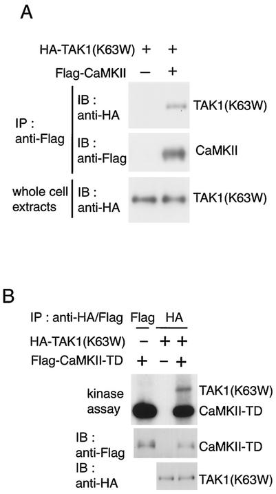
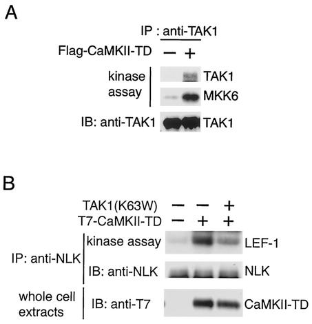
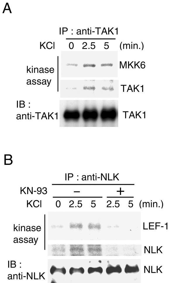
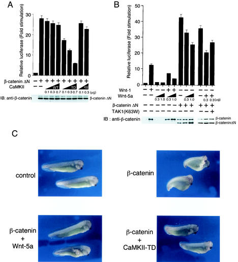
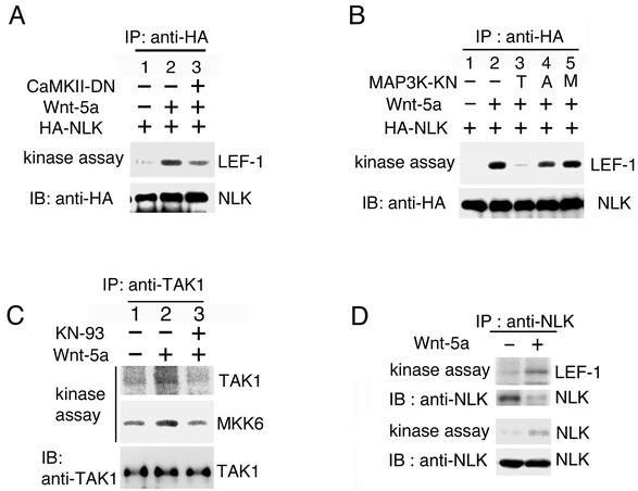
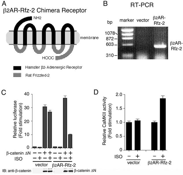
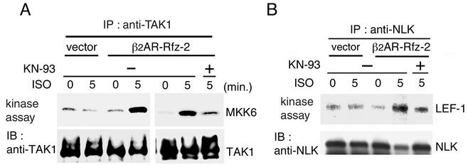
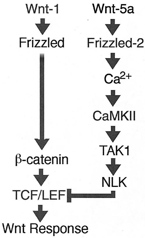
Wnt signaling controls a variety of developmental processes. The canonical Wnt/beta-catenin pathway functions to stabilize beta-catenin, and the noncanonical Wnt/Ca(2+) pathway activates Ca(2+)/calmodulin-dependent protein kinase II (CaMKII). In addition, the Wnt/Ca(2+) pathway activated by Wnt-5a antagonizes the Wnt/beta-catenin pathway via an unknown mechanism. The mitogen-activated protein kinase (MAPK) pathway composed of TAK1 MAPK kinase kinase and NLK MAPK also negatively regulates the canonical Wnt/beta-catenin signaling pathway. Here we show that activation of CaMKII induces stimulation of the TAK1-NLK pathway. Overexpression of Wnt-5a in HEK293 cells activates NLK through TAK1. Furthermore, by using a chimeric receptor (beta(2)AR-Rfz-2) containing the ligand-binding and transmembrane segments from the beta(2)-adrenergic receptor (beta(2)AR) and the cytoplasmic domains from rat Frizzled-2 (Rfz-2), stimulation with the beta-adrenergic agonist isoproterenol activates activities of endogenous CaMKII, TAK1, and NLK and inhibits beta-catenin-induced transcriptional activation. These results suggest that the TAK1-NLK MAPK cascade is activated by the noncanonical Wnt-5a/Ca(2+) pathway and antagonizes canonical Wnt/beta-catenin signaling.


Figures

FIG. 1.
FIG. 1.
CaMKII interacts with and phosphorylates TAK1. (A) Interaction between CaMKII and TAK1. 293 cells were transfected with Flag-CaMKII and HA-TAK1(K63W) as indicated. Cell extracts were immunoprecipitated (IP) with anti-Flag antibody. The immunoprecipitates were immunoblotted with anti-HA (top panel) and anti-Flag (middle panel) antibodies. Whole-cell extracts were immunoblotted (IB) with anti-HA antibody (bottom panel). (B) Phosphorylation of TAK1 by CaMKII. 293 cells were transfected with Flag-CaMKII(T286D) (TD) and HA-TAK1(K63W) as indicated. Immunoprecipitated complexes with anti-Flag or anti-HA antibody were incubated with [γ-32P]ATP and analyzed by autoradiography (top panel). The immunoprecipitates were immunoblotted with anti-Flag (middle panel) and anti-HA (bottom panel) antibodies.
FIG. 2.
FIG. 2.
Activation of TAK1 and NLK by CaMKII. (A) Activation of endogenous TAK1. 293 cells were transfected with Flag-CaMKII(T286D) (TD) as indicated. Endogenous TAK1 was immunoprecipitated (IP) with anti-TAK1 antibody. The immunoprecipitates were subjected to an in vitro phosphorylation assay by autophosphorylation of TAK1 (top panel) and bacterially expressed MKK6 as an exogenous substrate (middle panel). The immunoprecipitates were analyzed by immunoblotting (IB) with anti-TAK1 antibody (bottom panel). (B) Activation of endogenous NLK by CaMKII. 293 cells were transfected with expression plasmids encoding T7-CaMKII(T286D) (TD) and TAK1(K63W) as indicated. Endogenous NLK was immunoprecipitated with anti-NLK antibody. The immunoprecipitates were subjected to an in vitro phosphorylation assay with bacterially expressed LEF-1 as an exogenous substrate (top panel). The immunoprecipitates were analyzed by immunoblotting with anti-NLK antibody (middle panel). Whole-cell extracts were immunoblotted with anti-T7 antibody (bottom panel).
FIG. 3.
FIG. 3.
Calcium-induced activation of TAK1 and NLK. (A) Calcium-induced activation of TAK1. PC12 cells were treated with 75 mM KCl for the indicated periods. Cell extracts were immunoprecipitated (IP) with anti-TAK1 antibody. The immunoprecipitates were subjected to an in vitro phosphorylation assay with bacterially expressed MKK6 as an exogenous substrate (top panel) and autophosphorylation of TAK1 (middle panel). The immunoprecipitates were analyzed by immunoblotting (IB) with anti-TAK1 antibody (bottom panel). (B) Calcium-induced activation of NLK. PC12 cells pretreated with or without 20 μM KN-93 for 20 min were treated with 75 mM KCl for the indicated periods. Cell extracts were immunoprecipitated with anti-NLK antibody. The immunoprecipitates were subjected to an in vitro phosphorylation assay with bacterially expressed LEF-1 as an exogenous substrate (top panel) and autophosphorylation of NLK (middle panel). The immunoprecipitates were analyzed by immunoblotting with anti-NLK antibody (bottom panel).
FIG. 4.
FIG. 4.
Effects of CaMKII and Wnt-5A on Wnt/β-catenin signaling. (A) Effects of CaMKII on TCF-dependent reporter gene activity. 293 cells were transfected with luciferase reporter plasmid (0.1 μg), β-cateninΔN expression plasmid (0.5 μg), and the indicated amounts of plasmids encoding CaMKII (wild type [WT]), CaMKII(T286D) (TD), and CaMKII(K24 M) (KM). After 24 h of incubation, cells were harvested, and luciferase activity was measured. The values shown are the average of one representative experiment in which each transfection was performed in duplicate. Equal amounts of cell lysates were immunoblotted (IB) with anti-β-catenin antibody. (B) Effects of Wnt-1 and Wnt-5a on β-catenin stabilization. 293 cells were transfected with luciferase reporter (0.1 μg), β-cateninΔN (0.5 μg), Wnt-1 expression plasmid (0.5 μg), TAK1(K63W) (0.2 μg), and the indicated amounts of Wnt-5a expression plasmid. Cell lysates were used for measuring luciferase activity. Equal amounts of cell lysates were immunoblotted with anti-β-catenin antibody. (C) Effect of Wnt-5a and CaMKII on β-catenin-induced axis formation in Xenopus embryos. β-Catenin mRNA (100 pg) was injected into two ventral blastomeres at the four-cell stage with Wnt-5a or CaMKII(T286D) (TD) mRNA (5 pg) as indicated. Embryos were examined for axial duplications at the tadpole stage. Injection of β-catenin led to secondary axis formation with head in 52% of the injected embryos (n = 50), injection of β-catenin plus Wnt-5a had the same result in 0% of the embryos (n = 50), and injection with β-catenin plus CaMKII-TD had the same result in 14% of the embryos (n = 50).
FIG. 5.
FIG. 5.
Activation of TAK1 and NLK by Wnt-5a. (A and B) Activation of NLK by Wnt-5a is dependent on CaMKII and TAK1. 293 cells were transfected with the indicated expression plasmids. HA-NLK was immunoprecipitated (IP) with anti-HA antibody. The immunocomplexes were used for in vitro kinase assays with bacterially expressed LEF-1 as an exogenous substrate (upper panels) and immunoblotted (IB) with anti-HA antibody (lower panels). CaMKII-DN, CaMKII(K42 M) (1-271); T, TAK1(K63W); A, ASK1(K709 M); M, MTK1(K1371R). (C) Activation of endogenous TAK1 by Wnt-5a. 293 cells were transfected with Wnt-5a as indicated. Cells were treated with or without KN-93 (20 μM). Endogenous TAK1 was immunoprecipitated with anti-TAK1 antibody. The immunoprecipitates were subjected to an in vitro phosphorylation assay with autophosphorylation of TAK1 (top panel) and bacterially expressed MKK6 as an exogenous substrate (middle panel). The immunoprecipitates were analyzed by immunoblotting with anti-TAK1 antibody (bottom panel). (D) Activation of endogenous NLK by Wnt-5a. 293 cells were transfected with Wnt-5a as indicated. Endogenous NLK was immunoprecipitated with anti-NLK antibody. The immunoprecipitates were subjected to an in vitro phosphorylation assay with LEF-1 as a substrate (top panel) and autophosphorylation of NLK (third panel). The immunoprecipitates were analyzed by immunoblotting with anti-NLK antibody (second and bottom panels).
FIG. 6.
FIG. 6.
Activation of CaMKII by inducible Rfz-2. (A) Schematic representation of the β2AR-Rfz-2 construct. (B) RT-PCR of β2AR-Rfz-2 stably expressed in 293 cells. The RNA of 293 cells harboring the empty expression vector or the vector expressing β2AR-Rfz-2 chimera was reverse transcribed and amplified. The molecular markers indicate the relative size in base pairs of the amplified products. (C) Effect of β2AR-Rfz-2 on TCF-dependent reporter gene activity. Cells of 293-β2AR-Rfz-2 and 293 stably transfected with control vector were transfected with luciferase reporter plasmid (0.1 μg) and β-cateninΔN expression plasmid (0.5 μg) as indicated. Cells were treated with or without the β-adrenergic agonist ISO (100 μM), and luciferase activity was measured. The values shown are the average of one representative experiment in which each transfection was performed in duplicate. Equal amounts of cell lysates were immunoblotted (IB) with anti-β-catenin antibody. (D) Activation of endogenous CaMKII by β2AR-Rfz-2. Cells of 293-β2AR-Rfz-2 cells and 293 cells stably transfected with control vector were treated with or without ISO (50 μM) for 5 min. Cell extracts were assayed for CaMKII activity. The values shown are the average of one representative experiment in which each transfection was performed in duplicate.
FIG. 7.
FIG. 7.
Activation of TAK1 and NLK by inducible Rfz-2. Cells of 293-β2AR-Rfz-2 and 293 stably transfected with control vector were pretreated with or without KN-93 (50 μM) for 50 min. Then they were treated with ISO (50 μM) for the indicated time periods. Endogenous TAK1 (A) and NLK (B) were immunoprecipitated (IP) with anti-TAK1 and anti-NLK antibodies, respectively. The immunocomplexes were used for in vitro kinase assays (upper panels). The amounts of immunoprecipitated TAK1 and NLK were determined by immunoblotting (IB) with anti-TAK1 and anti-NLK antibodies, respectively (lower panels).
FIG. 8.
FIG. 8.
Model for interaction between the Wnt/β-catenin and Wnt/Ca2+ pathways. (See the text for details.)

Similar articles

Cited by

References

    1. Aberle, H., A. Bauer, J. Stappert, A. Kispert, and R. Kemler. 1997. β-Catenin is a target for the ubiquitin-proteasome pathway. EMBO J. 16:3797-3804. - PMC - PubMed
    1. Behrens, J., J. P. von Kries, M. Kuhl, L. Bruhn, D. Wedlich, R. Grosschedl, and W. Birchmeier. 1996. Functional interaction of β-catenin with the transcription factor LEF-1. Nature 382:638-642. - PubMed
    1. Cardigan, K. M., and R. Nusse. 1997. Wnt signaling: a common theme in animal development. Genes Dev. 11:3286-3305. - PubMed
    1. Du, S. J., S. M. Purcell, J. L. Christian, L. L. McGrew, and R. T. Moon. 1995. Identification of distinct classes and functional domains of Wnts through expression of wild-type and chimeric proteins in Xenopus embryos. Mol. Cell. Biol. 15:2625-2634. - PMC - PubMed
    1. Funayama, N., F. Fagotto, P. McCrea, and B. M. Gumbiner. 1995. Embryonic axis induction by the armadillo repeat domain of β-catenin: evidence for intracellular signaling. J. Cell Biol. 128:959-968. - PMC - PubMed

Publication types

MeSH terms

Substances