. 2003 Jan;23(1):140-9.
doi: 10.1128/MCB.23.1.140-149.2003.

Activating signal cointegrator 2 belongs to a novel steady-state complex that contains a subset of trithorax group proteins

Affiliations

Activating signal cointegrator 2 belongs to a novel steady-state complex that contains a subset of trithorax group proteins

Young-Hwa Goo et al. Mol Cell Biol. 2003 Jan.

Abstract

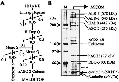
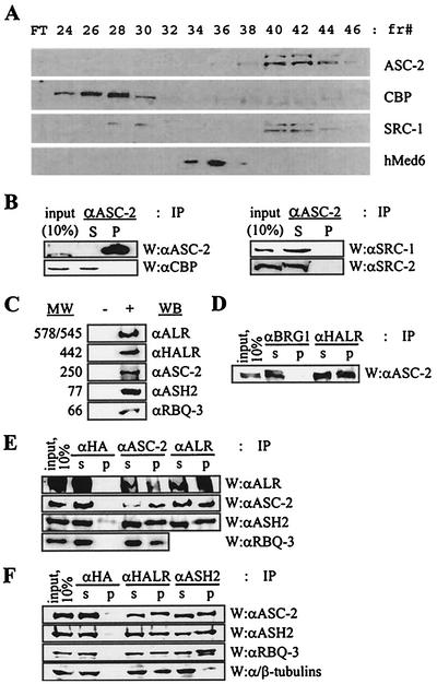
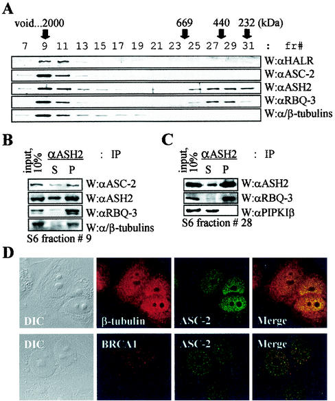
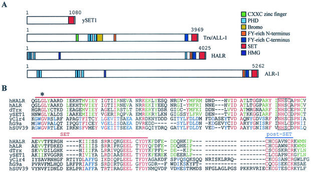
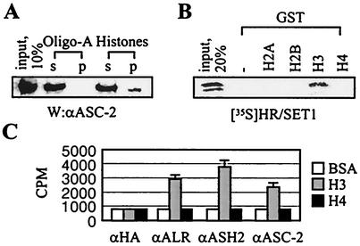
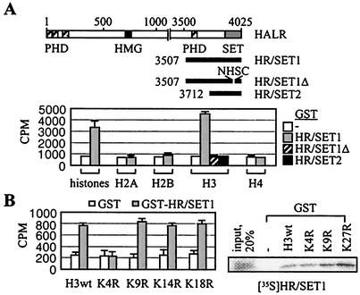
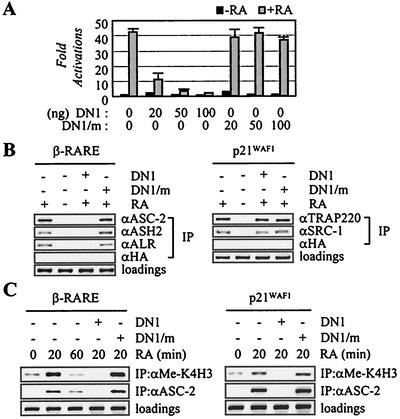
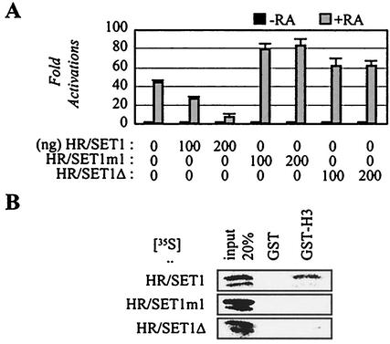
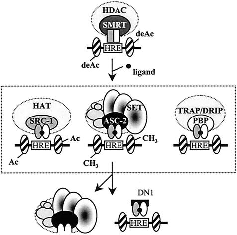
Many transcription coactivators interact with nuclear receptors in a ligand- and C-terminal transactivation function (AF2)-dependent manner. These include activating signal cointegrator 2 (ASC-2), a recently isolated transcriptional coactivator molecule, which is amplified in human cancers and stimulates transactivation by nuclear receptors and numerous other transcription factors. In this report, we show that ASC-2 belongs to a steady-state complex of approximately 2 MDa (ASC-2 complex [ASCOM]) in HeLa nuclei. ASCOM contains retinoblastoma-binding protein RBQ-3, alpha/beta-tubulins, and trithorax group proteins ALR-1, ALR-2, HALR, and ASH2. In particular, ALR-1/2 and HALR contain a highly conserved 130- to 140-amino-acid motif termed the SET domain, which was recently implicated in histone H3 lysine-specific methylation activities. Indeed, recombinant ALR-1, HALR, and immunopurified ASCOM exhibit very weak but specific H3-lysine 4 methylation activities in vitro, and transactivation by retinoic acid receptor appears to involve ligand-dependent recruitment of ASCOM and subsequent transient H3-lysine 4 methylation of the promoter region in vivo. Thus, ASCOM may represent a distinct coactivator complex of nuclear receptors. Further characterization of ASCOM will lead to a better understanding of how nuclear receptors and other transcription factors mediate transcriptional activation.


Figures

FIG. 1.
FIG. 1.
Purification of ASCOM. (A) The chromatography scheme for the purification of ASCOM from nuclear extract (HeLa NE) is shown. Numbers and parentheses indicate KCl molarity and pools, respectively. αASC-2, anti-ASC-2. (B) Mass spectrometric identification of polypeptides. Anti-ASC-2 antibody column eluates were run on an SDS-12% PAGE gel, silver stained, and analyzed by MALDI-TOF, with gene products and molecular masses (in parentheses) shown on the right. The masses of the marker proteins (M) are shown on the left, and asterisks indicate nonspecific bands. Antibody raised against AC22148, a protein of unknown function, revealed that it is a nonspecific contaminant. MALDI-TOF mass spectrometry analyses failed to unravel the identity of a protein of approximately 116 kDa (indicated Unknown).
FIG. 2.
FIG. 2.
Confirmation of the identity of the components of ASCOM. (A) HiTrap Q column fractions subjected to Western analysis with the indicated antibodies against ASC-2, CBP, SRC-1, and hMed6. FT, flowthrough; fr#, fraction number. (B) HiTrap Q fractions 40 to 44 were pooled and immunoprecipitated (IP) with anti-ASC-2 (αASC-2) antibody and Western (W) analyzed with the indicated antibodies. αCBP, anti-CBP; αSRC-1, anti-SRC-1; αSRC-2, anti-SRC-2. (C) Antibodies were generated against synthetic or recombinant polypeptides encoded by cDNAs isolated based on the MALDI-TOF mass spectrometry data of the purified proteins. + and −, HiTrap Q fractions containing ASCOM from HeLa nuclear extract (fractions 40 to 44) and unrelated fractions (30 to 34), respectively. Each antibody recognized a protein with the expected molecular weight (MW) (in thousands). αALR, anti-ALR; αHALR, anti-HALR; αASH2, anti-ASH2; αRBQ-3, anti-RBQ-3. (D, E, and F) HiTrap Q fractions of HeLa nuclear extract containing ASCOM (fractions 40 to 44) were immunoprecipitated with the indicated antibodies (IP), separated by SDS-4 to 6.5% PAGE, and probed with the indicated antibodies (W). αHA, anti-HA; αBRG1, anti-BRG1. S and s indicate supernatant, and P and p indicate precipitate. Ten percent of the total reaction mixture was loaded as input.
FIG. 3.
FIG. 3.
α/β-Tubulins in ASCOM. (A) For the size fractionation of ASCOM, HiTrapQ fractions 40 to 44 (shown in Fig. 2A) were loaded onto a Mono S column (HR5/5; Pharmacia). Immunoreactive fractions were pooled, applied to a Superose 6 column (HR10/30; Pharmacia), and analyzed by immunoblotting as indicated. fr#, fraction number; W, Western analysis; αHALR, anti-HALR; αASC-2, anti-ASC-2; αASH2, anti-ASH2; αRBQ-3, anti-RBQ-3. (B and C) Superose 6 fraction 9 (B) or 28 (C) containing ASCOM or a smaller complex of approximately 500 kDa was immunoprecipitated with the indicated antibodies (IP), separated by SDS-4 to 6.5% PAGE, and probed with indicated antibodies (W). S and P indicate supernatant and precipitate, respectively. Ten percent of the total reaction mixture was loaded as input. αPIPKIβ, anti-PIPKIβ. (D) Cells were treated with β-tubulin, BRCA1, and ASC-2 antibodies. Fluorescein isothiocyanate (green)- and tetramethyl rhodamine isothiocyanate (red)-conjugated antibodies were used to detect ASC-2 and β-tubulin/BRCA1, respectively. Note the colocalization of ASC-2 and β-tubulin. Similar results were also obtained with α-tubulin antibody (data not shown). DIC, differential interference contrast.
FIG. 4.
FIG. 4.
HALR/ALR SET domains. (A) Schematic representation of ySET1, hTrx/ALL-1, hHALR, and hALR-1. Various known protein motifs are color coded as indicated. (B) Amino acid alignment of SET and post-SET domains of hHALR, hALR, dTrx (accession no. AAF55041), ySET1 (accession no. AAB68867), S. pombe Clr4 (accession no. 060016), hG9a (accession no. S30385), and hSUV39 h1 (accession no. NP_003164). Amino acids conserved among all seven proteins; in hHALR, hALR, dTrx, and ySET1; and in yClr4, hG9a, and hSUV39 h1 are highlighted in red, green, and blue, respectively. Dashes indicate gaps in the alignment. The red and blue lines mark the extent of the SET and post-SET domains, respectively. The conserved block of four residues deleted in HR/SET1Δ is boxed, and the conserved glycine mutated to serine in HR/SET1m1 is marked with an asterisk.
FIG. 5.
FIG. 5.
H3 binding and methylation by ASCOM. (A) HiTrap Q column fractions containing ASCOM (fractions 40 to 44 in Fig. 2A) were incubated with either agarose beads coupled to histones or oligo(A) (Sigma), separated by SDS-7% PAGE, and probed with anti-ASC-2 (αASC-2) antibody (W). s and p indicate supernatant and precipitate, respectively. Ten percent of the total reaction mixture was loaded as input. (B) The GST pull-down experiments were done as previously described (17, 19) with GST alone or GST fusions to H2A, H2B, H3, and H4, and 20% of the total reaction mixture was loaded as input. (C) HiTrap Q column fractions containing ASCOM (fractions 40 to 44) were immunoprecipitated with the indicated antibodies and measured for methylation activities with bovine serum albumin, H3, or H4. αHA, anti-HA; αALR, anti-ALR; αASH2, anti-ASH2.
FIG. 6.
FIG. 6.
H3-K4-specific methylation by HALR. (A) Schematic representation of HALR and three C-terminal deletion fragments of HALR with four PHD fingers, an HMG-like domain, and the C-terminal SET domain. Histone methyltransferase activity was assayed by incubation of free histones in the presence of S-adenosyl-l-[methyl-3H]methionine, and incorporated radioactivity was determined by filter binding. (B) Histone methyltransferase activity was assayed by incubation of the indicated synthetic H3 peptides in the presence of S-adenosyl-l-[methyl-3H]methionine, and incorporated radioactivity was determined by filter binding. The GST pull-down experiments were done as previously described (17, 19) with GST fusions to the N-terminal 57 residues of human H3 (wild type [wt]) and point mutants, and 20% of the total reaction mixture was loaded as input.
FIG. 7.
FIG. 7.
Recruitment of ASCOM in RAR transactivation. (A) The retinoid-responsive β-RARE-LUC reporter construct was cotransfected into HeLa cells, along with lacZ expression vector (100 ng) and expression vectors for DN1 (100 ng) or DN1/m (100 ng). Closed and shaded boxes indicate the absence and presence of 0.1 μM 9-cis-retinoic acid (RA), respectively. Normalized luciferase expressions from triplicate samples were calculated relative to the lacZ expressions. Similar results were obtained with CV-1 cells (data not shown). (B and C) ASCOM recruitment to β-RARE and p21WAF1 and H3-K4 methylation. 293T cells were cotransfected with expression vectors for RAR (10 ng), DN1 (100 ng), and DN1/m (100 ng) either in the absence or in the presence of 0.1 μM 9-cis-RA, as indicated. Chromatin from these cells was isolated and immunoprecipitated with the indicated antibodies. The endogenous β-RARE or p21WAF1 region present in the immunoprecipitated samples was amplified by PCR, and input PCR is shown for the loading controls.
FIG. 8.
FIG. 8.
Inhibition of RAR transactivation by HALR-SET. (A) The retinoid-responsive β-RARE-LUC reporter construct was cotransfected into HeLa cells, along with the lacZ expression vector (100 ng) and expression vectors for HR/SET1, HR/SET1m1, and HR/SET1Δ, as indicated. Closed and shaded boxes indicate the absence and presence of 0.1 μM of 9-cis-retinoic acid (RA), respectively. Normalized luciferase expressions from triplicate samples were calculated relative to that of the lacZ expressions. Similar results were obtained with CV-1 cells (data not shown). (B) GST pull-down experiments were done as previously described (17, 19) with GST alone or a GST fusion to the N-terminal 57 residues of human H3, and 20% of the total reaction mixture was loaded as input.
FIG. 9.
FIG. 9.
Working model for ASCOM function. Upon ligand binding, nuclear receptors undergo a structural change, which signals the replacement of corepressor complexes by a series of distinct coactivator complexes. Three coactivator complexes that contain the well-defined LXXLL motif-based adaptor molecules (SRC-1, ASC-2, and PBP/TRAP220/DRIP205//TRIP2) are shown schematically. These and other coactivators can be selectively recruited to individual receptors under different promoter contexts and cellular conditions (see the text). The SET domains of HALR/ALR might, either directly or indirectly, be involved with methylating H3-K4 and/or other yet unknown substrates in vivo. DN1 competitively blocks receptors from recruiting ASCOM, but not other LXXLL-based complexes, and thus may specifically inhibit ASCOM-mediated H3-K4 methylation of the promoter region.

Similar articles

Cited by

References

    1. Ayton, P. M., and M. L. Cleary. 2001. Molecular mechanisms of leukemogenesis mediated by MLL fusion proteins. Oncogene 20:5695-5707. - PubMed
    1. Bernstam, V. A., R. H. Gray, and I. A. Bernstein. 1980. Effects of microtubule-disrupting drugs on protein and RNA synthesis in Physarum polycephalum amoebae. Arch. Microbiol. 128:34-40. - PubMed
    1. Caira, F., P. Antonson, M. Pelto-Huikko, E. Treuter, and J. A. Gustafsson. 2000. Cloning and characterization of RAP250, a novel nuclear receptor coactivator. J. Biol. Chem. 275:5308-5317. - PubMed
    1. Chen, D., H. Ma, H. Hong, S. S. Koh, S. M. Huang, B. T. Schurter, D. W. Aswad, and M. R. Stallcup. 1999. Regulation of transcription by a protein methyltransferase. Science 284:2174-2177. - PubMed
    1. Cremer, T., G. Kreth, H. Koester, R. H. Fink, R. Heintzmann, M. Cremer, I. Solovei, D. Zink, and C. Cremer. 2000. Chromosome territories, interchromatin domain compartment, and nuclear matrix: an integrated view of the functional nuclear architecture. Crit. Rev. Eukaryot. Gene Expr. 10:179-212. - PubMed

Publication types

MeSH terms

Substances