

Iron status in mice carrying a targeted disruption of lactoferrin
- PMID: 12482971
- PMCID: PMC140657
- DOI: 10.1128/MCB.23.1.178-185.2003
Iron status in mice carrying a targeted disruption of lactoferrin
Abstract
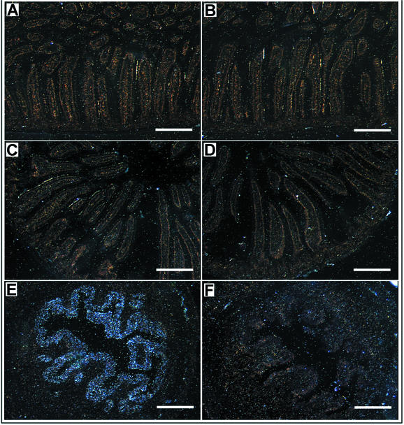
Lactoferrin is a member of the transferrin family of iron-binding glycoproteins present in milk, mucosal secretions, and the secondary granules of neutrophils. While several physiological functions have been proposed for lactoferrin, including the regulation of intestinal iron uptake, the exact function of this protein in vivo remains to be established. To directly assess the physiological functions of lactoferrin, we have generated lactoferrin knockout (LFKO(-/-)) mice by homologous gene targeting. LFKO(-/-) mice are viable and fertile, develop normally, and display no overt abnormalities. A comparison of the iron status of suckling offspring from LFKO(-/-) intercrosses and from wild-type (WT) intercrosses showed that lactoferrin is not essential for iron delivery during the postnatal period. Further, analysis of adult mice on a basal or a high-iron diet revealed no differences in transferrin saturation or tissue iron stores between WT and LFKO(-/-) mice on either diet, although the serum iron levels were slightly elevated in LFKO-/- mice on the basal diet. Consistent with the relatively normal iron status, in situ hybridization analysis demonstrated that lactoferrin is not expressed in the postnatal or adult intestine. Collectively, these results support the conclusion that lactoferrin does not play a major role in the regulation of iron homeostasis.
Figures


Similar articles
-
Lactoferrin: role in iron homeostasis and host defense against microbial infection.Biometals. 2004 Jun;17(3):203-8. doi: 10.1023/b:biom.0000027693.60932.26. Biometals. 2004. PMID: 15222466 Review.
-
Receptor-mediated binding of milk lactoferrin to nursing piglet enterocytes: a model for studies on absorption of lactoferrin-bound iron.J Pediatr Gastroenterol Nutr. 1995 Jul;21(1):37-43. doi: 10.1097/00005176-199507000-00006. J Pediatr Gastroenterol Nutr. 1995. PMID: 8576812
-
Does human lactoferrin in the milk of transgenic mice deliver iron to suckling neonates?Adv Exp Med Biol. 2001;501:233-9. doi: 10.1007/978-1-4615-1371-1_29. Adv Exp Med Biol. 2001. PMID: 11787686 Review.
-
Iron uptake from transferrin and lactoferrin by rat intestinal brush-border membrane vesicles.Am J Physiol. 1990 Apr;258(4 Pt 1):G535-41. doi: 10.1152/ajpgi.1990.258.4.G535. Am J Physiol. 1990. PMID: 2333967
-
Human lactoferrin in the milk of transgenic mice increases intestinal growth in ten-day-old suckling neonates.Adv Exp Med Biol. 2001;501:107-13. doi: 10.1007/978-1-4615-1371-1_13. Adv Exp Med Biol. 2001. PMID: 11787671
Cited by
-
Lactoferrin-iCre: a new mouse line to study uterine epithelial gene function.Endocrinology. 2014 Jul;155(7):2718-24. doi: 10.1210/en.2014-1265. Epub 2014 May 13. Endocrinology. 2014. PMID: 24823394 Free PMC article.
-
Iron: An Essential Element of Cancer Metabolism.Cells. 2020 Dec 3;9(12):2591. doi: 10.3390/cells9122591. Cells. 2020. PMID: 33287315 Free PMC article. Review.
-
Hepcidin messenger RNA expression in human lymphocytes.Immunology. 2010 Jun;130(2):217-30. doi: 10.1111/j.1365-2567.2009.03226.x. Epub 2010 Jan 19. Immunology. 2010. PMID: 20102409 Free PMC article.
-
A novel murine protein with no effect on iron homoeostasis is homologous with transferrin and is the putative inhibitor of carbonic anhydrase.Biochem J. 2007 Aug 15;406(1):85-95. doi: 10.1042/BJ20070384. Biochem J. 2007. PMID: 17511619 Free PMC article.
-
Iron homeostasis and the inflammatory response.Annu Rev Nutr. 2010 Aug 21;30:105-22. doi: 10.1146/annurev.nutr.012809.104804. Annu Rev Nutr. 2010. PMID: 20420524 Free PMC article. Review.
References
-
- Anderson, B. F., H. M. Baker, G. E. Norris, D. W. Rice, and E. N. Baker. 1989. Structure of human lactoferrin: crystallographic structure analysis and refinement at 2.8 A resolution. J. Mol. Biol. 209:711-734. - PubMed
-
- Andrews, N. C. 2000. Iron homeostasis: insights from genetics and animal models. Nat. Rev. Genet. 1:208-217. - PubMed
-
- Bailey, S., R. W. Evans, R. C. Garratt, B. Gorinsky, S. Hasnain, C. Horsburgh, H. Jhoti, P. F. Lindley, A. Mydin, R. Sarra, et al. 1988. Molecular structure of serum transferrin at 3.3-A resolution. Biochemistry 27:5804-5812. - PubMed
-
- Baranov, V. S., A. L. Schwartzman, V. N. Gorbunova, V. S. Gaitskhoki, N. B. Rubtsov, N. A. Timchenko, and S. A. Neifakh. 1987. Chromosomal localization of ceruloplasmin and transferrin genes in laboratory rats, mice and in man by hybridization with specific DNA probes. Chromosoma 96:60-66. - PubMed
Publication types
MeSH terms
Substances
LinkOut - more resources
Full Text Sources
Other Literature Sources
Medical
Molecular Biology Databases