. 2003 Jan;23(1):186-94.
doi: 10.1128/MCB.23.1.186-194.2003.

Proteolytic cleavage of MLL generates a complex of N- and C-terminal fragments that confers protein stability and subnuclear localization

Affiliations

Proteolytic cleavage of MLL generates a complex of N- and C-terminal fragments that confers protein stability and subnuclear localization

James J-D Hsieh et al. Mol Cell Biol. 2003 Jan.

Abstract

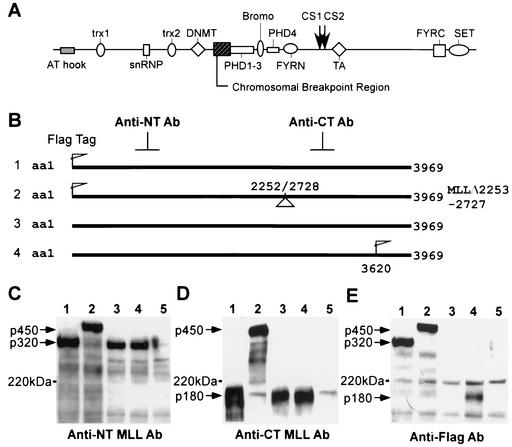
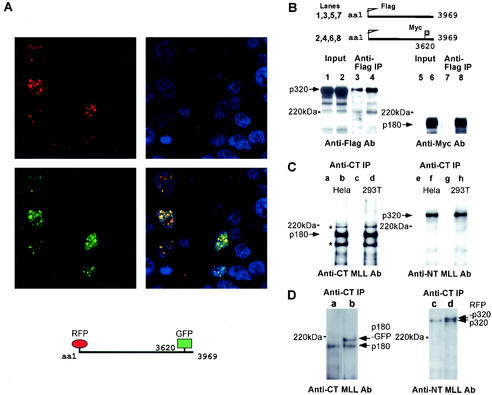
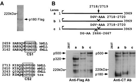
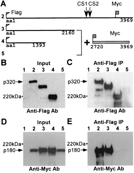
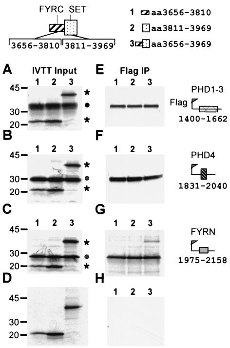
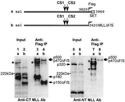
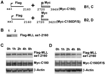
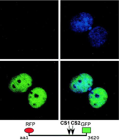
The mixed-lineage leukemia gene (MLL, ALL1, HRX) encodes a 3,969-amino-acid nuclear protein homologous to Drosophila trithorax and is required to maintain proper Hox gene expression. Chromosome translocations in human leukemia disrupt MLL (11q23), generating chimeric proteins between the N terminus of MLL and multiple translocation partners. Here we report that MLL is normally cleaved at two conserved sites (D/GADD and D/GVDD) and that mutation of these sites abolishes the proteolysis. MLL cleavage generates N-terminal p320 (N320) and C-terminal p180 (C180) fragments, which form a stable complex that localizes to a subnuclear compartment. The FYRN domain of N320 directly interacts with the FYRC and SET domains of C180. Disrupting the interaction between N320 and C180 leads to a marked decrease in the level of N320 and a redistribution of C180 to a diffuse nuclear pattern. These data suggest a model in which a dynamic post-cleavage association confers stability to N320 and correct nuclear sublocalization of the complex, to control the availability of N320 for target genes. This predicts that MLL fusion proteins of leukemia which would lose the ability to complex with C180 have their stability conferred instead by the fusion partners, thus providing one mechanism for altered target gene expression.


Figures

FIG. 1.
FIG. 1.
MLL is proteolytically cleaved into an N-terminal 320-kDa fragment, N320, and a C-terminal 180-kDa fragment, C180. (A) Conserved domain structure of MLL in relation to cleavage sites (CS1 and CS2) and the chromosomal breakpoint region. (B) MLL expression constructs used in the experiments below are numbered 1 to 4, corresponding to the lanes where they are expressed. (C) Western blot analysis using an Ab directed to an N-terminal region of MLL (anti-NT MLL) detects p320 in cells expressing Flag-tagged MLL (lane 1), untagged MLL (lane 3), and Myc-tagged MLL (lane 4); lane 2 contains the internal deletion mutant MLLΔ2253-2727 at ∼450 kDa; lane 5 contains whole-cell extracts purified from untransfected 293T cells as a control. (D) Anti-CT Ab detected a 180-kDa polypeptide in cells expressing Flag-tagged MLL (lane 1), untagged MLL (lane 3), and Myc-tagged MLL (lane 4), but the MLL deletion mutant was ∼450 kDa (lane 2). (E) Anti-Flag Ab Western blot analysis detected the p320 fragment (lane 1), the ∼450-kDa MLL deletion mutant (lane 2), and the C-terminal p180 fragment (lane 4).
FIG. 2.
FIG. 2.
Colocalization and association between the processed p320 and p180 fragments of MLL. (A) RFP-p320 and GFP-p180 colocalize to the punctate subnuclear compartment. Cells expressing dually fluorescent RFP-MLL-GFP were assessed by confocal microscopy for localization of the expected RFP-p320 (red, upper left panel) and GFP-p180 (green, lower left panel) fragments. The nuclei were stained with YO-PRO (blue, upper right panel). Colocalization of RFP and GFP resulted in yellow punctate subcellular fluorescence in the merged images (yellow, lower right panel). (B) Coimmunoprecipitation between Flag-p320 and Myc-p180. The N-terminally Flag-tagged MLL (Flag-MLL) or the N-terminally Flag- plus C-terminally Myc-tagged MLL (Flag-MLL-Myc) constructs were expressed in cells, solubilized, size fractionated in the corresponding lanes, and then subjected to immunoprecipitation (IP) using anti-Flag Ab. Western blots of input lysate or anti-Flag immunoprecipitates were developed with anti-Flag Ab or anti-Myc Ab. Flag-MLL-Myc was processed to Flag-p320 and Myc-p180 (lanes 2 and 6). Myc-p180 is efficiently coprecipitated with Flag-p320 (lane 8). (C) Endogenous MLL is cleaved to p320 and p180, which form a stable complex in cells. Cellular extracts from HeLa or 293T cells were incubated with (lane b, d, f, and h) or without (lane a, c, e, and g) anti-CT Ab and then precipitated with protein-A beads. Immunoprecipitates were subjected to SDS-PAGE and analyzed by Western blotting using either anti-NT or anti-CT Ab. Anti-CT Ab precipitated a 180-kDa protein from both cell lines tested (lanes b and d). Anti-NT Ab detected a 320-kDa protein that was coprecipitated with endogenous p180 (lanes f and h). The asterisks denote cross-reactive bands recognized by anti-CT Ab. (D) Dually fluorescent RFP-MLL-GFP is proteolytically processed as wt MLL. Cellular extracts from RFP-MLL-GFP-transfected 293T cells were immunoprecipitated with anti-CT Ab (lanes b and d). Anti-CT Ab Western blot analysis recognized C-180-GFP in addition to the endogenous C180 (lane b). RFP-N320 was detected as a slower-migrating band above the endogenous N320 (lane d). Lanes a and c contain anti-CT immunoprecipitates from untransfected 293T cells.
FIG. 3.
FIG. 3.
Identification and verification of two MLL cleavage sites. (A) C-terminally Flag-tagged MLL was expressed in 293T cells, and the processed Flag-C180 was purified using anti-Flag Ab. Captured protein was analyzed by SDS-PAGE, transferred to PVDF membranes, visualized with Coomassie blue (arrow), excised, and subjected to Edman degradation. This yielded a 12-aa sequence corresponding to residues 2719 to 2730 of MLL. Sequence alignment among hMLL (human), mMLL (murine), fMLL (Fugu rubripes), and Drosophila trx indicates conservation of D/GVDD cleavage site 2 (CS2). A second cleavage site, D/GADD (CS1), proximal to CS2 was subsequently identified but exists only in the vertebrate MLLs, not Drosophila trx. (B) MLL mutants possessing the indicated amino acid substitutions are shown in the upper panel and used in the corresponding lanes below. Expression of the indicated wt or mutant MLL proteins in 293T cells was assessed by either anti-Flag Ab (left panel) or anti-CT Ab (right panel) Western blotting. Alanine substitution of CS2 (2718DGV-AAA) failed to completely abolish the proteolytic cleavage of MLL (lane b, both panels). Mutation of both cleavage sites (2666DG-AA plus 2718DG-AAA) is required for complete abolition of MLL processing, as evidenced by the disappearance of both N320 and C180 and an increase in the amount of full-length p500 (lane c, both panels).
FIG. 4.
FIG. 4.
aa 1394 to 2160 of N320 mediate the interaction with C180. (A) Flag-tagged wt or truncated N-terminal MLL constructs were expressed in 293T cells either alone or in combination with Myc-tagged C180. (B to E) Lane 1 of each experiment contained extracts prepared from untransfected cells. Doubly tagged MLL (Flag-MLL-Myc) was in lane 2 throughout. (E) Anti-Flag immunoprecipitation (IP) followed by anti-Myc Western blot analysis indicated that MLL aa 1 to 2160 but not 1 to 1393 efficiently interact with Myc-p180 (lanes 3 and 4).
FIG. 5.
FIG. 5.
Direct physical interaction between the FYRN domain of N320 and the FYRC plus SET domains of C180. The top schematic depicts the conserved C-terminal domains of MLL that were in vitro translated in the presence of [35S]methionine and used in lanes 1 to 3, respectively. The side schematics depict Flag-tagged N-terminal domains that were35S labeled and used in panels A and E (PHD1 to PHD3), B and F (PHD4), and C and G (FYRN). The left four panels (A to D) show the input samples, while the right four panels (E to H) show the anti-Flag immunoprecipitation assays. Solid circles denote the positions of Flag-tagged N-terminal domains, while asterisks denote the positions of C-terminal domains. (G) The FYRN domain of N320 and the FYRC plus SET domains of C180 are the minimal interaction domains.
FIG. 6.
FIG. 6.
The interaction between N320 and C180 of MLL confers stability to the N320. Flag-tagged wt MLL and a C-terminal deletion lacking the FYRC and SET domains (MLLΔF/S) were expressed in 293 T cells used in lanes a and b, respectively. Input cellular extracts and anti-Flag immunoprecipitate (IP) were analyzed by Western blotting with the indicated Abs. MLL-FlagΔF/S was processed to N320 and C150ΔF/S (lanes 2 and 6). In the coimmunoprecipitation assays, C150ΔF/S failed to precipitate N320 (lane 8). The protein level of C150ΔF/S was comparable to that of C180 (lanes 1 to 4). In contrast, the level of N320 was markedly reduced when it could no longer associate with the C-terminal fragment (lanes 5 and 6). Small amounts of the newly synthesized, uncleaved MLL (∼500 kDa) and a few smaller migrating proteins, perhaps representing splice variants (asterisk), are noted.
FIG. 7.
FIG. 7.
The FYRC-plus-SET region of C180 protects N-terminal MLL from degradation. (A) Flag-tagged MLL aa 1 to 2140 was cotransfected with Myc-tagged C180 or C150ΔF/S in 293T cells. Cellular extracts coexpressing the MLL N terminus and C180 were used in panel B, lane 1, and panel C. Samples from extracts expressing the MLL N terminus and C150ΔF/S were analyzed in panel B, lane 2, and panel D. (B) Myc-tagged wt C terminus, C180 (lane 1) but not the truncated C150 (lane 2), increases the abundance of coexpressed N-terminal MLL, determined by anti-Flag Western blot analysis. (C and D) The degradation of N-terminal MLL in the presence of C180 (C, top panel) or C150ΔF/S (D, top panel) was compared. Cellular extracts obtained at the indicated time points after addition of 10 μg of cycloheximide per ml at 36 h following transfection were analyzed using anti-Flag Ab. The stable expression of C180 (C, middle panel) and C150ΔF/S (D, middle panel) was detected by anti-Myc Ab. Equal protein loading was determined by using anti-β actin Ab in both lower panels. The gel in panel D has been exposed five times longer than that in panel C to equilibrate the steady-state levels of Flag-MLL aa 1 to 2160.
FIG. 8.
FIG. 8.
Intact FYRC and SET domains are required for the punctate subnuclear localization of C180. Loss of the C180 interaction results in loss of RFP-N320 fluorescence. A dually fluorescent MLL mutant, with the FYRC and SET domains deleted (RFP-MLL-GFPΔF/S), was expressed, and the resultant RFP-N320 and GFP-C150ΔF/S fragments were assessed by confocal microscopy. GFP-C150ΔF/S was easily detected but had redistributed from the punctate nuclear pattern seen with wt C180 to a diffuse nuclear pattern (lower left panel). The same cells that expressed GFP-C180ΔF/S failed to exhibit detectable RFP-N320, as judged by the consistent lack of red fluorescence (upper left panel). The upper right panel shows the YO-PRO nuclear stain, while the lower right panel shows the merged image of RFP-N320 and GFP-C150ΔF/S.

Similar articles

Cited by

References

    1. Armstrong, S. A., J. E. Staunton, L. B. Silverman, R. Pieters, M. L. den Boer, M. D. Minden, S. E. Sallan, E. S. Lander, T. R. Golub, and S. J. Korsmeyer. 2002. MLL translocations specify a distinct gene expression profile that distinguishes a unique leukemia. Nat. Genet. 30:41-47. - PubMed
    1. Ayton, P. M., and M. L. Cleary. 2001. Molecular mechanisms of leukemogenesis mediated by MLL fusion proteins. Oncogene 20:5695-5707. - PubMed
    1. Breen, T. R., and P. J. Harte. 1993. Trithorax regulates multiple homeotic genes in the bithorax and Antennapedia complexes and exerts different tissue-specific, parasegment-specific and promoter-specific effects on each. Development 117:119-134. - PubMed
    1. Butler, L. H., R. Slany, X. Cui, M. L. Cleary, and D. Y. Mason. 1997. The HRX proto-oncogene product is widely expressed in human tissues and localizes to nuclear structures. Blood 89:3361-3370. - PubMed
    1. Caslini, C., A. Shilatifard, L. Yang, and J. L. Hess. 2000. The amino terminus of the mixed lineage leukemia protein (MLL) promotes cell cycle arrest and monocytic differentiation. Proc. Natl. Acad. Sci. USA 97:2797-2802. - PMC - PubMed

Publication types

MeSH terms