. 2003 Jan;23(1):259-71.
doi: 10.1128/MCB.23.1.259-271.2003.

Novel transcriptional potentiation of BETA2/NeuroD on the secretin gene promoter by the DNA-binding protein Finb/RREB-1

Affiliations

Novel transcriptional potentiation of BETA2/NeuroD on the secretin gene promoter by the DNA-binding protein Finb/RREB-1

Subir K Ray et al. Mol Cell Biol. 2003 Jan.

Abstract

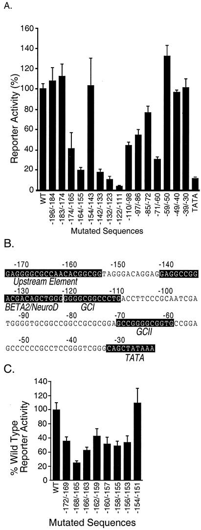
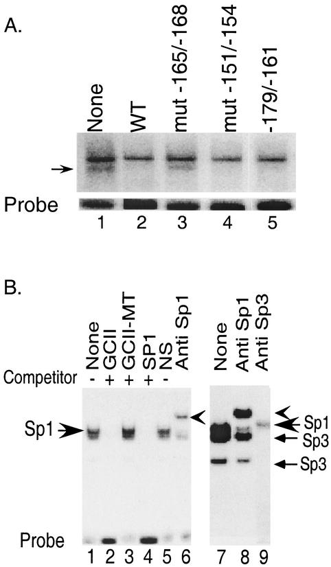
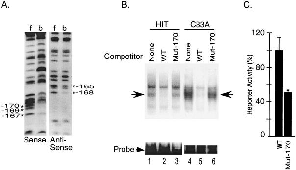
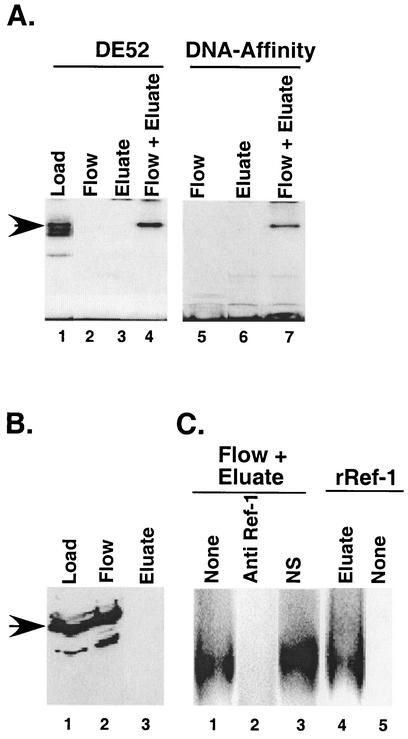
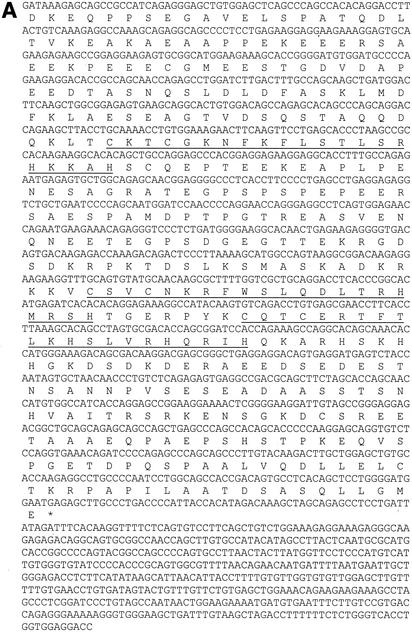
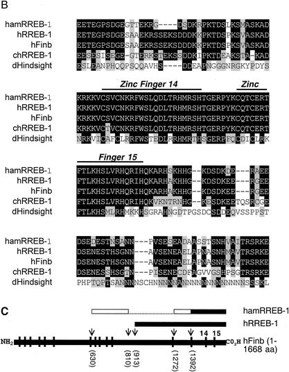
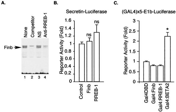
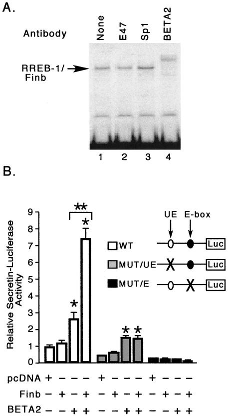
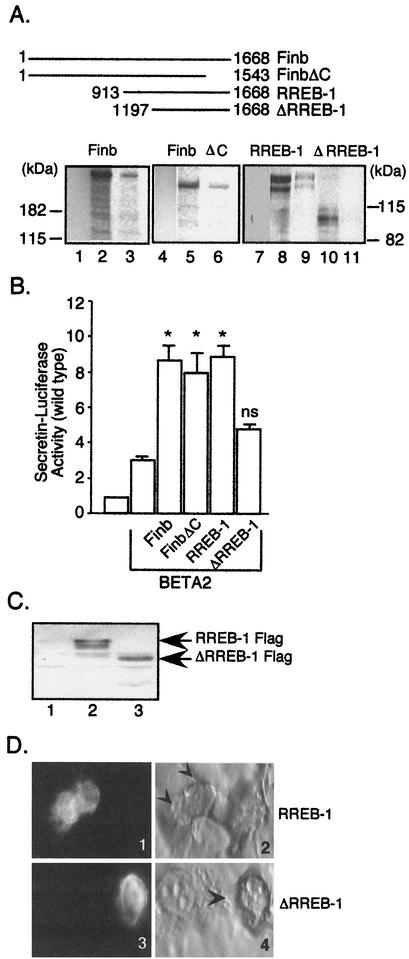
The basic helix-loop-helix protein BETA2/NeuroD activates transcription of the secretin gene and is essential for terminal differentiation of secretin-producing enteroendocrine cells. However, in heterodimeric complexes with its partner basic helix-loop-helix proteins, BETA2 does not appear to be a strong activator of transcription by itself. Mutational analysis of a proximal enhancer in the secretin gene identified several cis-acting elements in addition to the E-box binding site for BETA2. We identified by expression cloning the zinc finger protein RREB-1, also known to exist as a longer form, Finb, as the protein binding to one of the mutationally sensitive elements. Finb/RREB-1 lacks an intrinsic activation domain and by itself did not activate secretin gene transcription. Here we show that Finb/RREB-1 can associate with BETA2 to enhance its transcription-activating function. Both DNA binding and physical interaction of Finb/RREB-1 with BETA2 are required to potentiate transcription. Thus, Finb/RREB-1 does not function as a classical activator of transcription that recruits an activation domain to a DNA-protein complex. Finb/RREB-1 may be distinguished from coactivators, which increase transcription without sequence-specific DNA binding. We suggest that Finb/RREB-1 should be considered a potentiator of transcription, representing a distinct category of transcription-regulating proteins.


Figures

FIG. 1.
FIG. 1.
Mutational analysis of the secretin promoter. HIT cells were transiently transfected with a secretin-luciferase reporter gene (Materials and Methods) without (wild type [WT]) or with transversion mutations (10 to 13 bp) at different positions (shown by the numbers under each bar) including the TATA box (A) or with 4-bp transversion mutations at different positions (C). Luciferase activity was measured in cell extracts 16 h later. Results are expressed aspercentages relative to the wild-type reporter and are the means ± standard errors of the means (SEM) normalized for transfection efficiency for at least three independent experiments. (B) The mutationally sensitive sequences present in the promoter region are highlighted with a black background.
FIG. 2.
FIG. 2.
Characterization of factors binding to mutationally sensitive elements in the secretin gene enhancer. (A) A HindIII-EcoRI fragment containing sequence −174 to −146 was used as a probe in a gel shift assay with HIT cell nuclear extract. Arrowhead, specific DNA-protein complex. Lanes 2 to 5 included a 100-fold molar excess of unlabeled competitor from −174 to −146, with the indicated 4-bp block mutations in lanes 3 and 4. Lane 5 included a 100-fold molar excess of a competitor spanning −174 to −161. WT, wild type. (B) Left panel, HIT nuclear extract was incubated with a GCI probe in the absence (lane 1) or presence of different competitors at 100-fold molar excess (lanes 2 to 4) or in the presence of a nonimmune IgG (lane 5) or anti-Sp1 antibody (lane 6). The smaller arrow shows the slower mobility complex supershifted by the Sp1 antibody. Right panel, experimental conditions were as described for the left panel except that the electrophoresis was carried out to separate the individual complexes. Extracts were treated with anti-Sp1 (lane 8) or anti-Sp3 (lane 9) antibody.
FIG. 3.
FIG. 3.
Characterization of the factors binding to the upstream element in the secretin enhancer. (A) Partially methylated probes were incubated with HIT cell nuclear extract in an electrophoretic mobility shift assay. Following electrophoresis, free probe (f) and probe bound to proteins in the upstream element complex (b) were eluted from the gel, cleaved with piperidine, and resolved on an 8% sequencing gel to generate G ladders. *, G residues that are important for complex formation. (B) The upstream element probe was incubated in a gel shift reaction with the nuclear extract from HIT cells or C33A cells in the absence (lane 1) or presence of a 100-fold molar excess of the wild type (WT) (lane 2) or the Mut-170 (lane 3) competitor. Arrow, specific DNA-protein complex. (C) HIT cells were transiently transfected with a secretin-luciferase reporter plasmid (WT) or the same reporter with a point mutation at −170 (Mut-170). Luciferase activity was measured in cell extracts 20 h later. Results are expressed as percentages relative to the wild-type reporter and are the means ± SEM normalized for transfection efficiency for at least three independent experiments.
FIG. 4.
FIG. 4.
Stabilization of DNA-binding activity of the upstream element binding factor by Ref-1. (A) The DNA-binding activity was purified from C33A nuclear extracts. Aliquots from a heparin-agarose column salt eluate (lane 1) as well as the flowthrough and salt-eluted fractions of subsequent columns, either a DE52 column (lanes 2 to 4) or a DNA-affinity column (lanes 5 to 7), were tested for upstream element DNA-binding activity in a gel shift assay. The lost DNA-binding activity (lanes 2, 3, 5, and 6) was restored by mixing the two fractions (flow + eluate) (lanes 4 and 7). (B) Immunoblot analysis of proteins in the DE52 load, flow, and eluate fractions with anti-Ref-1 antibody. Proteins present in an equivalent volume of each column fraction were separated as described in Materials and Methods. Arrow, 38-kDa protein that reacts with the Ref-1 antibody. (C) An aliquot of the DE52 eluate fraction was mixed with the DE52 flow fraction that was untreated (lane 1), the flow fraction pretreated with 0.4 μg of anti-Ref-1 antibody (lane 2), a control antibody (lane 3), or a recombinant Ref-1 protein (∼200 ng) (lane 4) and was assayed for DNA-binding activity. Ref-1 alone was devoid of DNA-binding activity (lane 5).
FIG. 5.
FIG. 5.
The protein binding to the upstream element is the hamster homologue of human Finb/RREB-1. (A) The nucleotide and deduced amino acid (single-letter code) sequences of the identified cDNA clone are shown. The first 420-nucleotide sequence was obtained by 5′-RACE, whereas the rest was obtained from the identified cDNA clone. The zinc finger domains are underlined and an asterisk indicates the stop codon. (B) ClustalW alignment of hamster RREB-1/Finb (hamRREB-1) (residues 183 to 330) with the region containing zinc fingers 14 and 15 of human Finb (hFinb), human RREB-1 (hRREB-1), chicken RREB-1 (chRREB-1), and the Drosophila Hindsight protein (dHindsight). Identical amino acids are shown as white text on a black background, conservative substitutions are shown as black text on a gray background, and nonconserved residues are shown as black text on a white background. Gaps (-) were created to maximize the alignment. (C) Structural organization of hamster RREB-1, human RREB-1, and human Finb. Horizontal black bars denote sequenced (nucleotide) regions of each protein. The open bars indicate the homologous region of hamster RREB-1 identified by 5′-RACE and the dotted line areas have not been sequenced. Vertical bars indicate positions of zinc fingers in full-length human Finb. Arrows and numbers in parentheses show the positions of amino acids in each protein relative to the full-length protein.
FIG. 5.
FIG. 5.
The protein binding to the upstream element is the hamster homologue of human Finb/RREB-1. (A) The nucleotide and deduced amino acid (single-letter code) sequences of the identified cDNA clone are shown. The first 420-nucleotide sequence was obtained by 5′-RACE, whereas the rest was obtained from the identified cDNA clone. The zinc finger domains are underlined and an asterisk indicates the stop codon. (B) ClustalW alignment of hamster RREB-1/Finb (hamRREB-1) (residues 183 to 330) with the region containing zinc fingers 14 and 15 of human Finb (hFinb), human RREB-1 (hRREB-1), chicken RREB-1 (chRREB-1), and the Drosophila Hindsight protein (dHindsight). Identical amino acids are shown as white text on a black background, conservative substitutions are shown as black text on a gray background, and nonconserved residues are shown as black text on a white background. Gaps (-) were created to maximize the alignment. (C) Structural organization of hamster RREB-1, human RREB-1, and human Finb. Horizontal black bars denote sequenced (nucleotide) regions of each protein. The open bars indicate the homologous region of hamster RREB-1 identified by 5′-RACE and the dotted line areas have not been sequenced. Vertical bars indicate positions of zinc fingers in full-length human Finb. Arrows and numbers in parentheses show the positions of amino acids in each protein relative to the full-length protein.
FIG. 6.
FIG. 6.
Functional role for RREB-1/Finb. (A) Immunodepletion of RREB-1 diminishes the DNA-protein complex at the upstream element. Nuclear extracts from HIT cells were treated with protein A-Sepharose bound to either anti-RREB-1 (lane 4) or control IgG (lane 3). Note the loss of the specific band denoted by an arrow (lane 1). The specific band is competed out by unlabeled wild-type competitor (lane 2). (B) C33A cells were cotransfected with an equal amount (1 μg) of Finb or RREB-1 expression plasmid or empty vector (control) and a secretin reporter plasmid (0.25 μg). Results shown are means ± SEM for at least seven separate experiments. ns, not significantly different from control. (C) C33A cells were cotransfected with an equal amount (0.5 μg) of Gal4-Finb, Gal4-RREB-1, or Gal4-BETA2 expression plasmid or Gal4 DBD alone and an E1b-luciferase reporter plasmid containing five GAL4 binding sites. Results are shown as the means ± SEM for at least five separate experiments. *, significantly different (P < 0.001) from Gal4 DBD.
FIG. 7.
FIG. 7.
Finb/RREB-1 interacts with BETA2/NeuroD. (A) Heparin-agarose-purified binding activity from HIT cell nuclear extract was examined in a gel shift assay in the absence (lane 1) or presence of anti-E47 (lane 2), anti-Sp1 (lane 3), or anti-BETA2 (lane 4) antibody. (B) C33A cells were cotransfected with an equal amount (1 μg) of Finb expression plasmid or pcDNA (empty vector) in the absence or presence of BETA2 expression plasmid (0.05 μg) and a reporter plasmid (0.25 μg) containing the wild-type secretin promoter (white bars), the mutant promoter lacking the functional upstream element (gray bars), or the E-box element (black bars). Results are shown as the means ± SEM for at least three separate experiments. *, significantly different (P < 0.001) from the respective control (pcDNA); **, significantly different (P < 0.001) from BETA2 alone.
FIG. 8.
FIG. 8.
Functional cooperation between Finb/RREB-1 and BETA2 requires a BETA2 interaction domain in Finb. (A) The top panel shows the Finb and RREB-1 protein truncations relative to the full-length protein. Numbers indicate the positions of amino acids in each protein. The bottom panel shows in vitro-translated, 35S-labeled Finb/RREB-1 proteins examined for the ability to bind to BETA2 expressed as a GST fusion protein. Lanes 3, 6, 9, and 11, radiolabeled proteins captured by GST-BETA2 proteins; lanes 1, 4, and 7, proteins captured by GST alone. Lanes 2, 5, 8, and 10 show approximately 10% of the input protein applied to the affinity matrix. (B) C33A cells were cotransfected with a BETA2 expression plasmid, wild-type secretin reporter plasmid, and either Finb, FinbΔC, RREB-1, or ΔRREB-1 expression plasmid. Results are shown as the means ± SEM for at least five separate experiments normalized for transfection efficiency. ns, not significantly different versus BETA2 alone; *, P ≤ 0.001 versus BETA2 alone. (C) C33A cells were transfected with an equivalent amount of expression plasmid for RREB-1-FLAG (lane 2), ΔRREB-1-FLAG (lane 3), or pcDNA (lane 1). The expressed proteins were detected in the cell lysates by immunoblotting with a monoclonal anti-FLAG M2 antibody (Sigma). (D) Immunofluorescent staining of FLAG-tagged, transfected RREB-1. Staining of transfected RREB-1 (panel 1) and ΔRREB-1 (panel 3) is shown. The corresponding bright field views are shown in panels 2 and 4, respectively, with FLAG-stained cells denoted by arrowheads.

Similar articles

Cited by

References

    1. Dignam, J. D., R. M. Lebovitz, and R. G. Roeder. 1983. Accurate transcription initiation by RNA polymerase II in a soluble extract from isolated mammalian nuclei. Nucleic Acids Res. 11:1475-1489. - PMC - PubMed
    1. Dumonteil, E., B. Laser, I. Constant, and J. Philippe. 1998. Differential regulation of the glucagon and insulin I gene promoters by basic helix-loop-helix transcription factors E47 and Beta2. J. Biol. Chem. 273:19945-19954. - PubMed
    1. Edlund, T., M. D. Walker, P. J. Barr, and W. J. Rutter. 1985. Cell-specific expression of the rat insulin gene: evidence for role of two distinct 5′ flanking elements. Science 230:912-916. - PubMed
    1. Fried, M., and D. Crothers. 1983. CAP and RNA polymerase interactions with the lac promoter: binding stoichiometry and long range effects. Nucleic Acids Res. 11:141-158. - PMC - PubMed
    1. Fujimoto-Nishiyama, A., S. Ishii, S. Matsuda, J.-I. Inoue, and T. Yamamoto. 1997. A novel zinc finger protein, Finb, is a transcriptional activator and localized in nuclear bodies. Gene 195:267-275. - PubMed

Publication types

MeSH terms