. 2003 Jan;23(1):306-21.
doi: 10.1128/MCB.23.1.306-321.2003.

Two different Drosophila ADA2 homologues are present in distinct GCN5 histone acetyltransferase-containing complexes

Affiliations

Two different Drosophila ADA2 homologues are present in distinct GCN5 histone acetyltransferase-containing complexes

Selen Muratoglu et al. Mol Cell Biol. 2003 Jan.

Abstract

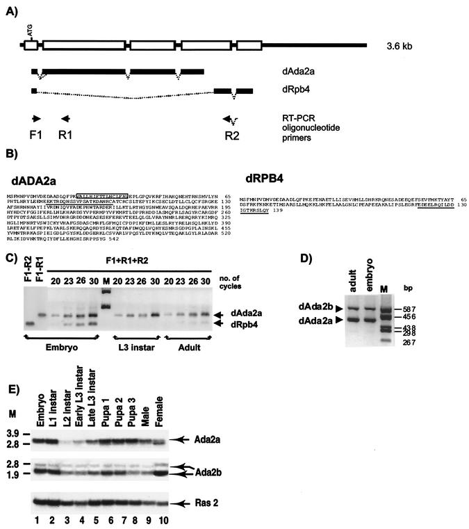
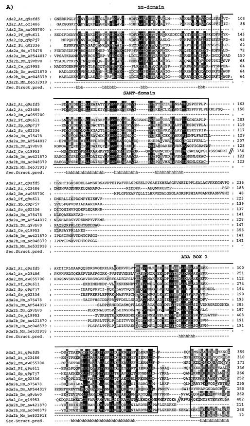
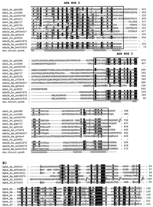
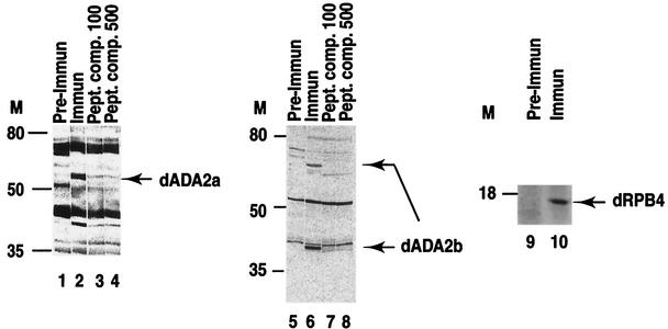
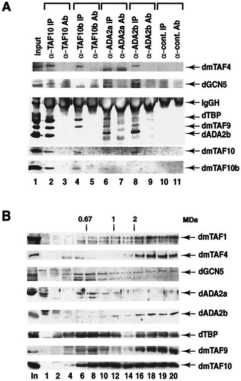
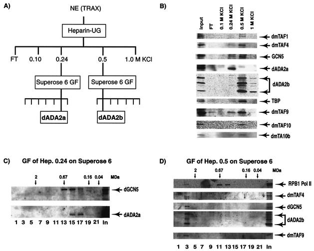
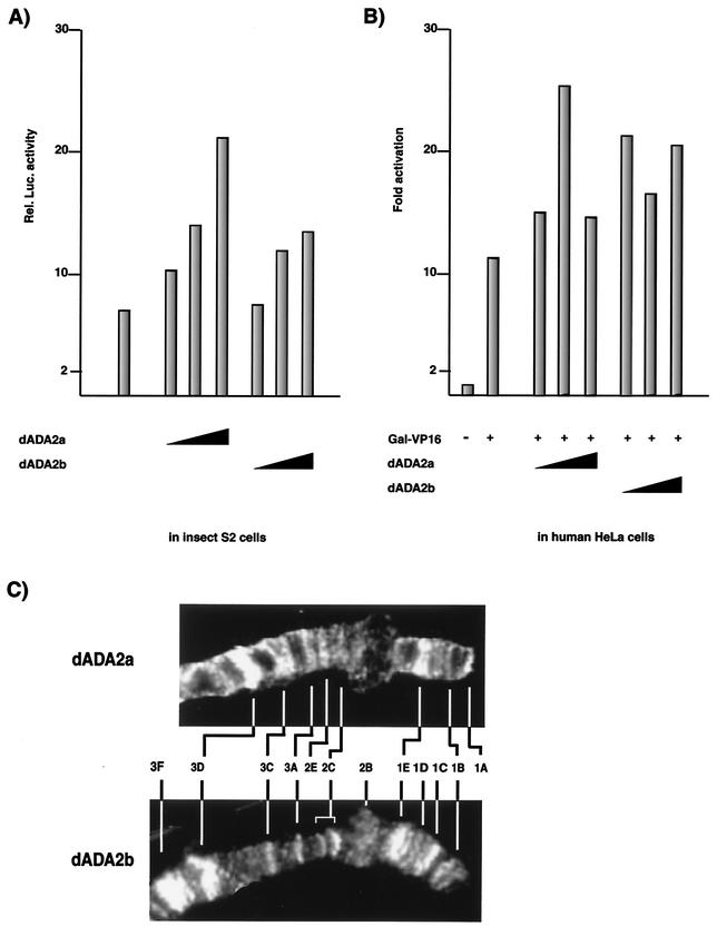
We have isolated a novel Drosophila (d) gene coding for two distinct proteins via alternative splicing: a homologue of the yeast adaptor protein ADA2, dADA2a, and a subunit of RNA polymerase II (Pol II), dRPB4. Moreover, we have identified another gene in the Drosophila genome encoding a second ADA2 homologue (dADA2b). The two dADA2 homologues, as well as many putative ADA2 homologues from different species, all contain, in addition to the ZZ and SANT domains, several evolutionarily conserved domains. The dada2a/rpb4 and dada2b genes are differentially expressed at various stages of Drosophila development. Both dADA2a and dADA2b interacted with the GCN5 histone acetyltransferase (HAT) in a yeast two-hybrid assay, and dADA2b, but not dADA2a, also interacted with Drosophila ADA3. Both dADA2s further potentiate transcriptional activation in insect and mammalian cells. Antibodies raised either against dADA2a or dADA2b both immunoprecipitated GCN5 as well as several Drosophila TATA binding protein-associated factors (TAFs). Moreover, following glycerol gradient sedimentation or chromatographic purification combined with gel filtration of Drosophila nuclear extracts, dADA2a and dGCN5 were detected in fractions with an apparent molecular mass of about 0.8 MDa whereas dADA2b was found in fractions corresponding to masses of at least 2 MDa, together with GCN5 and several Drosophila TAFs. Furthermore, in vivo the two dADA2 proteins showed different localizations on polytene X chromosomes. These results, taken together, suggest that the two Drosophila ADA2 homologues are present in distinct GCN5-containing HAT complexes.


Figures

FIG. 1.
FIG. 1.
The dAda2a/dRpb4 gene encodes two putative transcription factors, dADA2a and dRPB4. (A) Schematic representation of the dAda2a/dRpb4 gene structure. Exons are shown as boxes on the thick line representing the genomic sequence. The common ATG located in the first exon is indicated. The dAda2 and the dRpb4 transcripts produced by alternative splicing are indicated as bold lines under the genomic structure. F1, R1, and R2 show the locations of oligonucleotide primers used for RT-PCR analysis of the dAda2 or dRpb4 mRNA. (B) Deduced amino acid sequences of the Drosophila ADA2a and RPB4 proteins. The 15-amino-acid stretch that is generated by alternative 3′ splice site selection in certain dADA2a isoforms is boxed. The oligopeptides used for immunization to produce specific antibodies against dADA2a and dRPB4 are underlined. (C) RT-PCR analysis of total RNA prepared from Drosophila at different developmental stages, as indicated. The arrows show the dAda2a- and dRpb4-specific PCR products. Aliquots from each PCR amplification reaction were taken at 20, 23, 26, and 30 cycles, resolved on a 1.2% agarose gel, and visualized by ethidium bromide staining. M, molecular weight marker. (D) RT-PCR analysis using total RNA prepared from developmental stages, as indicated above the lanes. The dAda2a and dAda2b PCR products with different sizes are indicated. Aliquots from each PCR amplification reaction were resolved on a 2% agarose gel and visualized by ethidium bromide staining. M, molecular weight marker. (E) Northern blot analysis of dAda2a and dAda2b mRNAs during Drosophila development. cDNA fragments from either dADA2a or dADA2b cDNAs were labeled and hybridized with mRNA samples prepared from the indicated stages of the Oregon R strain. The same blot was also hybridized with the Ras2 probe as a control (53). M, molecular weight marker.
FIG. 2.
FIG. 2.
The novel dADA2s and dRPB4 are evolutionarily well conserved. ADA2 (A) and RPB4 (B) amino acid sequences from various species were extracted from the GenBank, EMBL, DDBJ, and Genome Sequencing Center databases by iterative searches using the PSI-BLAST program and hADA2a or yRPB4 as query. The one-letter amino acid code is used. The different ADA2 sequences are as follows: A. thaliana (At) (accession no. q9sfd5 and o23486), Z. mays (Zm) (accession no. aw055700), P. falciparum (Pf) (accession no. q9u611), S. pombe (Sp) (accession no. q9p7j7), S. cerevisiae (Sc) (accession no. q02336), H. sapiens (Hs) ADA2a (accession no. o75478), D. melanogaster (Dm) ADA2a (accession no. AF544017), D. melanogaster (Dm) ADA2b (accession no. q9vhv0), C. elegans (Ce) (accession no. q19953), D. rerio (Dr) (accession no. aw421870), H. sapiens (Hs) ADA2b (accession no. ac048379), M. musculus (Mm) (accession no. be532918). Residues with the same physicochemical properties conserved among the different ADA2 homologues at between 100 and 91% have a black background, those conserved at between 90 and 76% have a dark gray background, and those conserved at between 75 and 60% have a light gray background. The highly conserved ZZ and SANT domains are boxed. The amino acid positions in the different proteins are labeled on the left in the sequences represents the deletion of a nonconserved region in the corresponding protein. In panel A, the nonconserved N-terminal end of certain ADA2 factors is not represented. The conserved secondary structures predicted (Sec. Struct. Pred.) for the ADA2 proteins are labeled such that the positions of the α-helices (h) and the β-strands (b) are shown under the sequences. The amino acid sequence in dmADA2b that was used to generate the anti-dADA2b PAbs is underlined.
FIG. 2.
FIG. 2.
The novel dADA2s and dRPB4 are evolutionarily well conserved. ADA2 (A) and RPB4 (B) amino acid sequences from various species were extracted from the GenBank, EMBL, DDBJ, and Genome Sequencing Center databases by iterative searches using the PSI-BLAST program and hADA2a or yRPB4 as query. The one-letter amino acid code is used. The different ADA2 sequences are as follows: A. thaliana (At) (accession no. q9sfd5 and o23486), Z. mays (Zm) (accession no. aw055700), P. falciparum (Pf) (accession no. q9u611), S. pombe (Sp) (accession no. q9p7j7), S. cerevisiae (Sc) (accession no. q02336), H. sapiens (Hs) ADA2a (accession no. o75478), D. melanogaster (Dm) ADA2a (accession no. AF544017), D. melanogaster (Dm) ADA2b (accession no. q9vhv0), C. elegans (Ce) (accession no. q19953), D. rerio (Dr) (accession no. aw421870), H. sapiens (Hs) ADA2b (accession no. ac048379), M. musculus (Mm) (accession no. be532918). Residues with the same physicochemical properties conserved among the different ADA2 homologues at between 100 and 91% have a black background, those conserved at between 90 and 76% have a dark gray background, and those conserved at between 75 and 60% have a light gray background. The highly conserved ZZ and SANT domains are boxed. The amino acid positions in the different proteins are labeled on the left in the sequences represents the deletion of a nonconserved region in the corresponding protein. In panel A, the nonconserved N-terminal end of certain ADA2 factors is not represented. The conserved secondary structures predicted (Sec. Struct. Pred.) for the ADA2 proteins are labeled such that the positions of the α-helices (h) and the β-strands (b) are shown under the sequences. The amino acid sequence in dmADA2b that was used to generate the anti-dADA2b PAbs is underlined.
FIG. 3.
FIG. 3.
The two dADA2 homologues and dRPB4 are expressed in Drosophila. Drosophila embryo extracts were resolved by SDS-PAGE on 10% (ADA2a and ADA2b) or 15% (RPB4) polyacrylamide gels and blotted with purified anti-dADA2a, anti-dADA2b, or anti-dRPB4 PAbs (Immun) or with preimmune (Pre-Immun) sera taken from the corresponding rabbits before immunization. Molecular mass markers (M) are indicated in kilodaltons. To test the specificity of the sera raised against dADA2a and dADA2b, competition experiments with peptides used for the immunization (see Fig. 1B and 2) were done. Polyclonal sera raised against either ADA2a (500× dilution) or ADA2b (1,000× dilution) were preincubated with 100 μg (Pept. comp. 100; lanes 3 and 7) or 500 μg (Pept. comp. 500; lanes 4 and 8) of the corresponding peptides, respectively, prior to incubation with the Western blots.
FIG. 4.
FIG. 4.
dADA2a and dADA2b are subunits of different GCN5 HAT-containing multiprotein complexes. dADA2a and dADA2b coimmunoprecipitate with dGCN5 and with several different dTAFs. Proteins from nuclear extracts were immunoprecipitated with affinity-purified anti-dTAF10, anti-dTA10b, anti-dADA2a, and anti-dADA2b PAbs and a nonrelated PAb raised against a human protein, as indicated. The input nuclear fraction (input), the immunoprecipitated protein A-Sepharose antibody-bound proteins (IP), and the protein A-Sepharose bound PAbs alone (Ab) were resolved by SDS-PAGE (10 or 15% polyacrylamide) and transferred. The blots were then probed with antibodies raised against the indicated proteins. (B) Glycerol gradient sedimentation analysis of Drosophila embryo nuclear extract. A 200-μl volume of TRAX was loaded onto a 10 to 30% glycerol gradient and centrifuged for 4 h at 50 krpm, using an SW60 Ti rotor. The sedimentation standards, thyroglobulin (669 kDa), ferritin (440 kDa), catalase (232 kDa), lactate dehydrogenase (140 kDa), and albumin (67 kDa), were centrifuged in parallel gradients. Fractions of 200 μl were collected. Aliquots (20 μl) from the indicated fractions were resolved in parallel with the unfractioned nuclear extract (In) by SDS-PAGE (10% polyacrylamide), transferred to a filter, and probed with the indicated PAbs. The position of the 0.67-MDa molecular mass marker and some extrapolated molecular masses are indicated at the top. The numbers of the analyzed fractions are indicated at the bottom. Note that only the 42-kDa form of dADA2b is shown; however, the 70-kDa form showed exactly the same migration pattern.
FIG. 5.
FIG. 5.
dADA2a- and dADA2b-containing complexes are chromatographically separable. (A) The chromatography protocol used to separate the dADA2-containing complexes is outlined. All procedures were performed at 4°C. The KCI concentrations used in the heparin-Ultrogel (UG) elution are indicated. NE (TRAX), Drosophila 0- to 12-h embryo nuclear extract; GF, gel filtration. (B) Portions (10-μl) of the input nuclear extract and of the different fractions eluting from the heparin-Ultrogel column were tested by Western blot analysis for the presence of the different factors (as indicated). FT, flowthrough fraction. (C) The heparin 0.24 M KCl-derived fraction was further separated on a Superose 6 gel filtration column. Portions of the input (In) (10 μl) and of every second fraction (as indicated) eluting from the column (20 μl) were tested by Western blot analysis for the presence of the different factors (as indicated). (D) The heparin 0.5 M KCl-derived fraction was also separated on a Superose 6 gel filtration column. Portions of the input (In) (10 μl) and of every second fraction eluting from the column (20 μl) was tested by Western blot analysis for the presence of the different factors (as indicated). Pol II was monitored by using an antibody recognizing the C-terminal repeat of the largest Pol II subunit, RPB1. In panels C and D, the positions of known molecular mass markers are indicated above the panels.
FIG. 6.
FIG. 6.
dADA2a and dADA2b work as cofactors and localize to different but overlapping loci on the distal region of Drosophila X chromosome. dADA2a and dADA2b stimulate transcriptional activity in Drosophila S2 cells (A) and human HeLa cells (B). (A) Reporter plasmid containing a luciferase reporter under the control of an ecdysone-inducible promoter was cotransfected with increasing amounts (0.5, 1.0, and 2.0 μg) of recombinant plasmids expressing either dADA2a or dADA2b into Drosophila S2 cells. The luciferase activity of the cell extracts was measured 40 h after transfection. (B) HeLa cells were cotransfected with a luciferase reporter gene under the control of the rabbit β-globin promoter harboring one GAL4-binding site, a GAL-VP16 expression plasmid, and increasing amounts (50, 100, and 500 ng) of dADA2a and dADA2b expression plasmids (as indicated). Luciferase activity was measured. (C) Association of dADA2a and dADA2b with specific loci of the Drosophila polytene chromosome. Immunostaining of the polytene X chromosome from wild-type larvae (Oregon R) with antibodies raised against dADA2a (upper panel) and dADA2b (lower panel) and Cy3-conjugated secondary antibodies is shown. The labeled regions of chromosome X are indicated for both panels.

Similar articles

Cited by

References

    1. Aasland, R., A. F. Stewart, and T. Gibson. 1996. The SANT domain: a putative DNA-binding domain in the SWI-SNF and ADA complexes, the transcriptional co-repressor N-CoR and TFIIIB. Trends Biochem. Sci. 21:87-88. - PubMed
    1. Balaguer, P., A. M. Boussioux, E. Demirpence, and J. C. Nicolas. 2001. Reporter cell lines are useful tools for monitoring biological activity of nuclear receptor ligands. Luminescence 16:153-158. - PubMed
    1. Barlev, N. A., R. Candau, L. Wang, P. Darpino, N. Silverman, and S. L. Berger. 1995. Characterization of physical interactions of the putative transcriptional adaptor, ADA2, with acidic activation domains and TATA-binding protein. J. Biol. Chem. 270:19337-19344. - PubMed
    1. Baxevanis, A. D., and D. Landsman. 1998. Histone Sequence Database: new histone fold family members. Nucleic Acids Res. 26:372-375. - PMC - PubMed
    1. Belenkaya, T., A. Soldatov, E. Nabirochkina, I. Birjukova, S. Georgieva, and P. Georgiey. 1998. P-element insertion at the polyhomeotic gene leads to formation of a novel chimeric protein that negatively regulates yellow gene expression in P-element-induced alleles of Drosophila melanogaster. Genetics 150:687-697. - PMC - PubMed

Publication types

MeSH terms

Associated data