. 2003 Jan;23(1):414-23.
doi: 10.1128/MCB.23.1.414-423.2003.

A novel domain within the DEAD-box protein DP103 is essential for transcriptional repression and helicase activity

Affiliations

A novel domain within the DEAD-box protein DP103 is essential for transcriptional repression and helicase activity

Xiaomei Yan et al. Mol Cell Biol. 2003 Jan.

Abstract

Members of the DEAD-box family of helicases, distinguished by a core characteristic sequence of Asp-Glu-Ala-Asp, are expressed in a wide range of prokaryotes and eukaryotes and exhibit diverse cellular functions, including DNA transcription, recombination and repair, RNA processing, translation, and posttranslational regulation. Although ubiquitous, the function of most DEAD-box proteins is unknown. We and others have recently cloned DP103, which harbors conserved DEAD-box, helicase, and ATPase domains in its N terminus. DP103 (also termed Gemin3 and DDX20) interacts with SF-1, SMN, EBNA2, and EBNA3C in mammalian cells. Here we demonstrate that a discrete domain within the nonconserved C-terminal region of DP103 directly interacts with SF-1. This domain exhibits an autonomous repression function and is necessary and sufficient for repressing the transcriptional activity of SF-1. Furthermore, intact DP103 exhibits helicase activity. Importantly, the C-terminal domain is obligatory but not sufficient for this unwinding activity of DP103. Together, our results support a novel paradigm for transcriptional repression and demonstrate the bifunctional role of the C-terminal domain of DP103.


Figures

FIG. 1.
FIG. 1.
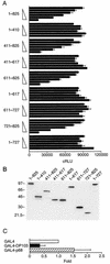
DP103 interacts with the PRD in SF-1 (aa 193 to 201) through aa 721 to 825. (A) Schematic diagram depicting the main transcriptional regulatory domains of SF-1. A mutated PRD, known to abrogate SF-1 repression, is shown. (B) A diagram of DP103, denoting eight highly conserved motifs within the N-terminal region of DEAD-box family proteins as well as the nonconserved C-terminal region. (C) DP103721-825 interacts with wild type SF-1 but not with PRD mutant SF-1. Interaction was detected by using a mammalian two-hybrid assay, with SF-1120-462 fused downstream from GAL4 and DP103 fragments (as shown) fused to the VP16 activation domain. Plasmids were transiently transfected into CV-1 cells along with the GAL4 reporter plasmid ΔGKI. Results represent three independent experiments performed in duplicate, expressed as fold activation over control in which the empty VP16 plasmid was used and normalized to β-galactosidase activity.
FIG. 2.
FIG. 2.
DP103721-825 physically interacts with SF-1's PRD (aa 193 to 201) in vitro. Wild-type SF-1 (W) and PRD mutant SF-1 (M) cloned in pBSK were expressed and labeled with [35S]methionine in vitro by using a TNT transcription-translation system. Labeled SF-1 was incubated with His-tagged DP103 fragments bound to Ni2+-NTA agarose resin. Bound SF-1 was detected by using SDS-10% PAGE and autoradiography. Input included 20% SF-1 that was used for each assay. The empty Ni2+-NTA agarose resin, reacted with wild-type or mutant SF-1, served as negative control.
FIG. 3.
FIG. 3.
DP103 harbors a repression domain at aa 721 to 825. (A) CV-1 cells were cotransfected with increasing amounts (0, 0.03, 0.1, 0.3, 1.0, and 3.0 μg) of either full-length DP103 or DP103 fragments, fused to GAL4, along with the reporter plasmid GAL4 × 5-tkLuc. Results (means ± SD) are expressed as cRLU normalized to β-galactosidase activity and represent three independent experiments, each performed in duplicate. (B) Western immunoblotting of the GAL4-DP103 fusion proteins analyzed in panel A, demonstrating equal cellular expression of transfected plasmids. Lysates (30 μg) were prepared from CV-1 cells that were transfected with 3 μg of each DP103 plasmid, separated by SDS-PAGE, and immunodetected by using anti-GAL4 antibody as described in Materials and Methods. (C) The DEAD-box protein p68 (3 μg, expressed as chimeric protein GAL4-p68) does not repress the reporter plasmid GAL4 × 5-tkLuc in CV-1 cells. Results are means of three independent experiments, each performed in duplicate, and are expressed as fold over control in which the empty GAL4 plasmid was used.
FIG. 4.
FIG. 4.
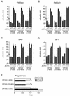
DP103721-825 represses the transcription of SF-1 target promoters. (A) The concentration-dependent influence of GAL4-fused full-length or truncated DP103 on the transcriptional activity of SF-1. JEG3 cells were cotransfected with 0.05 μg of CMV-SF-1 and either GAL4-DP1031-825, GAL4-DP103721-825, or GAL4-DP1031-727 (0, 0.1, 0.3, 1.0, and 3.0 μg), along with 0.5 μg of the SF-1 luciferase reporter plasmid S25. (B) The repression effect of DP103 is observed only in the presence of SF-1. Transfection was performed as described above, with 3 μg of GAL4-DP103721-825 or GAL4-DP1031-727. The empty expression vector CMV-neo or GAL4 was used as control for SF-1 and DP103, respectively. (C) Concentration-dependent influence of GAL4-fused full-length or truncated DP103 on the transcriptional activity of an SF-1-responsive rat P450scc reporter. Transfection was performed as described above, with 0.5 μg of the SF-1 luciferase reporter plasmid P450scc. (D) SF-1 is required for the repression effect of DP103. The repression was abrogated when the two SF-1 binding elements in the P450scc promoter were mutated. Transfection was performed as described above. Results (means ± SD) are expressed as cRLU normalized to β-galactosidase activity and represent three independent experiments, each performed in duplicate.
FIG. 5.
FIG. 5.
DP103721-825 diminishes the expression of SF-1 target genes in the adrenocortical line. Y1 cells were transfected with increasing amounts (0, 1.0, and 3.0 μg) of GAL4-DP1031-825, GAL4-DP103721-825, or GAL4-DP1031-727. Steroidogenesis was stimulated with 0.1 μM of ACTH or water vehicles 18 h after transfection. Total RNA was isolated and processed for quantitative RT-PCR as described in Materials and Methods. (A) Expression of P450scc mRNA. (B) Expression of P450c21 mRNA. (C) Expression of StAR mRNA. (D) GAL4-DP103 fusion proteins do not diminish the expression of SF-1. Results (means ± SD) are expressed as fold expression over control (no ACTH, no GAL4-DP103) and represent three independent experiments, each performed in duplicate. (E) Progesterone levels in media from cultured Y1 cells transfected with GAL4-DP103 plasmids (3 μg) and collected over 24 h prior to hormone determination. Results (means ± SD) are expressed as fold over control (GAL4) and represent three to five independent experiments, each performed in duplicate. An asterisk denotes a P value of <0.05 (Student's t test).
FIG. 6.
FIG. 6.
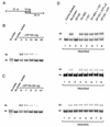
DP103 exhibits RNA helicase activity in vitro. Helicase assays (5′→3′) were performed under standard conditions as described in Materials and Methods, with 50 fmol of either 32P-labeled RNA/RNA, RNA/DNA, or DNA/DNA duplex substrates, along with His-tagged DP103 in the presence of ATP (3 mM) at 37°C. (A) The structure of the artificial RNA or DNA duplex substrates. (B) RNA helicase activity of DP103 is concentration dependent. A concentration range of full-length His-tagged DP103 (0, 3, 10, 30, and 50 ng) was examined. (C) A time course for the RNA helicase activity of full-length DP103 at the time points indicated. (D) Helicase assay with 50 ng of proteins as indicated, along with RNA/RNA (upper panel), RNA/DNA (middle panel), or DNA/DNA (lower panel) substrates. ATP (3 mM) was present unless indicated as being absent or replaced with ATP-γ-S (3 mM). All the helicase assays were performed for 15 min. Boiled double-stranded (ds) substrates or substrate incubated without protein at 37°C was used as control, indicating the position of released single-stranded (ss) or ds probe, respectively. 32P-labeled short ss probe was also included in the experiments depicted in panels B and C to highlight the position of released ssRNA or ssDNA.

Similar articles

Cited by

References

    1. Aratani, S., R. Fujii, T. Oishi, H. Fujita, T. Amano, T. Ohshima, M. Hagiwara, A. Fukamizu, and T. Nakajima. 2001. Dual roles of RNA helicase A in CREB-dependent transcription. Mol. Cell. Biol. 21:4460-4469. - PMC - PubMed
    1. Arenas, J. E., and J. N. Abelson. 1997. Prp43: an RNA helicase-like factor involved in spliceosome disassembly. Proc. Natl. Acad. Sci. USA 94:11798-11802. - PMC - PubMed
    1. Bland, M. L., C. A. Jamieson, S. F. Akana, S. R. Bornstein, G. Eisenhofer, M. F. Dallman, and H. A. Ingraham. 2000. Haploinsufficiency of steroidogenic factor-1 in mice disrupts adrenal development leading to an impaired stress response. Proc. Natl. Acad. Sci. USA 97:14488-14493. - PMC - PubMed
    1. Boddeker, N., K. Stade, and F. Franceschi. 1997. Characterization of DbpA, an Escherichia coli DEAD box protein with ATP independent RNA unwinding activity. Nucleic Acids Res. 25:537-545. - PMC - PubMed
    1. Campbell, L., K. M. Hunter, P. Mohaghegh, J. M. Tinsley, M. A. Brasch, and K. E. Davies. 2000. Direct interaction of Smn with dp103, a putative RNA helicase: a role for Smn in transcription regulation? Hum. Mol. Genet. 9:1093-1100. - PubMed

Publication types

MeSH terms

LinkOut - more resources