

Coronary arteriogenesis and differentiation of periarterial Purkinje fibers in the chick heart: is there a link?
- PMID: 12484610
- PMCID: PMC140288
Coronary arteriogenesis and differentiation of periarterial Purkinje fibers in the chick heart: is there a link?
Abstract
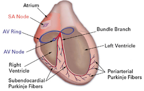
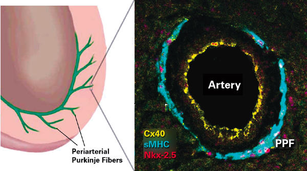
In the following review, we outline the cellular ontogeny and time course of coronary artery development within the vertebrate heart. Our eventual focus will be the potential role of arteriogenesis in the differentiation of a subset of specialized conduction cells in the chick heart. We begin by briefly outlining early heart formation, showing how the outermost layer of the looped, tube heart--the epicardium--is of extracardiac origin and provides the progenitor cells to the entire vascular bed. Subsequently, we summarize the events of coronary arterial development that follow epicardialization. Finally, we discuss work in the chick that indicates how arteries form pioneering, directional conduits through ventricular tissue, adjacent to which myocardial cells differentiate to form the most peripheral component of the avian conduction system--a network of periarterial Purkinje fibers.
Figures
Similar articles
-
The role of the epicardium and neural crest as extracardiac contributors to coronary vascular development.Tex Heart Inst J. 2002;29(4):255-61. Tex Heart Inst J. 2002. PMID: 12484609 Free PMC article.
-
Induction of Purkinje fiber differentiation by coronary arterialization.Proc Natl Acad Sci U S A. 1999 Nov 9;96(23):13214-8. doi: 10.1073/pnas.96.23.13214. Proc Natl Acad Sci U S A. 1999. PMID: 10557300 Free PMC article.
-
Coronary vascular development.Wien Klin Wochenschr. 2007;119(11-12 Suppl 1):4-5. Wien Klin Wochenschr. 2007. PMID: 19618587 No abstract available.
-
His-Purkinje lineages and development.Novartis Found Symp. 2003;250:110-22; discussion 122-4, 276-9. Novartis Found Symp. 2003. PMID: 12956326 Review.
-
Cellular precursors of the coronary arteries.Tex Heart Inst J. 2002;29(4):243-9. Tex Heart Inst J. 2002. PMID: 12484607 Free PMC article. Review.
Cited by
-
Prevalence and spectrum of coronary artery anomalies in patients with an isolated congenital left ventricular aneurysm or diverticulum.Clin Cardiol. 2011 Apr;34(4):226-32. doi: 10.1002/clc.20875. Epub 2011 Feb 11. Clin Cardiol. 2011. PMID: 21319171 Free PMC article.
-
Differentiation of cardiac Purkinje fibers requires precise spatiotemporal regulation of Nkx2-5 expression.Dev Dyn. 2006 Jan;235(1):38-49. doi: 10.1002/dvdy.20580. Dev Dyn. 2006. PMID: 16245335 Free PMC article.
-
Coronary artery anomalies. Part I: Recent insights from molecular embryology.Z Kardiol. 2004 Dec;93(12):929-37. doi: 10.1007/s00392-004-0152-7. Z Kardiol. 2004. PMID: 15599567 Review.
-
A quantitative structural and morphometric analysis of the Purkinje network and the Purkinje-myocardial junctions in pig hearts.J Anat. 2017 May;230(5):664-678. doi: 10.1111/joa.12594. Epub 2017 Mar 3. J Anat. 2017. PMID: 28256093 Free PMC article.
References
-
- Hamburger V, Hamilton HL. A series of normal stages in the development of the chick embryo. J Morphol 1951;88:49–92. - PubMed
-
- Rosenquist GC, DeHaan RL. Migration of precardiac cells in the chick embryo: a radiographic study. 1966. Carnegie Inst. Washington Publ. 625 (Contrib. To Embryol). 38, 111–21.
-
- Hatada Y, Stern CD. A fate map of the epiblast of the early chick embryo. Development 1994;120(10):2879–89. - PubMed
-
- Garcia-Martinez V, Schoenwolf GC. Primitive-streak origin of the cardiovascular system in avian embryos. Dev Biol 1993;159(2):706–19. - PubMed
-
- Rosenquist GC. A radioautographic study of labelled grafts in the chick blastoderm. 1966. Carnegie Inst. Washington Publ. 625 (Contrib. To Embryol). 38, 71–110.
Publication types
MeSH terms
Grants and funding
LinkOut - more resources
Full Text Sources