. 2002 Dec;7(12):1267-83.
doi: 10.1046/j.1365-2443.2002.00602.x.

A comparative cDNA microarray analysis reveals a spectrum of genes regulated by Pax6 in mouse lens

Affiliations

A comparative cDNA microarray analysis reveals a spectrum of genes regulated by Pax6 in mouse lens

Bharesh K Chauhan et al. Genes Cells. 2002 Dec.

Abstract

Background: Pax6 is a transcription factor that is required for induction, growth, and maintenance of the lens; however, few direct target genes of Pax6 are known.

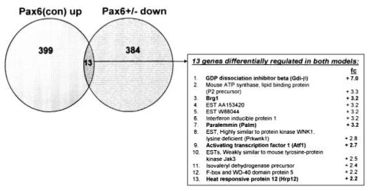
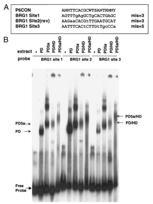
Results: In this report, we describe the results of a cDNA microarray analysis of lens transcripts from transgenic mice over-expressing Pax6 in lens fibre cells in order to narrow the field of potential direct Pax6 target genes. This study revealed that the transcript levels were significantly altered for 508 of the 9700 genes analysed, including five genes encoding the cell adhesion molecules beta1-integrin, JAM1, L1 CAM, NCAM-140 and neogenin. Notably, comparisons between the genes differentially expressed in Pax6 heterozygous and Pax6 over-expressing lenses identified 13 common genes, including paralemmin, GDIbeta, ATF1, Hrp12 and Brg1. Immunohistochemistry and Western blotting demonstrated that Brg1 is expressed in the embryonic and neonatal (2-week-old) but not in 14-week adult lenses, and confirmed altered expression in transgenic lenses over-expressing Pax6. Furthermore, EMSA demonstrated that the BRG1 promoter contains Pax6 binding sites, further supporting the proposition that it is directly regulated by Pax6.

Conclusions: These results provide a list of genes with possible roles in lens biology and cataracts that are directly or indirectly regulated by Pax6.


Figures

Figure 1
Figure 1
Venn diagram showing total numbers of genes down-regulated in Pax6 heterozygous lenses (Pax6+/-), and Pax6(con)-transgenic lenses [Pax6(con)]. Thirteen genes in the intersection (white) are displayed. Those chosen for further confirmations are noted in bold type. Fc (fold change), a ratio of transcript levels between Pax6(con) and wild-type lenses.
Figure 2
Figure 2
Confirmation of microarray results obtained from wild-type, Pax6(con) transgenic, and Pax6 heterozygous (Pax6+/-) lenses using semi-quantitative RT-PCR. (A) Control experiments showing that the expression levels of β5 tubulin (Tubb5) and uridine monophosphate kinase (UMK) were unchanged in Pax6(con) transgenic and Pax6+/- lenses, respectively, while Pax6/Pax6(5a) levels were increased in Pax6(con) lenses and decreased in Pax6+/- lenses. Lanes 1 and 2 represent total RNA isolated from wild-type (WT, FVB/N genotype, left, and NMRI genotype, right, respectively) lenses and lanes 3 and 4 represent total RNA isolated from Pax6(con) transgenic (left panel) and Pax6 heterozygous (right panel) lenses, respectively. The triangles indicate that two concentrations of RNA (50 ng in lanes 1 and 3 and 17 ng in lanes 2 and 4) were used for each assay. (B) Differential expression of paralemmin, GDPβ, ATF-1, and Hrp12 in Pax6(con) transgenic, Pax6 heterozygous (Pax6+/-) and normal (WT) lenses. Lanes 1-4 and triangles are described above. Size of specific PCR products is given in parentheses; the 100 bp DNA ladder is shown on the left.
Figure 3
Figure 3
Analysis of transcript levels of genes encoding Brg1 and Brm in wild-type, Pax6(con)-transgenic (Pax6(con)), Pax6 heterozygous (Pax6+/-), and Pax6(5a)-transgenic (5a) lenses using semi-quantitative RT-PCR. Total RNA was isolated from 2-week-old wild-type FVB/N (lanes 1 and 2, Duncan et al. 2002), 6 week NMRI (lanes 5 and 6, St-Onge et al. 1997), and 3-week-old FVB/N lenses (lanes 9 and 10, Duncan et al. 2000). These samples were compared with total RNA isolated from age matched Pax6(con) (lanes 3 and 4), Pax6 heterozygous (lanes 7 and 8), and Pax6(5a) lenses (lanes 11 and 12), respectively. The triangles indicate that two concentrations of RNA (50 ng in lanes 1, 3, 5, 7, 9 and 11 and 17 ng in lanes 2, 4, 6, 8, 10 and 12) were used for each assay. Size of specific PCR products is given in parentheses; the 100 bp DNA ladder is shown on the left.
Figure 4
Figure 4
Brg1 is dynamically expressed during early eye development and up-regulated in Pax6(con) transgenic lenses. (A) Western blot of Brg1 expression in 2 week Pax6(con) transgenic and wild-type mouse lens. (B) Western blot of Brg1 expression in newborn, 2 week, 6 week and 14 week wild-type mouse lenses. (C) Immunohistochemical staining of Brg1 in 2-week postnatal wild-type lens transition zone. (D) Immunohistochemical staining of Brg1 in 2-week Pax6(con) transgenic lens transition zone. (E) Immunohistochemical staining of Brg1 in 2-week Pax6(con) transgenic lens epithelium and central fibres. (F, G). Immunohistochemical staining of Brg1 in embryonic lens at 9.5 dpc (F, Brg1 only; G, 9.5 dpc merged image of Brg1 and nuclear counter stain) (H) Immunohistochemical staining of Brg1 in embryonic lens at 16.5 dpc. Abbreviations: kDa, kilodaltons; WT, wild-type; TG, Pax6(con)-transgenic; e, lens epithelium; lp, lens placode; ov, optic vesicle; sf, secondary fibres; t, transition zone/bow region;wt, wild-type. Red—Brg1, Green—DNA. All immunohistochemistry images are approximately 200×.
Figure 5
Figure 5
Interaction of recombinant Pax6 proteins with human BRG1 candidate binding sites 1, 2, and 3. (A) An alignment of sites 1, 2 and 3 with P6CON (5′-ANNTTCACGCWTSANTKMNY-3′) (Epstein et al. 1994a). Conserved bases (upper case letters) and non-conserved bases (lower case letters) nucleotides, W = A/T, S = G/C, K = G/T, M = A/C, Y = T,C, N = A/C/G/T. Number of mismatches (mis) between the P6CON and the individual sequences is given. (B) EMSA showing binding of extracts containing GST-fusions of Pax6 PD, PD5a, PD/HD and PD5a/HD (Duncan et al. 2000) to sites 1, 2 and 3. Arrows indicate positions of PD, PD5a, PD/HD, and PD(5a)/HD monomeric complexes.
Figure 6
Figure 6
Semi-quantitative RT-PCR analysis of structural protein and cell adhesion molecule encoding genes identified as differentiatially expressed in Pax6(con) transgenic lenses by microarray analysis. (A) Experiments showing expression levels of αB- and γS-crystallins and calpastatin. Lanes 1 and 2 represent total RNA isolated from wild-type (WT, FVB/N background) lenses and lanes 3 and 4 represent total RNA isolated from Pax6(con) transgenic lenses. The triangles indicate two concentrations of RNA (50 ng in lanes 1 and 3 and 17 ng in lanes 2 and 4) were used for each assay. (B) Differential expression of β1-integrin, N-CAM, L1 CAM, JAM1 and neogenin in Pax6(con) transgenic and normal (WT) lenses. Lanes 1-4 and triangles are described above. Size of the specific PCR products is given in parentheses; the 100 bp DNA ladder is shown on the left.
Figure 7
Figure 7
Analysis of transcript levels of Brg1, ATF1, L1 CAM, neogenin, and calpastatin in wild-type and Pax6 heterozygous cerebellum. (A) Amplification of ‘reference’ transcripts encoding GAPDH. Lanes 1 and 2 represent total RNA isolated from wild-type (WT, NMRI genotype) and lanes 3 and 4 represent total RNA isolated from Pax6 heterozygous cerebellum. The triangles indicate two concentrations of RNA (50 ng in lanes 1 and 3 and 17 ng in lanes 2 and 4) were used for each assay. (B) Differential expression of Brg1, ATF1, L1 CAM, neogenin and calpastatin. Lanes 1-4 and triangles are described above. Size of specific PCR products is given in parentheses; the 100 bp DNA ladder is shown on the left.

Similar articles

Cited by

References

    1. Ashery-Padan R, Marquardt T, Zhou X, Gruss P. Pax6 activity in the lens primordium is required for lens formation and for correct placement of a single retina in the eye. Genes Dev. 2000;14:2701–2711. - PMC - PubMed
    1. Baulmann DC, Ohlmann A, Flugel-Koch C, Goswami S, Cvekl A, Tamm ER. Pax6 heterozygous eyes show defects in chamber angle differentiation that are associated with a wide spectrum of the other anterior eye segment abnormalities. Mech. Dev. 2002;118:3–17. - PubMed
    1. Belecky-Adams, Tomarev S, Li HS, et al. Pax-6, Prox 1, and Chx10 homeobox gene expression correlates with phenotypic fate of retinal precursor cells. Invest. Ophthalmol. Vis. Sci. 1997;38:1293–1303. - PubMed
    1. Bernier G, Vukovichn W, Neidhardt L, Herrmann BG, Gruss P. Isolation and characterization of a downstream target of Pax6 in the mammalian retinal primordium. Development. 2001;128:3987–3994. - PubMed
    1. Bultman S, Gebuhr T, Yee D, et al. A Brg1 null mutation in the mouse reveals functional differences among mammalian SWI/SNF complexes. Mol. Cell. 2000;6:1287–1295. - PubMed

Publication types

MeSH terms

Associated data