

Long-term evolution of the acute tubular necrosis (ATN) induced by glycerol: role of myofibroblasts and macrophages
- PMID: 12485461
- PMCID: PMC2517681
- DOI: 10.1046/j.1365-2613.2002.00223.x
Long-term evolution of the acute tubular necrosis (ATN) induced by glycerol: role of myofibroblasts and macrophages
Abstract
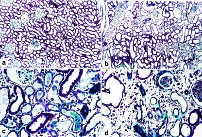
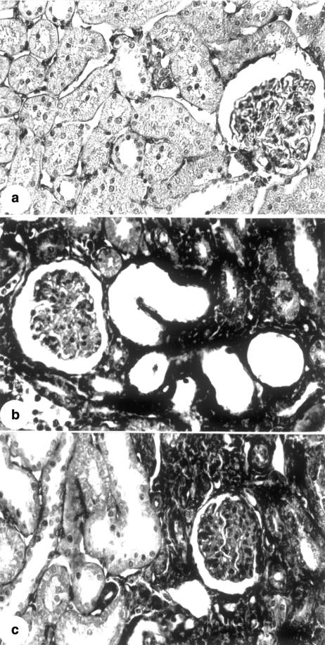
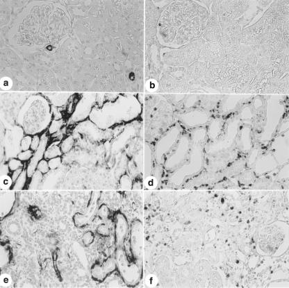
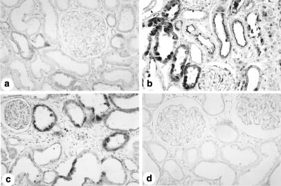
Late structural changes such as interstitial fibrosis in the renal cortex and tubular atrophy have been detected after severe acute tubular necrosis (ATN). The aim of this study was to investigate the expression of fibronectin, alpha-smooth muscle actin and macrophages during the evolution of the ATN induced by glycerol and their relationship with the late structural changes observed in the kidneys of these animals. Forty-nine male Wistar rats were injected with a 50% glycerol solution, 8 mL/kg (4 mL/kg applied i.m. to each hind leg) and 14 with 0.15 m NaCl solution. Before glycerol injection on day 1, water was removed for 17 h. Blood and urine samples were collected 1 day after the injection to quantify sodium and creatinine. The animals were killed 5, 30 and 60 days after the injections and the kidneys removed for histological and immunohistochemical studies. The results of the histological and immunohistochemical studies were scored according to the extent of lesion or staining in the cortical tubulointerstitium, respectively. The percentage of tubulointerstitial lesions was determined by morphometry. Glycerol-injected rats presented a transitory increase in plasma creatinine levels and in fractional sodium excretion. The immunohistochemical studies showed increased fibronectin, alpha-smooth muscle actin (alpha-SM-actin), TGF-beta and ED-1 (macrophages) staining in the renal cortex from rats killed 5, 30 and 60 days after glycerol injection (P < 0.05) compared to control. The animals killed on day 30 and 60 also presented chronic lesions (fibrosis, tubular dilatation and atrophy) in the renal cortex, despite the recovery of renal function. Macrophages, TGF-beta and myofibroblasts may have contributed to the development of renal fibrosis in these rats.
Figures
Similar articles
-
Angiotensin II and endothelin in the renal cortex during the evolution of glycerol-induced acute tubular necrosis.Ren Fail. 2005;27(6):757-62. doi: 10.1080/08860220500244781. Ren Fail. 2005. PMID: 16350830
-
Role of myofibroblasts, macrophages, transforming growth factor-beta endothelin, angiotensin-II, and fibronectin in the progression of tubulointerstitial nephritis induced by gentamicin.J Nephrol. 2002 Nov-Dec;15(6):633-42. J Nephrol. 2002. PMID: 12495276
-
Inhibition of nuclear factor-kappa B activation reduces glycerol-induced renal injury.J Nephrol. 2006 Jul-Aug;19(4):439-48. J Nephrol. 2006. PMID: 17048201
-
Interstitial alterations in renal cortex in acute tubular necrosis (ATN) post-renal transplantation and in patients with ATN not related to renal transplant.Clin Transplant. 2004 Apr;18(2):156-65. doi: 10.1046/j.1399-0012.2003.00140.x. Clin Transplant. 2004. PMID: 15016130
-
Effects of resveratrol on glycerol-induced renal injury.Life Sci. 2007 Aug 2;81(8):647-56. doi: 10.1016/j.lfs.2007.06.032. Epub 2007 Jul 24. Life Sci. 2007. PMID: 17698148
Cited by
-
Vitamin D and Acute Kidney Injury: A Reciprocal Relationship.Biomolecules. 2025 Apr 15;15(4):586. doi: 10.3390/biom15040586. Biomolecules. 2025. PMID: 40305356 Free PMC article. Review.
-
Inhalation of 4% and 67% hydrogen ameliorates oxidative stress, inflammation, apoptosis, and necroptosis in a rat model of glycerol-induced acute kidney injury.Med Gas Res. 2023 Apr-Jun;13(2):78-88. doi: 10.4103/2045-9912.345169. Med Gas Res. 2023. PMID: 36204787 Free PMC article.
-
Experimental models of acute kidney injury for translational research.Nat Rev Nephrol. 2022 May;18(5):277-293. doi: 10.1038/s41581-022-00539-2. Epub 2022 Feb 16. Nat Rev Nephrol. 2022. PMID: 35173348 Review.
-
Histone acetyltransferase PCAF regulates inflammatory molecules in the development of renal injury.Epigenetics. 2015;10(1):62-72. doi: 10.4161/15592294.2014.990780. Epub 2015 Jan 20. Epigenetics. 2015. PMID: 25496441 Free PMC article.
-
D-Limonene Alleviates Acute Kidney Injury Following Gentamicin Administration in Rats: Role of NF-κB Pathway, Mitochondrial Apoptosis, Oxidative Stress, and PCNA.Oxid Med Cell Longev. 2021 Jan 13;2021:6670007. doi: 10.1155/2021/6670007. eCollection 2021. Oxid Med Cell Longev. 2021. PMID: 33510839 Free PMC article.
References
-
- Baroni EA, Costa RS, Da Silva CGA, Coimbra TM. Heparin treatment reduces glomerular injury in rats with adriamycin-induced nephropathy but does not modify tubulointerstitial damage or the renal production of transforming growth factor-beta. Nephron. 2000;84:248–257. - PubMed
-
- Basile DP, Rovak JM, Martin DR, Hammerman MR. Increased transforming growth factor-(1 expression in regenerating rat tubules following ischemic injury. Am. J. Physiol. 1996;39:F500–F509. - PubMed
-
- Border WA, Noble NA. Interactions of transforming growth factor-beta and angiotensin II in renal fibrosis. Hypertension. 1998;31(Suppl.):181–188. - PubMed
-
- Brophy D, Najarian JS, Kjellstrand CM. Acute tubule necrosis after renal transplantation. Transplantation. 1980;29:245–248. - PubMed
Publication types
MeSH terms
Substances
LinkOut - more resources
Full Text Sources
