

Diffusion tensor imaging and its application to neuropsychiatric disorders
- PMID: 12485979
- PMCID: PMC2853779
- DOI: 10.1080/10673220216231
Diffusion tensor imaging and its application to neuropsychiatric disorders
Abstract
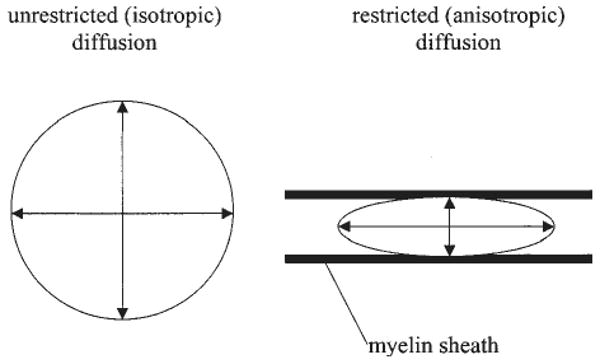
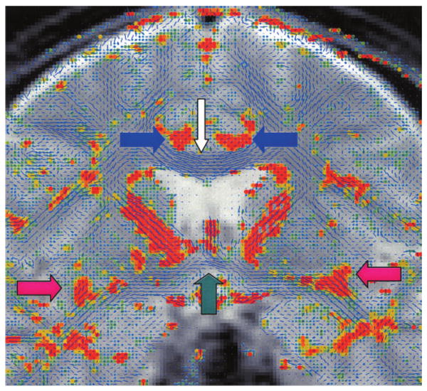
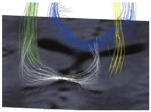
Magnetic resonance diffusion tensor imaging (DTI) is a new technique that can be used to visualize and measure the diffusion of water in brain tissue; it is particularly useful for evaluating white matter abnormalities. In this paper, we review research studies that have applied DTI for the purpose of understanding neuropsychiatric disorders. We begin with a discussion of the principles involved in DTI, followed by a historical overview of magnetic resonance diffusion-weighted imaging and DTI and a brief description of several different methods of image acquisition and quantitative analysis. We then review the application of this technique to clinical populations. We include all studies published in English from January 1996 through March 2002 on this topic, located by searching PubMed and Medline on the key words "diffusion tensor imaging" and "MRI." Finally, we consider potential future uses of DTI, including fiber tracking and surgical planning and follow-up.
Figures
References
-
- Beaulieu C, Allen PS. Determinants of anisotropic water diffusion in nerves. Magn Reson Med. 1994;31:394–400. - PubMed
-
- Wimberger DM, Roberts TP, Barkovich AJ, Prayer LM, Moseley ME, Kucharczyk J. Identification of “premyelination” by diffusion-weighted MRI. J Comput Assist Tomogr. 1995;19:28–33. - PubMed
-
- Hüppi PS, Maier SE, Peled S, Zientara GP, Barnes PD, Jolesz FA, et al. Microstructural development of human newborn cerebral white matter assessed in vivo by diffusion tensor magnetic resonance imaging. Pediatr Res. 1998;44:584–90. - PubMed
-
- Le Bihan D, Breton E, Lallemand D, Grenier P, Cabanis E, Laval-Jeantet M. MR imaging of intravoxel incoherent motions: application to diffusion and perfusion in neurologic disorders. Radiology. 1986;161:401–7. - PubMed
-
- Pierpaoli C, Jezzard P, Basser PJ, Barnett A, Di Chiro G. Diffusion tensor MR imaging of the human brain. Radiology. 1996;201:637–48. - PubMed
Publication types
MeSH terms
Grants and funding
- R01 MH040799/MH/NIMH NIH HHS/United States
- R01 MH050740/MH/NIMH NIH HHS/United States
- R01 RR011747/RR/NCRR NIH HHS/United States
- R01 MH 50747/MH/NIMH NIH HHS/United States
- K02 MH001110/MH/NIMH NIH HHS/United States
- K02 MH 01110/MH/NIMH NIH HHS/United States
- P41 RR013218/RR/NCRR NIH HHS/United States
- R01 MH 40799/MH/NIMH NIH HHS/United States
- P41 RR 13218/RR/NCRR NIH HHS/United States
- R01 NS039335/NS/NINDS NIH HHS/United States
- R01 NS 39335/NS/NINDS NIH HHS/United States
- R01 RR 11747/RR/NCRR NIH HHS/United States
LinkOut - more resources
Full Text Sources
Medical