. 2002 Dec 16;21(24):6660-72.
doi: 10.1093/emboj/cdf619.

A novel variant of the immunoglobulin fold in surface adhesins of Staphylococcus aureus: crystal structure of the fibrinogen-binding MSCRAMM, clumping factor A

Affiliations

A novel variant of the immunoglobulin fold in surface adhesins of Staphylococcus aureus: crystal structure of the fibrinogen-binding MSCRAMM, clumping factor A

Champion C S Deivanayagam et al. EMBO J. .

Abstract

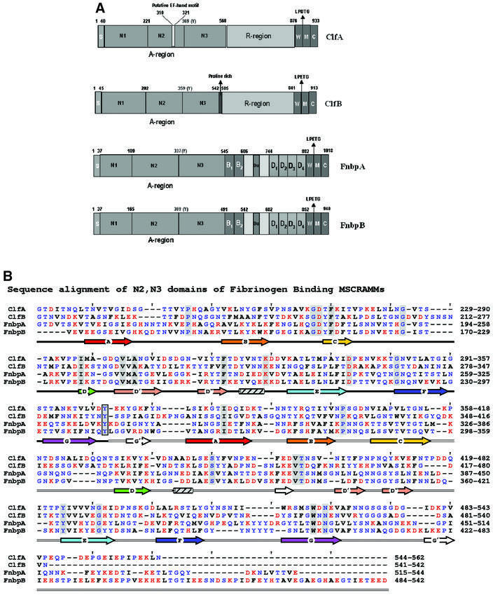
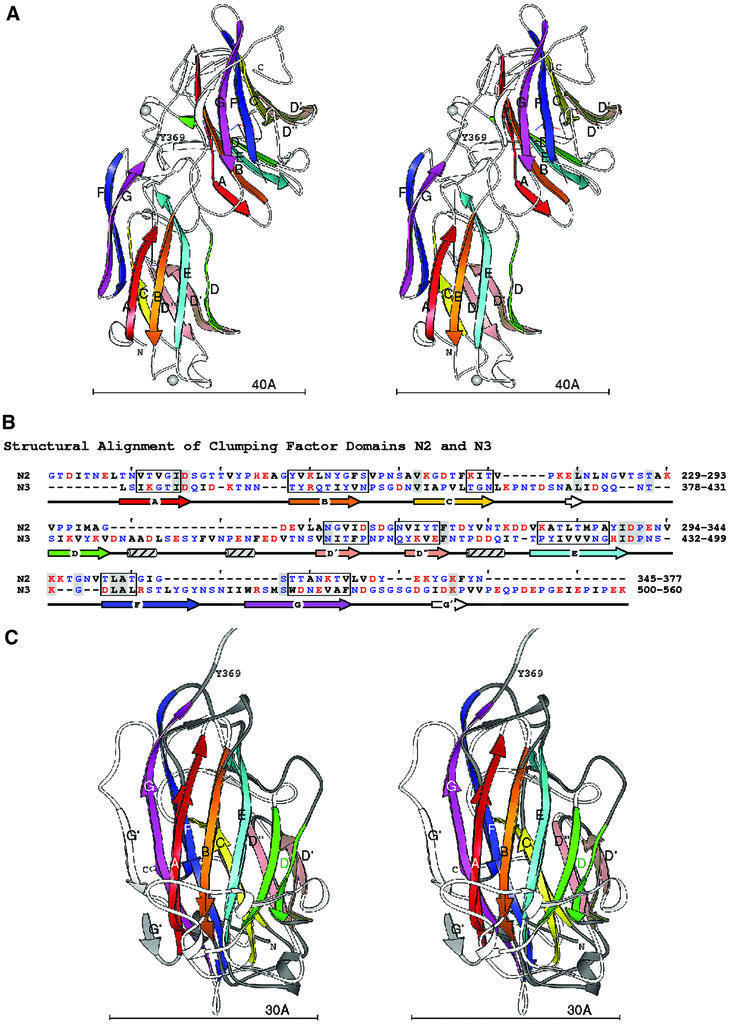
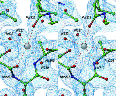
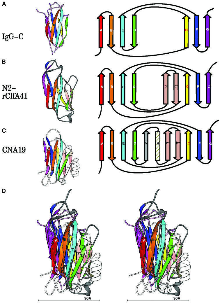
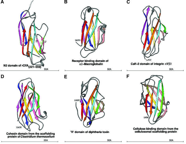
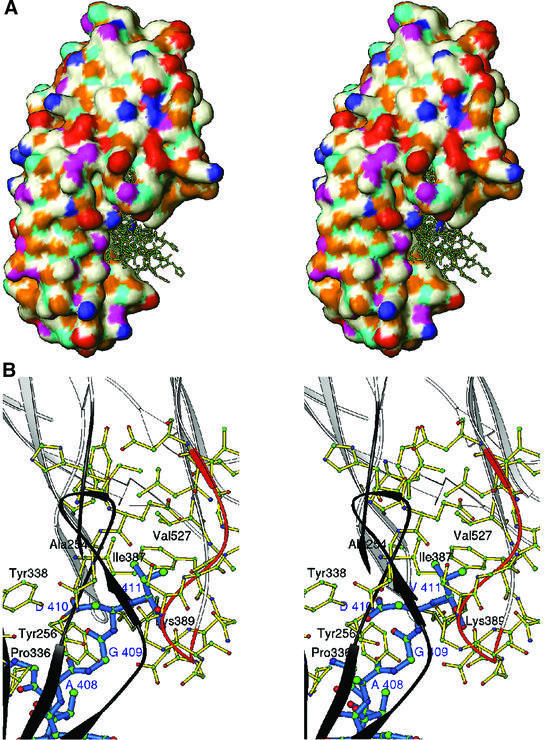
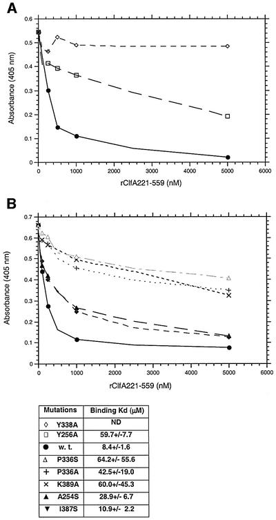
We report here the crystal structure of the minimal ligand-binding segment of the Staphylococcus aureus MSCRAMM, clumping factor A. This fibrinogen-binding segment contains two similarly folded domains. The fold observed is a new variant of the immunoglobulin motif that we have called DE-variant or the DEv-IgG fold. This subgroup includes the ligand-binding domain of the collagen-binding S.aureus MSCRAMM CNA, and many other structures previously classified as jelly rolls. Structure predictions suggest that the four fibrinogen-binding S.aureus MSCRAMMs identified so far would also contain the same DEv-IgG fold. A systematic docking search using the C-terminal region of the fibrinogen gamma-chain as a probe suggested that a hydrophobic pocket formed between the two DEv-IgG domains of the clumping factor as the ligand-binding site. Mutagenic substitution of residues Tyr256, Pro336, Tyr338 and Lys389 in the clumping factor, which are proposed to contact the terminal residues (408)AGDV(411) of the gamma-chain, resulted in proteins with no or markedly reduced affinity for fibrinogen.


Figures

None
Fig. 1. The Fg-binding MSCRAMMs of S.aureus: ClfA, ClfB, FnbpA and FnbpB. (A) The four Fg-binding MSCRAMMs of S.aureus identified so far have a common structural organization including a signal peptide(s) followed by the N-terminal ligand binding A region in which three subdomains, N1, N2 and N3, can be identified. The boundary between N2 and N3 is indicated by a conserved Tyr residue. At the C-terminus, the cell wall-binding region (W), the membrane-spanning domains (M) and the charged C-terminus (C) are present. ClfA has a unique putative EF-hand in the ligand binding A region and a ‘DS’ dipeptide repeat R region. ClfB is similar to ClfA and contains an additional proline-rich segment linking the A- and the R- regions. FnbpA and FnbpB contain the unique fibronectin-binding D repeats and B repeats of unknown function. (B) Sequence alignment of the N-terminal two-thirds of rClfA(221–559) with corresponding regions of ClfB, FnbpA and FnbpB. Red, blue and black letters represent charged, polar and hydrophobic residues, respectively. Identical residues are shaded and a conserved Tyr residue in the connector between the N2 and N3 domains is boxed. The secondary structural elements are colored in rainbow fashion similar to Figures 2, 4 and 5.
None
Fig. 2. Domain structure of rClfA(221–559). (A) Stereo ribbon diagram of the ClfA crystal structure. In the two independent domains, the β-strands A–G are colored in rainbow fashion. The N- and C-termini and the boundary between the domains indicated by Y369 are also labeled. Each domain has a coordinate axis defined by the relative orientation of the sheets. The long, black axis is the average sheet direction. The silver spheres represent the metal ions. (B) Structure-based sequence alignment of the ClfA N2 and N3 domains. The strands A–G are marked with arrows colored as in the ribbon diagrams. Ranges of residues where all Cα superpose within 1.5 Å are boxed. Hydrophobic residues are black, polar are blue and charged are red. Identical residues are highlighted with gray. (C) Superposition of the ClfA N2 and N3 domains. Stereo ribbon diagrams of the two domains are overlaid. β-strands A–G are labeled and colored as above. The coil regions of the first domain are dark gray, the coil linking the two domains is light gray and the coils of the second domain are shown in white. The average sheet direction is aligned with the y-axis, and the average direction between adjacent strands with the x-axis.
None
Fig. 3. Structure of metal binding site M1. A representative 2fofc electron density map (rendered from a normalized map at 1.0σ level) displays octahedral coordination of the metal binding site M1, where three main-chain carbonyl oxygen atoms from Asn267, Ala269 and Val323 contribute together with three water molecules (W622, W627 and W719) to the coordination geometry.
None
Fig. 4. Topology of the IgG and MSCRAMM domains. Ribbon diagrams of the labeled strands A–G colored in rainbow fashion as in Figure 2. The corresponding topology diagram of the structure is shown to the right. (A) IgG-C domain. (B) ClfA N2 domain [rClfA(221–559)]. (C) CNA19 domain (CNA169–318). (D) Superposition of IgG, rClfA(221–559)-N2 and CNA169–318.
None
Fig. 5. Ribbon diagram of crystal structures with the DEv-IgG fold colored in rainbow fashion as in Figures 2 and 4. (A) N2 domain of ClfA. (B) Receptor-binding domain of α2-macroglobulin (PDB code: 1AYO, Z-score: 8.7, r.m.s.d.: 2.8, equiv. resid: 133). (C) Calf-2 domain of extracellular segment of the integrin αVβ3 (PDB Code: 1JV2, Z-score: 8.4, r.m.s.d.: 12.3, equiv. resid: 149). (D) Cohesin domain from the scaffolding protein of Clostridium thermocellum (PDB Code: 1AOH, Z-score: 7.6, r.m.s.d.: 3.4, equiv. resid: 123). (E) The R-domain of diphtheria toxin (PDB Code: 1DDT, Z-score: 7.2, r.m.s.d.: 4.2, equiv. resid: 129). (F) The cellulosomal scaffolding protein (PDB Code: 1NBC, Z-score: 7.0, r.m.s.d.: 3.0, equiv. resid: 109). Z-scores, r.m.s.d. values and equiv. resid. are from DALI.
None
Fig. 6. (A) Stereo surface plot showing seven solutions of the Fg γ-chain peptide docked into a hydrophobic pocket between N2 and N3 domains. The hydrophobic, polar, positive and negative residues are shown in white, magenta, blue and red, respectively. Cyan and gold represent hydrogen bond donors and acceptors, respectively. (B) Stereo ribbon diagram showing the interactions of residues 408AGDV411 (blue) of the γ-chain of Fg with residues from both the N2 (dark gray) and N3 (white) domains of rClfA(221–559). The ribbon of the glycine-rich region 532GSGSGDGI539 is colored in orange.
None
Fig. 7. (A) The relative affinities of rClfA221–559w.t. (closed circles), rClfA221–559Y256→A (open squares) and rClfA221–559Y338→A (open diamonds) for Fg were examined in an inhibition ELISA-type assay (Smith et al., 1993). Biotin-labeled rClfA221–559 was incubated with the indicated increasing concentration of unlabeled ClfA proteins in Fg-coated microtiter wells. The amount of labeled ClfA protein bound to the immobilized Fg was then quantitated. The Kd of each protein was calculated by determining the affinity of the proteins for a fluorescein-labeled 17-amino-acid-long synthetic peptide representing the C-terminus of the Fg γ-chain using fluorescence polarization. (B) The relative affinities of rClfA221–559A254→S (upright closed triangles), rClfA221–559I387→S (inverted closed triangles), rClfA221–559 K389→A (×), rClfA221–559P336→A (+), rClfA221–559P336→S (open upright triangles) for Fg were examined in the same way as above. Kd was also calculated as indicated.

References

    1. Adams P.D., Pannu,N.S., Read,R.J. and Brünger,A.T. (1997) Cross-validated maximum likelihood enhances crystallographic simulated annealing refinement Proc. Natl Acad. Sci. USA, 94, 5018–5023. - PMC - PubMed
    1. Blanchet C., Combet,C., Geourjon,C. and Deleage,G. (2000) MPSA: integrated system for multiple protein sequence analysis with client/server capabilities. Bioinformatics, 16, 286–287. - PubMed
    1. Bork P., Holm,L. and Sander,C. (1994) The immunoglobulin superfamily domains in cell adhesion molecules and surface receptors belong to a new structural set, which is close to that containing the variable domains. J. Mol. Biol., 242, 309–320. - PubMed
    1. Branden C. and Tooze,J. (1999) Introduction to Protein Structure, 2nd edn. Garland Publishing, New York, NY.
    1. Brünger A.T. (1992) Free R value: a novel statistical quantity for assessing the accuracy of crystal structures. Nature, 355, 472–475. - PubMed

Publication types

MeSH terms

Associated data

LinkOut - more resources