. 2002 Dec 16;21(24):6689-99.
doi: 10.1093/emboj/cdf689.

Myosin VIIa, harmonin and cadherin 23, three Usher I gene products that cooperate to shape the sensory hair cell bundle

Affiliations

Myosin VIIa, harmonin and cadherin 23, three Usher I gene products that cooperate to shape the sensory hair cell bundle

Batiste Boëda et al. EMBO J. .

Abstract

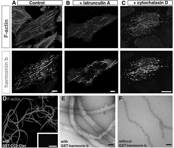
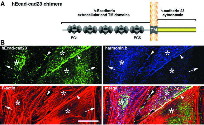
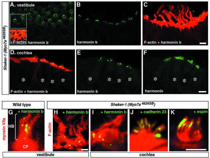
Deaf-blindness in three distinct genetic forms of Usher type I syndrome (USH1) is caused by defects in myosin VIIa, harmonin and cadherin 23. Despite being critical for hearing, the functions of these proteins in the inner ear remain elusive. Here we show that harmonin, a PDZ domain-containing protein, and cadherin 23 are both present in the growing stereocilia and that they bind to each other. Moreover, we demonstrate that harmonin b is an F-actin-bundling protein, which is thus likely to anchor cadherin 23 to the stereocilia microfilaments, thereby identifying a novel anchorage mode of the cadherins to the actin cytoskeleton. Moreover, harmonin b interacts directly with myosin VIIa, and is absent from the disorganized hair bundles of myosin VIIa mutant mice, suggesting that myosin VIIa conveys harmonin b along the actin core of the developing stereocilia. We propose that the shaping of the hair bundle relies on a functional unit composed of myosin VIIa, harmonin b and cadherin 23 that is essential to ensure the cohesion of the stereocilia.


Figures

None
Fig. 1. (A) Predicted structures of harmonin a, b and c isoforms. Class a isoforms contain three PDZ domains and one coiled-coil (CC1) domain. The longest isoforms are grouped into class b; they contain an additional coiled-coil domain (CC2), and a proline, serine, threonine (PST)-rich region. Class c isoforms are the shortest; they contain only the first two PDZ domains and the first coiled-coil domain. (B) Schematic representation of an inner ear sensory hair cell. At the apical surface of the hair cell, a number of microvilli-like structures, called stereocilia, form the hair bundle. Each stereocilium contains a core of actin filaments (shown in red in the right panel). The central actin filaments of all the stereocilia insert into the cuticular plate, i.e. a dense meshwork of actin filaments lying beneath the apical cell surface. (C–F) Harmonin b in the vestibular hair cells (mouse). (C and D) At E14, harmonin b isoforms are only detected at the apical surface of the hair cells, whereas myosin VIIa staining delineates the whole cells. In a few cells, harmonin b immunoreactivity appears as a circle of bead-like foci located around the periphery of the cuticular plate (D and insets). At P8, harmonin b is located at the tips of the stereocilia (E, and detail in F), whereas myosin VIIa is present in both the hair cell bodies and stereocilia (E and F). (G–I) Harmonin in the cochlear hair cells (rat). (G) Schematic representation of the cochlear auditory organ (organ of Corti). It is made up of sensory cells (in red), namely the single row of inner hair cells (ihc) and the three rows of outer hair cells (ohc), and various types of supporting cells (in blue). (H and I) Distribution of harmonin isoforms in the rat organ of Corti at P4. Harmonin b is only detected in the hair bundle (H) of the sensory cells whereas, using the NW2 pan-harmonin antibody, harmonin isoforms a, b and c are detected in the cuticular plate as well as in the apical hair bundle (I). Asterisks indicate hair cell bodies. Bars: 10 µm in (C–E) and (H–I); and 5 µm in (F).
None
Fig. 2. Harmonin b induces F-actin bundling in HeLa cells. (A–D) HeLa cells were transiently transfected with either harmonin a (A) or harmonin b (B–D) cDNAs for 20 h. (A) Harmonin a is distributed throughout the cell body. (BD) In contrast, overexpression of harmonin b (green) leads to the formation of harmonin b-immunoreactive long curvy bundles that co-localize with actin filaments (red). (EJ) HeLa cells transfected with harmonin b and collected 6 h after transfection. At this stage, most harmonin b labelling is present almost exclusively at the cell plasma membrane–substrate interface. A triple staining with rhodamine–phalloidin (red), anti-vinculin (green) and anti-harmonin b (blue) antibodies reveals that harmonin b decorates the extremities of actin stress fibres (violet in E–G). The enrichment of harmonin b labelling (blue) occurs close to the focal adhesion plaques visualized by anti-vinculin (green) staining (H–J). (F), (G), (I) and (J) are higher magnification views of the areas boxed in (E) and (H), respectively. Harmonin b and F-actin are both enriched at the distal ends of actin fibres (violet in F and G), and they slightly overlapped with the vinculin staining (I and J). Bars: 10 µm.
None
Fig. 3. (A–C) Harmonin b induces a resistance of actin filament to latrunculin A and cytochalasin D. (A) Transfected HeLa cells labelled for F-actin (red) and harmonin b (green). Actin stress fibres are observed in transfected cells that produce harmonin b as well as in the surrounding untransfected cells. (B and C) The disruptive effect of latrunculin A (B) and cytochalasin D (C) on actin filaments is not observed in transfected cells that produce harmonin b. With either of these drugs, both the cortical actin filaments and the harmonin b-unlabelled stress fibres were disrupted, whereas the har monin b-associated actin fibres were unaffected. (D–F) Harmonin b is an actin-bundling protein. Light microscopy (E) and electron microscopy (F and G) demonstration of F-actin bundle formation. (D) In the presence of GST-tagged CC2-Cter harmonin fragment, large and long F-actin bundles are observed. No bundled F-actin is observed in the absence of this protein (inset in D). In vitro electron microscopy analysis showed that actin filaments assemble into bundles in the presence (E), but not in the absence (F) of harmonin b. Bars: 10 µm in (A–C), 5 µm in (D) and 60 nm in (E) and (F).
None
Fig. 4. Cadherin 23 in the differentiating hair cells (mouse). (A and B) In the vestibule, at E14, cadherin 23 is only detected at the apical surface of the hair cells (A), whereas myosin VIIa is detected in the entire hair cells (B). Up to E16, cadherin 23 delineates the entire length of the hair bundle, as shown by double staining for cadherin 23 and F-actin (C). (DF) In a tangential section of a P8 vestibular macula, cadherin 23 (green) is also detected in the hair bundles, mainly at the tips of the stereocilia, visualized by F-actin staining (red). (G and H) Electron microscopy of a hair bundle from a P3 mouse cochlear hair cell. Cadherin 23 is detected between adjacent stereocilia, especially at their apical ends, as shown at higher magnification (arrowheads in H). Bars: 20 µm in (A) and (B); 3 µm in (C); 10 µm in (D); and (E), 5 µm in (F) and 100 nm in (G) and (H).
None
Fig. 5. Harmonin b co-localizes with hEcad–cad23 in co-transfected HeLa cells. (A) Schematic diagram of the hEcad–cad23 chimera: it is composed of the five extracellular cadherin repeats (EC) and the transmembrane domain (TM) of human Ecadherin (hEcad) fused to the human cadherin 23 cytodomain (cad23). (B) HeLa cells were transiently transfected with either hEcad–cad23 or harmonin b cDNAs for 20 h. The hEcad–cad23 chimeric protein is targeted to cell–cell contacts (green; revealed by Pcad-C) essentially at the junctions between two transfected cells (arrowheads). Harmonin b (V5-tagged) is also recruited to these contacts, where it co-localized with the hEcad–cad23 chimera and with actin filaments (red). Neither the hEcad–cad23 chimera nor harmonin b is enriched at cell–cell contacts established with untransfected cells (arrows). In the cell cytoplasm, the presence of the hEcad–cad23 protein shifts the filamentous pattern usually observed with harmonin b, to punctiform actin-rich structures. Asterisks indicate HeLa cell bodies. Bar: 20 µm.
None
Fig. 6. Harmonin binds to cadherin 23 and myosin VIIa. (A) Pull-down assay. Extracts of transfected HEK293 cells producing either harmonin a, harmonin b or the myosin VIIa tail were incubated with immobilized GST-tagged cadherin 23 cytodomain or GST alone. The harmonins a and b bind to the GST-tagged cadherin 23 cytodomain but not to GST alone. The myosin VIIa tail does not bind to cadherin 23. (B–D) In vitro binding assays. (B) Characterization of the harmonin–cadherin 23 interaction domain. Different harmonin fragments were incubated with immobilized biotin-tagged cadherin 23 cytodomain. Only the PDZ1–PDZ2 peptide (amino acids 138–403) of harmonin and the PDZ2 domain alone (amino acids 189–307) bound to the cadherin 23 cytodomain. Biotin-tagged CAT (chloramphenicol acetyltransferase) was used as a negative control. (C) The C-terminal MyTH4 + FERM repeat of myosin VIIa (myosin VIIa-Cter) is incubated with different GST-tagged proteins. The myosin VIIa-Cter interacts with GST-tagged harmonin a, and with GST–MyRIP (used as a positive control), whereas it fails to bind to GST. The ezrin FERM domain (used as a negative control) does not bind to GST–harmonin a. (D) Characterization of the harmonin–myosin VIIa interaction domain. The myosin VIIa-Cter was incubated with different immobilized GST-tagged harmonin fragments. The PDZ1 domain of harmonin, but neither PDZ2 nor PDZ3, binds to myosin VIIa. (E) Schematic diagram illustrating how harmonin b could interact with myosin VIIa, cadherin 23 and F-actin. Acronyms: FERM (4.1, ezrin, radixin, moesin); MyTH4 (myosin tail homology 4); SH3 (src homology-3), IQ (isoleucine–glutamine motifs); EC (extracellular cadherin repeats).
None
Fig. 7. Myosin VIIa is required for the proper targeting of harmonin b. Vestibular (A–C) and cochlear (D–F) hair cells from shaker-1 Myo7a4626SB mice at P6. (AC) Harmonin b (green) decorates the apical surface of the hair cells, around the cuticular plate. Harmonin b is not detected in the hair bundles visualized by F-actin staining (red). (DF) In the cochlea as well (asterisks over hair cell bodies), harmonin b fails to reach the hair bundle and punctiform harmonin b staining is observed around and within the hair cell cuticular plate (D and E). (F) In an adjacent section labelled with the NW2 antibody to harmonins a, b, and c, all harmonin isoforms are detected simultaneously. In addition to the harmonin b staining revealed in (E), a diffuse labelling corresponding to harmonins a and c is observed in the cuticular plate and the hair bundle as well. (G–K) Higher magnification views of vestibular (G and H) and cochlear (IK) hair bundles from P2 mice. (G) In wild-type mice, myosin VIIa (red) is present throughout the cell, including the cuticular plate (CP) and the overlying hair bundle (HB). Harmonin b (yellow) is located mainly into the hair bundle. (H–K) In contrast, in shaker-1 Myo7a4626SB mice, harmonin b is organized mainly in a circle of bead-like foci located between the actin-rich cuticular plate and the actin of the circumferential belt (H and I), whereas cadherin 23 (J) and espin (K), a putative actin filament cross-linking protein of the stereocilia, are properly targeted to the hair bundle. Bars: 10 µm in (A–F); and 5 µm in (G–K).

References

    1. Ahmed Z.M., Riazuddin,S., Bernstein,S.L., Ahmed,Z., Khan,S., Griffith,A.J., Morell,R.J., Friedman,T.B. and Wilcox,E.R. (2001) Mutations of the protocadherin gene PCDH15 cause Usher syndrome type 1F. Am. J. Hum. Genet., 69, 25–34. - PMC - PubMed
    1. Alagramam K.N. et al. (2001a) Mutations in the novel protocadherin PCDH15 cause Usher syndrome type 1F. Hum. Mol. Genet., 10, 1709–1718. - PubMed
    1. Alagramam K.N., Murcia,C.L., Kwon,H.Y., Pawlowski,K.S., Wright,C.G. and Woychik,R.P. (2001b) The mouse Ames waltzer hearing-loss mutant is caused by mutation of Pcdh15, a novel protocadherin gene. Nat. Genet., 27, 99–102. - PubMed
    1. Angst B.D., Marcozzi,C. and Magee,A.I. (2001) The cadherin superfamily. J. Cell Sci., 114, 625–626. - PubMed
    1. Berg J.S., Powell,B.C. and Cheney,R.E. (2001) A millennial myosin census. Mol. Biol. Cell, 12, 780–794. - PMC - PubMed

Publication types

MeSH terms