

Is protein disulfide isomerase a redox-dependent molecular chaperone?
- PMID: 12485997
- PMCID: PMC139105
- DOI: 10.1093/emboj/cdf685
Is protein disulfide isomerase a redox-dependent molecular chaperone?
Abstract
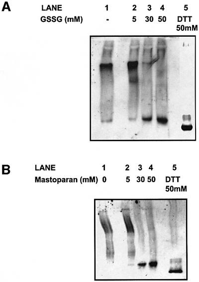
Protein disulfide isomerase (PDI) is a multifunctional protein catalysing the formation of disulfide bonds, acting as a molecular chaperone and being a component of the enzymes prolyl 4-hydroxylase (P4H) and microsomal triglyceride transfer protein. The role of PDI as a molecular chaperone or polypeptide-binding protein is mediated primarily through an interaction of substrates with its b' domain. It has been suggested that this binding is regulated by the redox state of PDI, with association requiring the presence of glutathione, and dissociation the presence of glutathione disulfide. To determine whether this is the case, we investigated the ability of PDI to bind to a folding polypeptide chain within a functionally intact endoplasmic reticulum and to be dissociated from the alpha-subunit of P4H in vitro in the presence of reducing or oxidizing agents. Our results clearly demonstrate that binding of PDI to these polypeptides is not regulated by its redox state. We also demonstrate that the dissociation of PDI from substrates observed in the presence of glutathione disulfide can be explained by competition for the peptide-binding site on PDI.
Figures


Similar articles
-
Protein disulfide isomerase: the multifunctional redox chaperone of the endoplasmic reticulum.Semin Cell Dev Biol. 1999 Oct;10(5):481-93. doi: 10.1006/scdb.1999.0319. Semin Cell Dev Biol. 1999. PMID: 10597631 Review.
-
Protein disulfide isomerase acts as a molecular chaperone during the assembly of procollagen.J Biol Chem. 1998 Apr 17;273(16):9637-43. doi: 10.1074/jbc.273.16.9637. J Biol Chem. 1998. PMID: 9545296
-
Molecular characterization of the principal substrate binding site of the ubiquitous folding catalyst protein disulfide isomerase.J Biol Chem. 2004 Mar 12;279(11):10374-81. doi: 10.1074/jbc.M312193200. Epub 2003 Dec 18. J Biol Chem. 2004. PMID: 14684740
-
Protein disulfide-isomerase, a folding catalyst and a redox-regulated chaperone.Free Radic Biol Med. 2015 Jun;83:305-13. doi: 10.1016/j.freeradbiomed.2015.02.007. Epub 2015 Feb 17. Free Radic Biol Med. 2015. PMID: 25697778 Review.
-
Procollagen binds to both prolyl 4-hydroxylase/protein disulfide isomerase and HSP47 within the endoplasmic reticulum in the absence of ascorbate.FEBS Lett. 2000 Jan 21;466(1):19-25. doi: 10.1016/s0014-5793(99)01713-5. FEBS Lett. 2000. PMID: 10648804
Cited by
-
Celiac anti-type 2 transglutaminase antibodies induce phosphoproteome modification in intestinal epithelial Caco-2 cells.PLoS One. 2013 Dec 31;8(12):e84403. doi: 10.1371/journal.pone.0084403. eCollection 2013. PLoS One. 2013. PMID: 24391952 Free PMC article.
-
Potential Application of Ixeris dentata in the Prevention and Treatment of Aging-Induced Dry Mouth.Nutrients. 2018 Dec 15;10(12):1989. doi: 10.3390/nu10121989. Nutrients. 2018. PMID: 30558302 Free PMC article.
-
Specific proteins of the trapezius muscle correlate with pain intensity and sensitivity - an explorative multivariate proteomic study of the trapezius muscle in women with chronic widespread pain.J Pain Res. 2016 Jun 2;9:345-56. doi: 10.2147/JPR.S102275. eCollection 2016. J Pain Res. 2016. PMID: 27330327 Free PMC article.
-
Comparison of protein expression profiles of different stages of lymph nodes metastasis in breast cancer.Int J Biol Sci. 2012;8(3):353-62. doi: 10.7150/ijbs.3157. Epub 2012 Feb 20. Int J Biol Sci. 2012. PMID: 22393307 Free PMC article.
-
Pancreas-specific protein disulfide isomerase has a cell type-specific expression in various mouse tissues and is absent in human pancreatic adenocarcinoma cells: implications for its functions.J Mol Histol. 2009 Jun;40(3):189-99. doi: 10.1007/s10735-009-9230-5. Epub 2009 Oct 11. J Mol Histol. 2009. PMID: 19821078
References
-
- Bottomley M.J., Batten,M.R., Lumb,R.A. and Bulleid,N.J. (2001) Quality control in the endoplasmic reticulum of unassembled procollagen C-propeptides. Curr. Biol., 11, 1–5. - PubMed
-
- Cai H., Wang,C.-C. and Tsou,C.-L. (1994) Chaperone-like activity of protein disulfide isomerase in the refolding of a protein with no disulfide bonds. J. Biol. Chem., 269, 24550–24552. - PubMed
-
- Chessler S.D. and Byers,P.H. (1992) Defective folding and stable association with protein disulfide isomerase/prolyl hydroxylase of type I procollagen with a deletion in the proα2(I) chain that preserves the gly-X-Y repeat pattern. J. Biol. Chem., 267, 7751–7757. - PubMed
-
- Chessler S.D. and Byers,P.H. (1993) BiP binds type I procollagen proαchains with mutations in the carboxy-terminal propeptide synthesized by cells from patients with osteogenesis imperfecta. J. Biol. Chem., 268, 18226–18233. - PubMed
Publication types
MeSH terms
Substances
LinkOut - more resources
Full Text Sources
Other Literature Sources
Molecular Biology Databases
