. 2002 Dec 16;21(24):6811-9.
doi: 10.1093/emboj/cdf669.

Differential acetylation of Tat coordinates its interaction with the co-activators cyclin T1 and PCAF

Affiliations

Differential acetylation of Tat coordinates its interaction with the co-activators cyclin T1 and PCAF

Vanessa Brès et al. EMBO J. .

Abstract

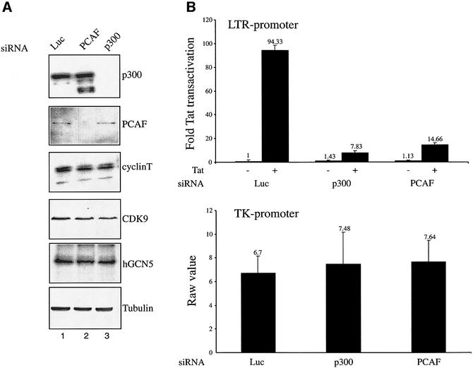
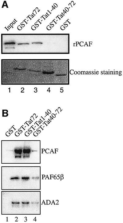
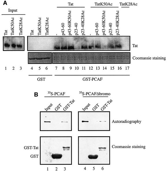
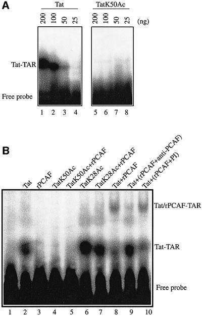
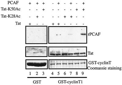
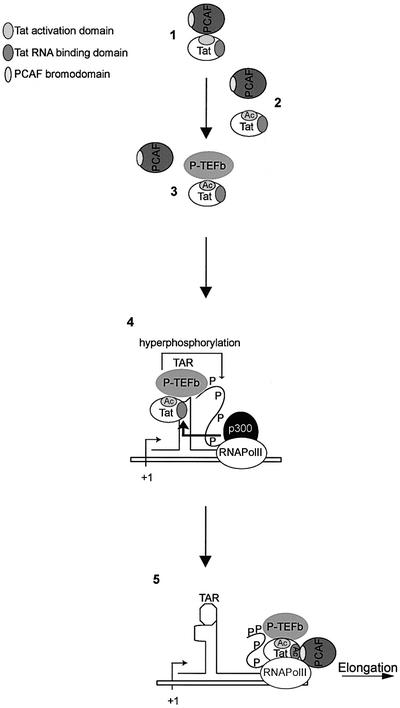
The HIV-1 transactivator protein, Tat, is an atypical transcriptional activator that functions through binding, not to DNA, but to a short leader RNA, TAR. Although details of its functional mechanism are still unknown, emerging findings suggest that Tat serves primarily to adapt co-activator complexes such as p300, PCAF and P-TEFb to the HIV-1 long terminal repeat. Hence, an understanding of how Tat interacts with these cofactors is crucial. It has recently been shown that acetylation at a single lysine, residue 50, regulated the association of Tat with PCAF. Here, we report that in the absence of Tat acetylation, PCAF binds to amino acids 20-40 within Tat. Interestingly, acetylation of Tat at Lys28 abrogates Tat-PCAF interaction. Acetylation at Lys50 creates a new site for binding to PCAF and dictates the formation of a ternary complex of Tat-PCAF-P-TEFb. Thus, differential lysine acetylation of Tat coordinates the interactions with its co-activators, cyclin T1 and PCAF. Our results may help in understanding the ordered recruitment of Tat co-activators to the HIV-1 promoter.


Figures

None
Fig. 1. Endogenous p300 and PCAF are required for Tat-mediated transactivation of the HIV-1 LTR. (A) HeLa P4 cells containing the lacZ gene under the control of an integrated HIV-1 LTR were transfected with siRNA specific for p300, PCAF or luciferase. Western blotting analyses of p300, PCAF, hGCN5, cyclin T1, CDK9 and tubulin expression in HeLa P4 cells transfected with the indicated siRNA were performed. (B) Top panel: Tat-mediated transactivation of the LTR was analyzed 24 h post-transfection with 30 ng of a Tat expression plasmid. Fold Tat transactivation was calculated relative to transfection in the absence of Tat expression plasmid. Bottom panel: luciferase activity measured from an internal control plasmid encoding Renilla under the control of the TK promoter.
None
Fig. 2. Amino acids 1–40 within full-length non-acetylated Tat protein bind both free rPCAF and complex-associated PCAF. (A) Amino acids 1–40 of Tat bind PCAF. GST, GST–Tat1–72, GST–Tat1–40 or GST–Tat40–72 was incubated separately with recombinant, Flag-tagged PCAF (25 ng). The beads were extensively washed in buffer containing 0.25 M KCl and resuspended in Laemmli buffer. The presence of rPCAF was assessed by western blotting using anti-Flag M2 antibody (top panel). Input is shown in lane 1. Coomassie Blue stainings of GST and GST fusion proteins are shown (bottom panel). (B) Tat amino acids 1–40 bind complex-associated PCAF. GST, GST–Tat1–72, GST–Tat1–40 or GST–Tat40–72 was incubated with purified, nuclear, PCAF complex. The beads were extensively washed in buffer containing 0.25 M KCl and resuspended in Laemmli buffer. The presence of PCAF, PAF65β and ADA2 was assessed by western blotting.
None
Fig. 3. Acetylation at Lys28 and Lys50 of Tat dictates the region of Tat interacting with PCAF. (A) GST (lanes 4–6) or GST–PCAF (lanes 7–17) beads were incubated with 100 ng of chemically synthesized Tat1–86 protein in non-acetylated (Tat), acetylated at Lys50 (TatK50Ac) or acetylated at Lys28 (TatK28Ac) forms for 1 h at 4°C. Additionally, GST–PCAF–beads were incubated separately with 1 µg of peptides corresponding to amino acids 43–60 (p43–60, lanes 8 and 13; p43–60K50Ac, lanes 9 and 14) and amino acids 23–40 (p23–40, lanes 10 and 15; p23–40K28Ac, lanes 11 and 16) for 1 h at 4°C prior to incubation with the indicated Tat proteins. After incubation, the beads were pelleted, extensively washed in buffer containing 0.25 M KCl and resuspended in Laemmli buffer. The presence of Tat was assessed by western blotting using anti-Tat antibody (top panel). Coomassie Blue stainings are shown (bottom panel). Lanes 1–3 correspond to the input materials. (B) GST or GST–Tat was incubated with in vitro translated, [35S]methionine/cysteine-labeled PCAF or PCAFΔbromo for 1 h at 4°C in binding buffer containing 0.25 M KCl. After incubation, the beads were pelleted and extensively washed in buffer containing 0.25 M KCl, and resuspended in Laemmli buffer. Bound materials were separated by SDS–PAGE and analyzed by Coomassie Blue staining and autoradiography. Lanes 1 and 4 correspond to the input materials.
None
Fig. 4. Tat acetylation regulates formation of a Tat–TAR–PCAF complex. (A) Acetylation of Tat at Lys50 is sufficient to abrogate binding to TAR RNA. 32P-labeled TAR RNA was incubated with increasing amounts of chemically synthesized Tat protein, which was either non-acetylated (lanes 1–4) or acetylated at Lys50, Tat K50Ac (lanes 5–8). Tat–TAR complexes were analyzed by non-denaturing acrylamide gel electrophoresis. (B) PCAF binds a Tat–TAR complex. 32P-labeled TAR RNA was incubated with 100 ng of chemically synthesized Tat protein either unmodified (lane 2), acetylated at Lys50, Tat K50Ac (lane 4), or acetylated at Lys28, TatK28Ac (lane 6), alone or with 10 ng of rPCAF (lanes 5, 7 and 8) or rPCAF that had been previously immunodepleted with anti-PCAF polyclonal antibody (lane 9) or control pre-immune serum (lane 10). Lane 1 corresponds to 32P-labeled TAR RNA alone and lane 3 corresponds to TAR RNA incubated with rPCAF.
None
Fig. 5. Formation of the cyclin T1–Tat–PCAF complex is regulated by acetylation of Tat Lys50. GST (lanes 1–3) or GST–cyclin 1 (lanes 4–9) beads were incubated separately with 100 ng of Tat (lanes 1 and 4), TatK28Ac (lanes 2 and 6) or TatK50Ac (lanes 3 and 8). GST–cyclin T1 was incubated first with either Tat (lane 5), TatK28Ac (lane 7) or TatK50Ac (lane 9) for 1 h at 4°C, washed, and further incubated with 25 ng of rPCAF for an additional hour at 4°C. After extensive washing of beads, the presence of Tat and rPCAF was assessed by western blotting using anti-Flag (top panel) or anti-Tat (middle panel) antibody. Coomassie Blue stainings are shown (bottom panel).
None
Fig. 6. Proposed model for the regulation of Tat transcriptional activity by p300 and PCAF. (1) PCAF interacts with Tat amino acids 20–40. (2) PCAF acetylates Tat at Lys28 and dissociates from Tat. (3) P-TEFb associates with Tat. (4) P-TEFb–Tat complex binds TAR RNA, and p300 acetylates Tat at Lys50. (5) Tat dissociates from TAR RNA and PCAF interacts with K50-acetylated TAR RNA-binding domain of Tat. Tat–P-TEFb–PCAF complex associates with the transcription elongation complex.

References

    1. Ait-Si-Ali S. et al. (1998) Histone acetyltransferase activity of CBP is controlled by cycle-dependent kinases and oncoprotein E1A. Nature, 396, 184–186. - PubMed
    1. Barany G. and Merrifield,R.B. (1979) The Peptide. In Gross,E. and Meinhofer,J. (eds), Solid phase peptide synthesis, Vol. 2. Academic Press, New York, NY, pp. 1–284.
    1. Beckett D. (2001) Regulated assembly of transcription factors and control of transcription initiation. J. Mol. Biol., 314, 335–352. - PubMed
    1. Benkirane M., Chun,R.F., Xiao,H., Ogryzko,V.V., Howard,B.H., Nakatani,Y. and Jeang,K.T. (1998) Activation of integrated provirus requires histone acetyltransferase. p300 and P/CAF are coactivators for HIV-1 Tat. J. Biol. Chem., 273, 24898–24905. - PubMed
    1. Berkhout B., Silverman,R.H. and Jeang,K.T. (1989) Tat trans-activates the human immunodeficiency virus through a nascent RNA target. Cell, 59, 273–282. - PubMed

Publication types

MeSH terms