. 2002 Dec 16;21(24):6865-73.
doi: 10.1093/emboj/cdf692.

The DNA chaperone HMGB1 facilitates ACF/CHRAC-dependent nucleosome sliding

Affiliations

The DNA chaperone HMGB1 facilitates ACF/CHRAC-dependent nucleosome sliding

Tiziana Bonaldi et al. EMBO J. .

Abstract

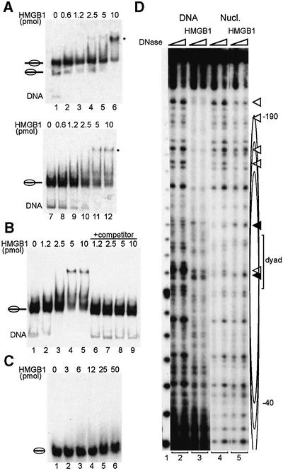
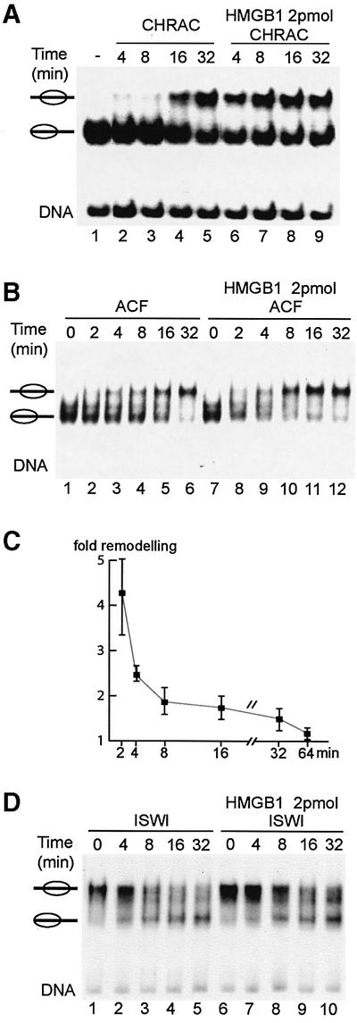
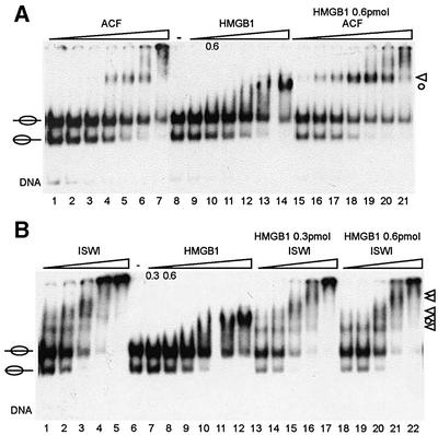
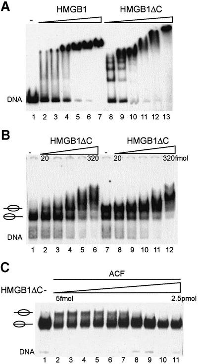
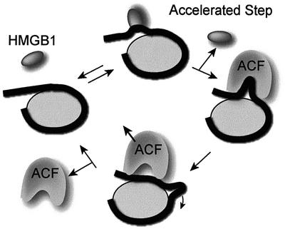
Nucleosome remodelling complexes CHRAC and ACF contribute to chromatin dynamics by converting chemical energy into sliding of histone octamers on DNA. Their shared ATPase subunit ISWI binds DNA at the sites of entry into the nucleosome. A prevalent model assumes that DNA distortions catalysed by ISWI are converted into relocation of DNA relative to a histone octamer. HMGB1, one of the most abundant nuclear non-histone proteins, binds with preference to distorted DNA. We have now found that transient interaction of HMGB1 with nucleosomal linker DNA overlapping ISWI-binding sites enhances the ability of ACF to bind nucleosomal DNA and accelerates the sliding activity of limiting concentrations of remodelling factor. By contrast, an HMGB1 mutant with increased binding affinity was inhibitory. These observations are consistent with a role for HMGB1 as a DNA chaperone facilitating the rate-limiting DNA distortion during nucleosome remodelling.


Figures

None
Fig. 1. Specific interaction of HMGB1 with nucleosomes exhibiting protruding DNA. (A) Increasing amounts of full-length HMGB1 (as indicated) were incubated with nucleosomes (60 fmol) positioned at the centre (lanes 1–6) or at the end (lanes 7–12) of the 248 bp rDNA fragment. The complexes were resolved by electrophoresis on a native polyacrylamide gel. An autoradiograph of the gel is shown. The major nucleosome–HMGB1 complex is marked by an asterisk. Nucleosomes are schematized by ellipses with protruding DNA. (B) HMGB1 binding to the nucleosome does not disrupt the histone octamer. HMGB1 binding was analysed as described in (A) without (lanes 2–5) and with the addition of competitor DNA (lanes 6–9) prior to gel loading. (C) Nucleosomes (60 fmol) assembled on a 146 bp DNA fragment were incubated with increasing amounts of HMGB1 as indicated and analysed by electromobility shift assay as above. (D) DNA (lanes 2 and 3) or purified nucleosomes (lanes 4 and 5) were incubated with HMGB1. DNase I-treated free DNA, nucleosomes and nucleosome–HMGB1 complexes were resolved by PAGE, the DNA isolated from the corresponding bands and analysed on sequencing gels. The cluster of central nucleosome positions is indicated by ellipses, and the area of perturbation of histone–DNA interaction upon HMGB1 binding is marked by white (DNase protection) or black triangles (DNase hypersensitivity). A 10 bp DNA ladder was used as size marker (lane 1).
None
Fig. 2. HMGB1 stimulates CHRAC- and ACF-mediated nucleosome remodelling. (A) Nucleosomes positioned at the end of the DNA fragment (lane 1) were incubated with 5 fmol of native CHRAC and ATP, in the absence (lanes 2–5) or presence of 2 pmol of HMGB1 (lanes 6–9). The reactions were incubated at 16°C and stopped at the indicated time points by the addition of competitor DNA. Nucleosome positions were subsequently analysed by electrophoresis. Nucleosomes are schematized by ellipses with protruding DNA. (B) Nucleosome remodelling assay, as described in (A), using 5 fmol of recombinant ACF in the absence (lanes 1–6) or presence of 2 pmol of HMGB1 (lanes 7–12). (C) Quantification of the HMGB1 effect on nucleosome remodelling. The ratio of nucleosomes moved in the presence of HMGB1 versus nucleosomes moved in the absence of HMGB1 was calculated for four independent experiments and displayed in a graph. (D) Nucleosomes positioned at the centre of the DNA fragment were incubated with 30 fmol of ISWI and ATP, in the absence (lanes 1–5) or presence of 2 pmol of HMGB1 (lanes 6–10) and assayed as described in (A).
None
Fig. 3. HMGB1 promotes the formation of a ACF–nucleosome complex. (A) Nucleosomes preferentially positioned at the centre of the DNA fragment were incubated with increasing amounts of ACF (lanes 1–7; 15, 30, 60, 120, 240, 480 and 960 fmol), HMGB1 (lanes 9–14; 0.3, 0.6, 1.2, 2.4, 4.8 and 9.6 pmol) and increasing amounts of ACF in the presence of 0.6 pmol HMGB1 (lanes 15–21). Complexes were analysed in EMSAs, as before. Nucleosome–ACF complexes are marked by a triangle and nucleosome–HMGB1 complexes are indicated by a circle. (B) Positioned nucleosomes were incubated with increasing amounts of ISWI (lanes 1–5; 60, 120, 240, 480 and 960 fmol), HMGB1 (lanes 7–12, 0.3, 0.6, 1.2, 2.4, 4.8 and 9.6 pmol) and ISWI with fixed amounts of HMGB1 (lanes 13–17, 0.3 pmol HMGB1; lanes 18–22, 0.6 pmol HMGB1). Nucleosome– ISWI complexes are marked by triangles and the nucleosome– HMGB1 complex is indicated by a circle.
None
Fig. 4. HMGB1ΔC inhibits ACF-mediated nucleosome remodelling. (A) Comparison of HMGB1 and HMGB1ΔC binding to the 248bp rDNA fragment. The DNA fragment was incubated with increasing amounts of HMGB1 (lanes 2–7; 1.5, 2.3, 3.4, 5, 7.6 and 11 pmol) and HMGB1ΔC (lanes 8–13; 50, 75, 112, 169, 253 and 380 fmol). HMG–DNA complexes were separated on native polyacrylamide gels. (B) HMGB1ΔC interacts with nucleosomes positioned at the border or the centre of the rDNA fragment. Positioned nucleosomes were incubated with increasing amounts of HMGB1ΔC (20, 40, 80, 160 and 320 fmol). (C) ACF-mediated nucleosome remodelling is inhibited by the addition of HMGB1ΔC. Nucleosomes positioned at the border of the DNA fragment were incubated with ACF, ATP and increasing amounts of HMGB1ΔC (lanes 2–11; 5, 10, 20, 40, 80, 160, 320, 640, 1240 and 2560 fmol). Remodelling reactions were stopped by the addition of competitor DNA and nucleosome positions were analysed on a 4.5% polyacrylamide gel.
None
Fig. 5. Model showing the stimulatory effect of HMGB1 on ACF-mediated nucleosome remodelling. The wrapping of DNA (black line) around a histone octamer (grey sphere) is shown sideways. See text for details.

References

    1. Aalfs J.D. and Kingston,R.E. (2000) What does ‘chromatin remodeling’ mean? Trends Biochem. Sci., 25, 548–555. - PubMed
    1. Aidinis V., Bonaldi,T., Beltrame,M., Santagata,S., Bianchi,M.E. and Spanopoulou,E. (1999) The RAG1 homeodomain recruits HMG1 and HMG2 to facilitate recombination signal sequence binding and to enhance the intrinsic DNA-bending activity of RAG1-RAG2. Mol. Cell. Biol., 19, 6532–6542. - PMC - PubMed
    1. An W., van Holde,K. and Zlatanova,J. (1998) The non-histone chromatin protein HMG1 protects linker DNA on the side opposite to that protected by linker histones. J. Biol. Chem., 273, 26289–26291. - PubMed
    1. Beard P. (1978) Mobility of histones on the chromosome of simian virus 40. Cell, 15, 955–967. - PubMed
    1. Becker P.B. and Hörz,W. (2002) ATP-dependent nucleosome remodelling. Annu. Rev. Biochem., 71, 247–273. - PubMed

Publication types