. 2002 Dec 16;21(24):6915-24.
doi: 10.1093/emboj/cdf678.

Human Dcp2: a catalytically active mRNA decapping enzyme located in specific cytoplasmic structures

Affiliations

Human Dcp2: a catalytically active mRNA decapping enzyme located in specific cytoplasmic structures

Erwin van Dijk et al. EMBO J. .

Abstract

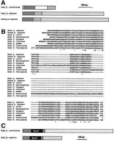
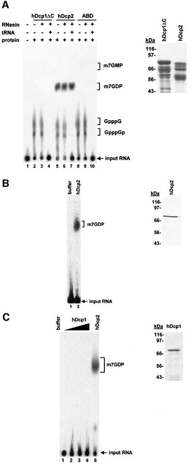
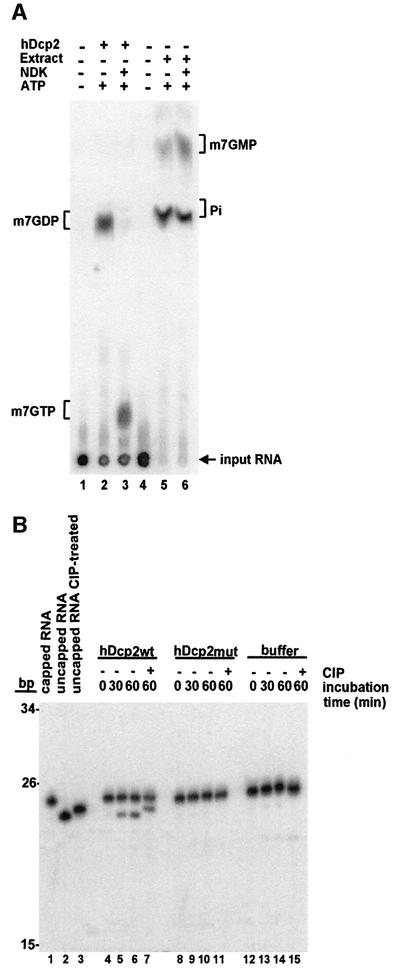
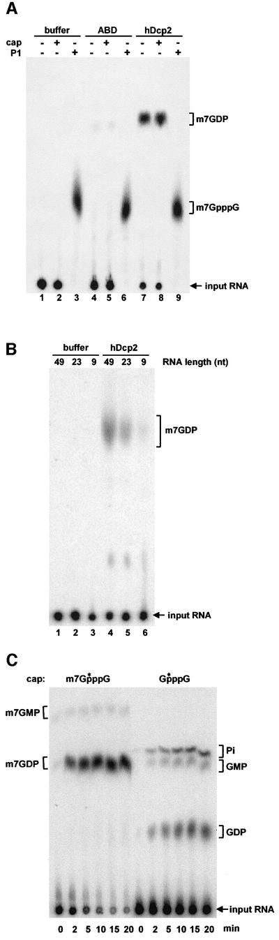
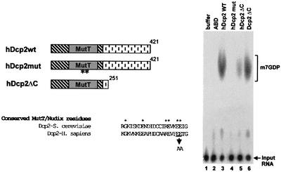
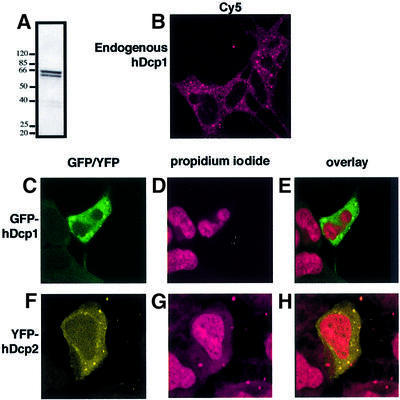
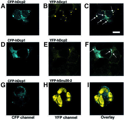
We have cloned cDNAs for the human homologues of the yeast Dcp1 and Dcp2 factors involved in the major (5'-3') and NMD mRNA decay pathways. While yeast Dcp1 has been reported to be the decapping enzyme, we show that recombinant human Dcp2 (hDcp2) is enzymatically active. Dcp2 activity appears evolutionarily conserved. Mutational and biochemical analyses indicate that the hDcp2 MutT/Nudix domain mediates this activity. hDcp2 generates m7GDP and 5'-phosphorylated mRNAs that are 5'-3' exonuclease substrates. Corresponding decay intermediates are present in human cells showing the relevance of this activity. hDcp1 and hDcp2 co-localize in cell cytoplasm, consistent with a role in mRNA decay. Interestingly, these two proteins show a non-uniform distribution, accumulating in specific foci.


Figures

None
Fig. 1. Sequence similarities and domain structures of yeast Dcp1 and Dcp2 and human homologues. (A) Schematic representation of the structure of yeast Dcp1 and the human homologues hDcp1 and hDcp1b. The two shaded areas in Dcp1 are conserved between yeast and human, yeast-specific sequences are indicated in white. The long C-terminal tails in hDcp1 and hDcp1b that are not present in yeast Dcp1 and show little sequence similarity between the two human proteins are shown as distinct stippled areas. (B) Sequence alignment of the conserved domain of Dcp1 from a variety of eukaryotic species. Identical residues are indicated by a ‘#’ while similar residues are indicated by an asterisk (*). (C) Schematic structures of yeast and human Dcp2. The highly conserved MutT(/Nudix) domain is indicated in grey. The regions marked with diagonal stripes are conserved between yeast and human, while the C-terminal tails are divergent (stippled).
None
Fig. 2. hDcp2 has decapping activity. (A) Analysis of decapping activity from purified His6–GST-tagged recombinant proteins hDcp1ΔC (lanes 2–4), hDcp2 (lanes 5–7) and the control protein ABD (a fragment of human α-actinin) (lanes 8–10) (2 µg each). Cap-labelled RNA was incubated with the indicated proteins as described in Materials and methods. The presence or absence of protein, RNasin (lanes 3, 4, 6, 7, 9 and 10) or tRNA (lanes 4, 7 and 10) in the reactions is indicated by (+) and (–) symbols above the lanes, respectively. Lane 1 contains a control reaction with buffer alone. The reaction products were separated by PEI cellulose TLC along with unlabelled standards; their positions of migration are indicated on the right. The position of the input RNA, which remained at the origin of loading, is also indicated. A Coomassie Blue-stained gel containing His6–GST-tagged hDcp1ΔC and hDcp2 is shown on the right. (B) Decapping activity of purified GST–hDcp2-His6. Cap-labelled RNA was incubated with buffer alone (lane 1) or 100 ng of full-length GST–hDcp2-His6 purified by two successive affinity purification steps (lane 2). The purified GST–hDcp2-His6 protein detected by Coomassie Blue staining is shown on the right. (C) Full-length in vitro translated Dcp1 is not active for decapping. Various amounts of purified His6-tagged hDcp1 protein (30–90 ng) were assayed for decapping (lanes 2–4). hDcp2 was used as a positive control (lane 5) while enzyme storage buffer was used as a negative control (lane 1) for the decapping reaction. An aliquot of the in vitro translated, purified His6-Dcp1 was fractionated by gel electrophoresis and stained with Coomassie Blue. This protein contains only a His6 tag and thus migrates at a position lower than the truncated protein expressed in E.coli (A) that contains in addition a GST tag.
None
Fig. 3. Decapping by hDcp2 generates m7GDP and a 5′-phosphorylated downstream product. (A) Cap-labelled RNA (lanes 1 and 4) was incubated with recombinant hDcp2 (lanes 2 and 3) or HeLa cell extract as a source of DcpS (lanes 5 and 6) and the products were separated by TLC. Both the hDcp2 and the DcpS reactions were treated with nucleotide diphosphate kinase (NDK), which specifically converts diphosphates into triphosphates in the presence of ATP. The migration positions of unlabelled standards are indicated on the sides. Lanes 1 and 4 contain control reactions with buffer alone. (B) Analysis of the downstream product of decapping. Internally labelled capped RNA (23 nt) was incubated with recombinant hDcp2wt (lanes 4–7), hDcp2mut (lanes 8–11) or buffer alone (lanes 12–15). Aliquots were taken after 0, 30 and 60 min, and reaction products were separated on a 7 M urea–8% polyacrylamide denaturing gel. Part of the 60 min aliquots was treated with calf intestinal phosphatase (CIP) and run on the gel along with the other samples (lanes 7, 11 and 15). Lanes 1 and 2 contain untreated capped and uncapped RNA, respectively. Lane 3 contains uncapped RNA treated with CIP. Note that CIP treatment results in a slower migration because of the loss of a negative charge. The migration positions of fragments from a DNA size marker are indicated on the left.
None
Fig. 4. Requirements for decapping by hDcp2. (A) Cap-labelled RNA was incubated with hDcp2 in the absence (–) or presence (+) of a 1000-fold molar excess unlabelled m7GpppG cap analog (lanes 7 and 8, respectively). Cap-labelled RNA digested with nuclease P1 as a source of radiolabelled free cap (m7GpppG) was also incubated with hDcp2 (lane 9). As controls, the same reactions were performed with buffer alone (lanes 1–3) or ABD (lanes 4–6). The positions of the input RNA and unlabelled standards are indicated on the right. (B) Inefficient decapping of short RNAs. 49, 23 and 9 nt long RNAs were incubated with hDcp2 (lanes 4–6) or buffer (lanes 1–3). Input RNAs and m7GDP locations are indicated. (C) In a time-course experiment, cap-labelled RNA with either a methylated or an unmethylated cap structure was incubated with hDcp2. Aliquots were taken from the reactions after 0, 2, 5, 10, 15 and 20 min and run on a TLC plate. The presence or absence of a methyl group on the cap is indicated above the lanes, the asterisk marks the radiolabelled phosphate. Positions of unlabelled standards and the input RNA are indicated on the sides. With both methylated and unmethylated substrates, some of the diphosphate products were converted to monophosphate and free phosphate due to the presence of contaminating phosphatase that was more active on the unmethylated substrate. This contaminating activity was not detected in other hDcp2 preparations.
None
Fig. 5. The MutT/Nudix domain in hDcp2 is essential for decapping. On the left, the schematic structures of hDcp2wt and two derivatives, hDcp2mut and hDcp2ΔC, are shown. In hDcp2mut, two highly conserved glutamic acid residues in the MutT/Nudix domain (E147 and E148) were mutated to alanine. hDcp2ΔC contains a C-terminal truncation of 170 amino acids. An alignment of a fragment of the MutT domain of yeast Dcp2 and human Dcp2 is shown together with the amino acids mutated in hDcp2mut. hDcp2wt, hDcp2mut and hDcp2ΔC (2 µg) were incubated with cap-labelled RNA and the reaction products were separated by TLC (lanes 3, 4 and 5, respectively; high amounts of protein were used to ensure detection of weak activities). Mutating the MutT/Nudix domain reduced activity to background levels while deleting the C terminus caused only a moderate reduction in hDcp2 activity. A recombinant fragment of yeast Dcp2 (lacking the C-terminal 657 amino acids) was also active (lane 6) demonstrating that this property is evolutionarily conserved. Control reactions were performed with buffer alone and ABD (lanes 1 and 2).
None
Fig. 6. hDcp1 and hDcp2 localize to dots in human cell cytoplasm. (A) Western blot demonstrating the specific recognition of hDcp1 in human cell extracts by the corresponding anti-serum. (B) Indirect immunoflorescence of HEK293 cells using the antiserum directed against hDcp1 and Cy5 labelled secondary antibodies. (CH) Analysis of HEK293 cells transfected with a GFP-hDcp1 expression plasmid (C–E) or a YFP-hDcp2 expression plasmid (F–H). (C and F) Visualiza tion of GFP-hDcp1 and YFP-hDcp2. (D and G) Propidium iodide staining of the nuclei of transfected cells. (E and H) Overlay of the protein and the propidium iodide signals.
None
Fig. 7. hDcp1 and hDcp2 co-localize. HEK293 cells stably expressing YFP-hDcp1 (top row; A–C) or YFP-hDcp2 (middle row; D–F) were transiently transfected respectively with CFP-hDcp2 or CFP-hDcp1 expression contructs. (A) CFP-hDcp2 signal in a YFP-hDcp1-expressing cell. (B) YFP-hDcp1 signal in the stably transfected cell line. (C) Overlay of CFP-hDcp2 and YFP-hDcp1 signals. The dots containing both CFP-hDcp2 and YFP-hDcp1 appear as white and are indicated by arrows. (D) CFP-hDcp1 signal in a YFP-hDcp2-expressing cell. (E) YFP-hDcp2-signal in the stably transfected cell line. (F) Overlay of CFP-hDcp1 and YFP-hDcp2 signals. The dots that contain both CFP-hDcp1 and YFP-hDcp2 appear as white and are indicated by arrows. As a control for CFP/YFP aggregation and for the lack of cross-contamination of CFP and YFP signals we co-expressed CFP-hDcp1 with the YFP-hSnu30-2 splicing factor. (G) CFP-hDcp1 signal. (H) YFP-hSnu30-2-signal. (I) Overlay of CFP-hDcp1 and YFP-hSnu30-2 signals. The bar depicted in (C) indicates a size of 8 µm.

References

    1. Bai R.Y., Koester,C., Ouyang,T., Hahn,S.A., Hammerschmidt,M., Peschel,C. and Duyster,J. (2002) SMIF, a Smad4-interacting protein that functions as a co-activator in TGFβ signalling. Nat. Cell Biol., 4, 181–190. - PubMed
    1. Bashkirov V.I., Scherthan,H., Solinger,J.A., Buerstedde,J.M. and Heyer,W.D. (1997) A mouse cytoplasmic exoribonuclease (mXRN1p) with preference for G4 tetraplex substrates. J. Cell Biol., 136, 761–773. - PMC - PubMed
    1. Beelman C.A., Stevens,A., Caponigro,G., LaGrandeur,T.E., Hatfield,L., Fortner,D.M. and Parker,R. (1996) An essential component of the decapping enzyme required for normal rates of mRNA turnover. Nature, 382, 642–646. - PubMed
    1. Bessman M.J., Frick,D.N. and O’Handley,S.F. (1996) The MutT proteins or ‘Nudix’ hydrolases, a family of versatile, widely distributed, ‘housecleaning’ enzymes. J. Biol. Chem., 271, 25059–25062. - PubMed
    1. Bousquet-Antonelli C., Presutti,C. and Tollervey,D. (2000) Identification of a regulated pathway for nuclear pre-mRNA turnover. Cell, 102, 765–775. - PubMed

Publication types

MeSH terms