. 2003 Jan;185(1):107-14.
doi: 10.1128/JB.185.1.107-114.2003.

Inactivation of the selB gene in Methanococcus maripaludis: effect on synthesis of selenoproteins and their sulfur-containing homologs

Affiliations

Inactivation of the selB gene in Methanococcus maripaludis: effect on synthesis of selenoproteins and their sulfur-containing homologs

Michael Rother et al. J Bacteriol. 2003 Jan.

Abstract

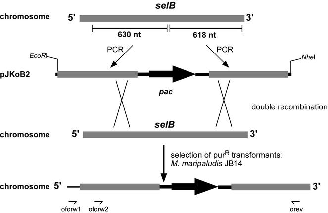
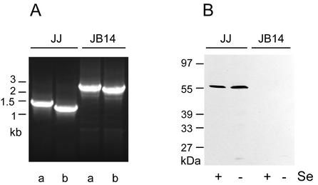
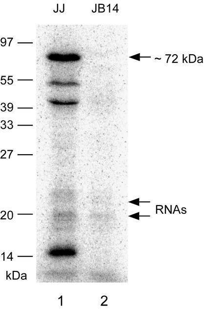
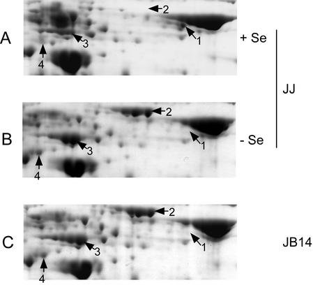
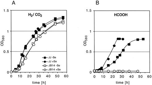
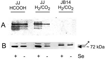
The genome of Methanococcus maripaludis harbors genes for at least six selenocysteine-containing proteins and also for homologs that contain a cysteine codon in the position of the UGA selenocysteine codon. To investigate the synthesis and function of both the Se and the S forms, a mutant with an inactivated selB gene was constructed and analyzed. The mutant was unable to synthesize any of the selenoproteins, thus proving that the gene product is the archaeal translation factor (aSelB) specialized for selenocysteine insertion. The wild-type form of M. maripaludis repressed the synthesis of the S forms of selenoproteins, i.e., the selenium-independent alternative system, in selenium-enriched medium, but the mutant did not. We concluded that free selenium is not involved in regulation but rather a successional compound such as selenocysteyl-tRNA or some selenoprotein. Apart from the S forms, several enzymes from the general methanogenic route were affected by selenium supplementation of the wild type or by the selB mutation. Although the growth of M. maripaludis on H(2)/CO(2) is only marginally affected by the selB lesion, the gene is indispensable for growth on formate because M. maripaludis possesses only a selenocysteine-containing formate dehydrogenase.


Figures

FIG. 1.
FIG. 1.
Strategy for the disruption of the selB gene of M. maripaludis JJ. pJKoB2 was constructed by flanking the pac resistance cassette with sequences of selB generated by PCR. M. maripaludis JJ was transformed with linear DNA (pac-containing EcoRI-NheI fragment of pJKoB2) to obtain puromycin-resistant transformants through double recombination events.
FIG. 2.
FIG. 2.
Analysis of strain JB14. (A) PCR products generated with the primer pairs oforw1-orev (lanes a) and oforw2-orev (lanes b) by using genomic DNA of M. maripaludis JJ and JB14, respectively, as templates; numbers give the size of the DNA standards in kilobase pairs. (B) Immunoblotting analysis of M. maripaludis JJ and JB14. Cells were grown in media supplemented with 10 μM selenite (+Se) or without selenium supplementation (−Se), respectively. Extracts were separated by SDS-PAGE, blotted onto a nitrocellulose membrane, and probed with antibodies directed against aSelB from M. jannaschii. Molecular mass standards are indicated on the left.
FIG. 3.
FIG. 3.
Selenoprotein synthesis by M. maripaludis JB14. Autoradiograph of a SDS-12% PAGE gel after electrophoresis of cell lysates from 75Se-labeled M. maripaludis JJ (lane 1) and JB14 (lane 2), respectively. The migration positions of the standard proteins are indicated on the left; the positions of the putative FdhA selenoprotein and of 75Se-labeled RNAs are also indicated (see the text for details).
FIG. 4.
FIG. 4.
Selenium-dependent protein synthesis by M. maripaludis. Cells of M. maripaludis were grown on H2/CO2 with (+Se) or without (−Se) selenite (10 μM) supplementation. Extracts were separated by 2D-PAGE, and the proteins were stained with Coomassie blue. (A and B) Sections of 2D gels with separated proteins from M. maripaludis JJ; (C) section of a 2D gel with separated proteins of M. maripaludis JB14 grown in medium containing 10 μM selenite. Arrows indicate the proteins that were subjected to N-terminal sequencing.
FIG. 5.
FIG. 5.
Selenium dependence of growth. (A) Growth of M. maripaludis JJ (solid symbols) and JB14 (open symbols) cultivated on H2/CO2 with (circles) or without (squares) selenite (10 μM). (B) Same as panel A, but with cultivation on formate.
FIG. 6.
FIG. 6.
Selenium dependence of the formation of FDH by M. maripaludis. (A) FDH activity staining of extracts from M. maripaludis JJ and JB14 after nondenaturing PAGE. Cells were grown on H2/CO2 or formate and with (+Se) or without (−Se) selenite (10 μM). (B) Immunoblotting analysis of FDH. The extracts in panel A were separated by SDS-PAGE, blotted onto a nitrocellulose membrane, and probed with antibodies directed against FDH from Methanobacterium formicicum. The arrow at 72 kDa indicates the position of the putative FdhA selenoprotein (see Fig. 3); the asterisk indicates a protein of 75 to 80 kDa, the synthesis of which is stimulated by selenium starvation (see the text for details).

Similar articles

Cited by

References

    1. Altschul, S. F., T. L. Madden, A. A. Schaffer, J. Zhang, Z. Zhang, W. Miller, and D. J. Lipman. 1997. Gapped BLAST and PSI-BLAST: a new generation of protein database search programs. Nucleic Acids Res. 25:3389-3402. - PMC - PubMed
    1. Ausubel, F. M., R. Brent, R. E. Kingston, D. D. Moore, J. G. Seidmann, J. A. Smith, and K. Struhl. 1997. Current protocols in molecular biology. John Wiley & Sons, Inc., New York, N.Y.
    1. Axley, M. J., A. Böck, and T. C. Stadtman. 1991. Catalytic properties of an Escherichia coli formate dehydrogenase mutant in which sulfur replaces selenium. Proc. Natl. Acad. Sci. USA 88:8450-8454. - PMC - PubMed
    1. Berghöfer, Y., and A. Klein. 1995. Insertional mutations in the hydrogenase vhc and frc operons encoding selenium free hydrogenases in Methanococcus voltae. Appl. Environ. Microbiol. 61:1770-1775. - PMC - PubMed
    1. Berry, M. J., J. D. Kieffer, and P. R. Larsen. 1991. Evidence that cysteine, not selenocysteine, is in the catalytic site of type II iodothyronine deiodinase. Endocrinology 129:550-552. - PubMed

Publication types

MeSH terms

LinkOut - more resources