. 2003 Jan;185(1):165-74.
doi: 10.1128/JB.185.1.165-174.2003.

Subunit topology of two 20S proteasomes from Haloferax volcanii

Affiliations

Subunit topology of two 20S proteasomes from Haloferax volcanii

Steven J Kaczowka et al. J Bacteriol. 2003 Jan.

Abstract

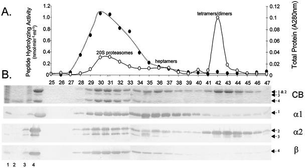
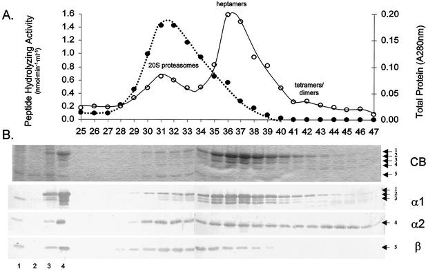
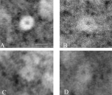
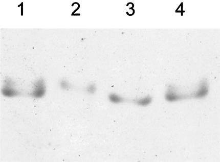
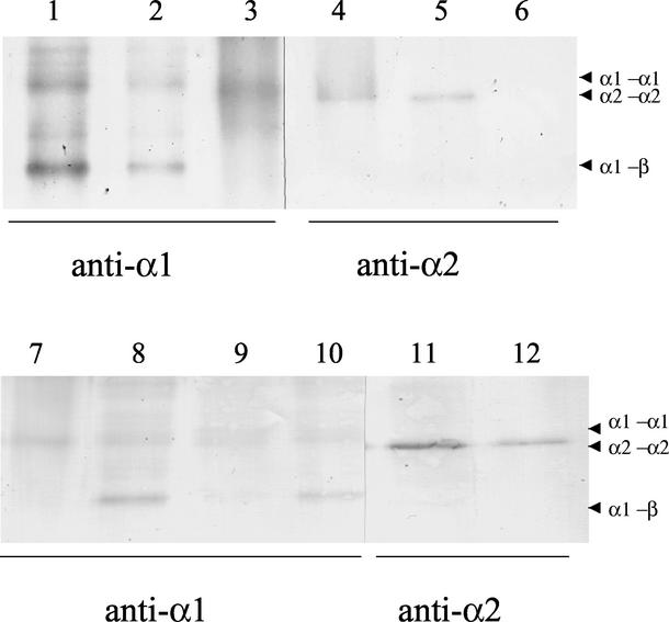
Haloferax volcanii, a halophilic archaeon, synthesizes three different proteins (alpha1, alpha2, and beta) which are classified in the 20S proteasome superfamily. The alpha1 and beta proteins alone form active 20S proteasomes; the role of alpha2, however, is not clear. To address this, alpha2 was synthesized with an epitope tag and purified by affinity chromatography from recombinant H. volcanii. The alpha2 protein copurified with alpha1 and beta in a complex with an overall structure and peptide-hydrolyzing activity comparable to those of the previously described alpha1-beta proteasome. Supplementing buffers with 10 mM CaCl(2) stabilized the halophilic proteasomes in the absence of salt and enabled them to be separated by native gel electrophoresis. This facilitated the discovery that wild-type H. volcanii synthesizes more than one type of 20S proteasome. Two 20S proteasomes, the alpha1-beta and alpha1-alpha2-beta proteasomes, were identified during stationary phase. Cross-linking of these enzymes, coupled with available structural information, suggested that the alpha1-beta proteasome was a symmetrical cylinder with alpha1 rings on each end. In contrast, the alpha1-alpha2-beta proteasome appeared to be asymmetrical with homo-oligomeric alpha1 and alpha2 rings positioned on separate ends. Inter-alpha-subunit contacts were only detected when the ratio of alpha1 to alpha2 was perturbed in the cell using recombinant technology. These results support a model that the ratio of alpha proteins may modulate the composition and subunit topology of 20S proteasomes in the cell.


Figures

FIG. 1.
FIG. 1.
Association of the α1, α2, and β proteasomal proteins as demonstrated by purification of α2-His-containing complexes. (A) Proteins were purified from recombinant H. volcanii(pJAM205) using Ni2+-Sepharose and Superose 6 gel filtration. Gel filtration fraction numbers are indicated on the x axis. Total protein measured by A280 (○) and N-Suc-LLVY-Amc-hydrolyzing activity (•) are indicated. (B) Proteins were separated by reducing 12% PAGE with SDS and analyzed by Coomassie blue (CB) staining or Western blotting using antibodies raised against α1, α2, and β as indicated on the right. H. volcanii(pBAP5010) cell lysate (lane 1) and Ni2+-purified fractions (lane 2) were included as controls. H. volcanii(pJAM205) cell lysate (lane 3), Ni2+-purified fractions (lane 4), and Superose 6 gel filtration fractions 25 to 47 are shown. Arrows to the right indicate α1 (band 1)-, α2 (bands 2 and 3)-, and β (band 4)-specific proteins.
FIG. 2.
FIG. 2.
Association of the α1, α2, and β proteasomal proteins as demonstrated by purification of α1-His-containing complexes. The figure is similar to Fig. 1, except plasmid pJAM204 was substituted for pJAM205. (A) Proteins purified by gel filtration. (B) Proteins separated by SDS-PAGE. Arrows to the right indicate α1 (bands 1 to 3)-, α2 (band 4)-, and β (band 5)-specific proteins.
FIG. 3.
FIG. 3.
Recombinant α1 and α2 proteins form ring structures. Transmission electron micrographs of negatively stained proteins are shown. An end-on view of a 20S proteasome (A) purified from H. volcanii(pJAM204) is included for comparison. (B and C) Typical end-on view of α1 (B) and α2 (C) purified from recombinant E. coli. (D) Typical end-on view of ring observed for the 200-kDa fraction purified from H. volcanii(pJAM204) which contains α1-His, α1, and α2 proteins. Bar, 12 nm. Samples were prepared and viewed on a Zeiss EM-10CA transmission electron microscope as previously described (33).
FIG. 4.
FIG. 4.
Native gel of H. volcanii 20S proteasomes reveals two distinct complexes. Lanes 2 and 3, α1-α2-β and α1-β proteasomes purified from H. volcanii WFD11 and psmC-1, respectively; lanes 1 and 4, equimolar mixture of the two proteasomes. Native gels containing 5% polyacrylamide were pre-equilibrated and run with 25 mM Tris-192 mM glycine buffer (pH 8.3) supplemented with 10 mM CaCl2. Proteins were stained with Coomassie blue R-250.
FIG. 5.
FIG. 5.
Cross-linking of H. volcanii 20S proteasomes reveals α1-α1 and α2-α2 contacts. The 20S proteasomes (300 nM) and α proteins (10 μM) were incubated (10 to 60 min, 37°C) in buffer B containing 10% glycerol and 0.22% glutaraldehyde. The cross-linking reaction was quenched with 1× 2 M Tris-Cl at pH 8.5 followed by precipitation with 10% trichloroacetic acid. Products and molecular mass standards (see Materials and Methods) were separated by reducing 7.5% PAGE with SDS. Specific antigens were detected by chromogenic Western blotting using antibodies raised against α1 (lanes 1 to 3 and 7 to 10), α2 (lanes 4 to 6 and 11 and 12), and βΔ-His (data not shown). Samples included the α1-β proteasome from H. volcanii psmC-1 (lanes 1, 6, and 8), the α1-α2-β proteasome from H. volcanii WFD11 (lanes 2, 5, 10, and 11), α1 (lanes 3 and 7) and α2 (lane 4) from recombinant E. coli(pJAM618) and E. coli(pJAM619), and β-His-containing proteasomes from H. volcanii(pJAM202) (lanes 9 and 12). Arrowheads indicate putative dimers.

Similar articles

Cited by

References

    1. Akopian, T. N., A. F. Kisselev, and A. L. Goldberg. 1997. Processive degradation of proteins and other catalytic properties of the proteasome from Thermoplasma acidophilum. J. Biol. Chem. 272:1791-1798. - PubMed
    1. Baumeister, W., J. Walz, F. Zühl, and E. Seemüller. 1998. The proteasome: paradigm of a self-compartmentalizing protease. Cell 92:367-380. - PubMed
    1. Betlach, M., J. Friedman, H. W. Boyer, and F. Pfeifer. 1984. Characterization of a halobacterial gene affecting bacterio-opsin gene expression. Nucleic Acids Res. 12:7949-7959. - PMC - PubMed
    1. Blanck, A., and D. Oesterhelt. 1987. The halo-opsin gene. II. Sequence, primary structure of halorhodopsin and comparison with bacteriorhodopsin. EMBO J. 6:265-273. - PMC - PubMed
    1. Brosius, J., M. L. Palmer, P. J. Kennedy, and H. F. Noller. 1978. Complete nucleotide sequence of a 16S ribosomal RNA gene from Escherichia coli. Proc. Natl. Acad. Sci. USA 75:4801-4805. - PMC - PubMed

Publication types

MeSH terms

LinkOut - more resources