. 2003 Jan;185(1):196-203.
doi: 10.1128/JB.185.1.196-203.2003.

Recruitment of MinC, an inhibitor of Z-ring formation, to the membrane in Escherichia coli: role of MinD and MinE

Affiliations

Recruitment of MinC, an inhibitor of Z-ring formation, to the membrane in Escherichia coli: role of MinD and MinE

Zonglin Hu et al. J Bacteriol. 2003 Jan.

Abstract

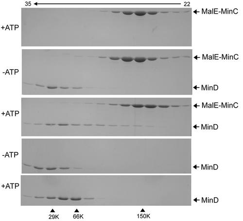
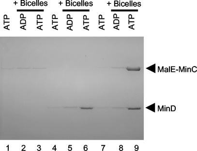
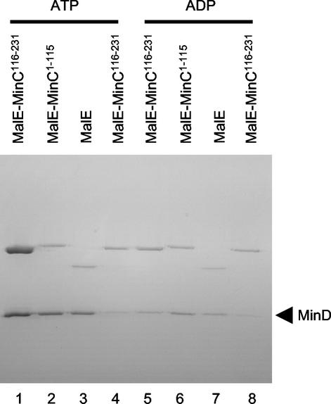
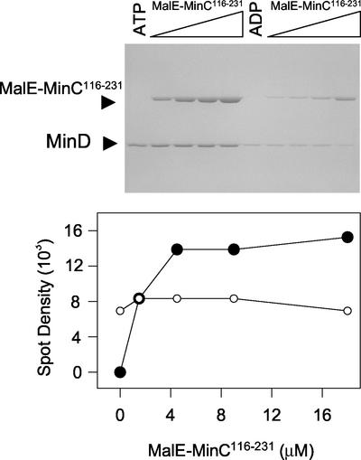
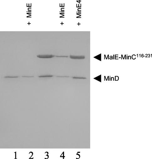
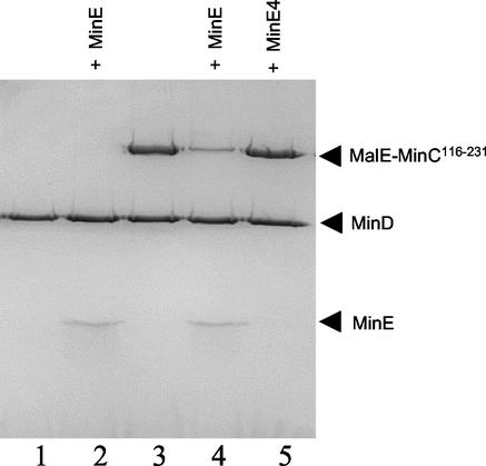
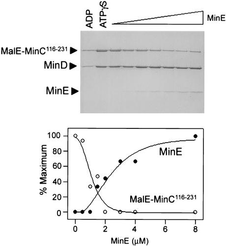
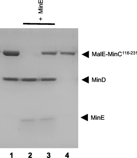
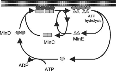
In Escherichia coli, the min system prevents division away from midcell through topological regulation of MinC, an inhibitor of Z-ring formation. The topological regulation involves oscillation of MinC between the poles of the cell under the direction of the MinDE oscillator. Since the mechanism of MinC involvement in the oscillation is unknown, we investigated the interaction of MinC with the other Min proteins. We observed that MinD dimerized in the presence of ATP and interacted with MinC. In the presence of a phospholipid bilayer, MinD bound to the bilayer and recruited MinC in an ATP-dependent manner. Addition of MinE to the MinCD-bilayer complex resulted in release of both MinC and MinD. The release of MinC did not require ATP hydrolysis, indicating that MinE could displace MinC from the MinD-bilayer complex. In contrast, MinC was unable to displace MinE bound to the MinD-bilayer complex. These results suggest that MinE induces a conformational change in MinD bound to the bilayer that results in the release of MinC. Also, it is argued that binding of MinD to the membrane activates MinC.


Figures

FIG. 1.
FIG. 1.
Size-exclusion chromatography of MalE-MinC and MinD. Proteins were incubated with or without nucleotide and analyzed by FPLC on a Superose 6 column. The elution buffer contained the same nucleotide as the preincubation mix at 0.5 mM. Aliquots of fractions were analyzed by SDS-PAGE. Top panel, MalE-MinC with ATP; second panel, MalE-MinC and MinD without nucleotide; third panel, MalE-MinC and MinD in the presence of ATP; fourth panel, MinD without nucleotide (the profile with ADP was the same); and last panel, MinD in the presence of ATP. The fraction numbers are indicated at the top. The size standards used were carbonic anhydrase (29 kDa), bovine serum albumin (66 kDa), and alcohol dehydrogenase (150 kDa).
FIG. 2.
FIG. 2.
MinD recruits MalE-MinC to bicelles in the presence of ATP. MalE-MinC (3 μM) and MinD (4 μM) were incubated separately or together with or without bicelles in the presence of ATP or ADP. The reaction mixtures were immediately centrifuged, and the pellets were analyzed by SDS-PAGE. MalE-MinC was added to lanes 1 to 3, MinD was added to lanes 4 to 6, and both MalE-MinC and MinD were added to lanes 7 to 9. The samples with bicelles and nucleotide added are indicated at the top of the figure.
FIG. 3.
FIG. 3.
MinD recruits MinC to bicelles through the C-terminal domain of MinC. MinD (4 μM) was incubated with bicelles and ATP. Various MalE fusions (4 μM) were added, the samples were immediately centrifuged, and the pellets were analyzed by SDS-PAGE. The specific MalE fusion is indicated at the top of the figure. The nucleotide used in the reaction is also indicated at the top of the figure. In lanes 4 and 8, MinD K16Q, which binds poorly to vesicles (12), was used in place of MinD.
FIG. 4.
FIG. 4.
Saturable binding of MalE-MinC116-231 to the MinD-bicelle complex. MinD (4 μM) was incubated with bicelles in the presence of ATP or ADP. Increasing concentrations of MalE-MinC116-231 were added, the reactions were immediately centrifuged, and pellets were analyzed by SDS-PAGE (top panel). The amounts of MinD and MalE-MinC116-231 in the pellet were determined by densitometry. The amount of MalE-MinC116-231 and MinD in the pellet in the presence of ADP (lanes 6 to 10) was subtracted from the values obtained in the presence of ATP (lanes 1 to 5) and plotted (bottom panel). It was determined that the ratio of the spot densities for an equimolar mixture of MalE-MinC116-231 and MinD was 1.8 (described in Materials and Methods). This ratio is similar to that obtained from averaging the ratios of the spot densities of MalE-MinC to MinD in the last three lanes, 1.9, in which MalE-MinC116-231 is saturating.
FIG. 5.
FIG. 5.
MinE removes MinC along with MinD from bicelles. MinD (4 μM), bicelles, and ATP were incubated with (lanes 3 to 5) or without MalE-MinC116-231 (lanes 1 and 2) (3 μM). After 1 min, MinE (4 μM) was added to lanes 2 and 4, and MinE4 (4 μM) was added to lane 5. The reaction mixtures were immediately centrifuged, and the pellets were analyzed by SDS-PAGE.
FIG. 6.
FIG. 6.
MinE displaces MinC from the MinCD-bicelle complex in the absence of ATP hydrolysis. MinD (4 μM) and bicelles were incubated in the presence of ATPγS. After 1 min, MalE-MinC116-231 (3 μM) was added to lanes 3 to 5. After an additional minute, MinE (lanes 2 and 4) or MinE4 (lane 5) was added (4 μM). The samples were immediately centrifuged and analyzed by SDS-PAGE.
FIG. 7.
FIG. 7.
Titration of the MinCD-bicelle complex with MinE. MinD (4 μM) and MinC116-231 (3 μM) were incubated with bicelles and ATPγS. After 1 min, MinE (0 to 12 μM) was added, the reaction mixtures were centrifuged immediately, and the pellets were analyzed by SDS-PAGE. The relative amounts of MinC, MinD, and MinE bound to bicelles were determined by densitometry and plotted after subtracting the amounts in the pellet observed with ADP (lane 1). The error for the amount of MinD in the pellet with ATPγS was 10%. For MinE, the amount in the last lane was set at 100%, whereas with MalE-MinC116-231, the amount in lane 2 (absence of MinE) was set at 100%. The ratio of the spot densities of MinD to MinE in the last lane, where MinE is in excess of MinD, is 4.2. This value is similar to that of a control, in which the ratio of spot densities for equimolar mixtures of MinD and MinE was found to be 4.0.
FIG. 8.
FIG. 8.
MinC cannot displace MinE bound to MinD-bicelle complex. MinD (4 μM) was incubated with bicelles in the presence of ATPγS. After 1 min, MalE-MinC116-231 (8 μM) (lane 1) or MinE (4 μM) (lane 2) was added. In another reaction, MinD and MinE were incubated with ATPγS for 1 min, and then MalE-MinC was added (lane 3). A control (lane 4) contained only MalE-MinC116-231 (8 μM) and bicelles. The reaction mixtures were centrifuged, and the pellets were analyzed by SDS-PAGE.
FIG. 9.
FIG. 9.
Model for regulation of the reversible interaction of Min proteins with the membrane. In the presence of ATP, MinD dimerizes and binds to MinC and the membrane. Upon binding to the membrane, MinD undergoes conformational changes leading to its assembly into filaments (12) and increased affinity for MinE. Binding of MinE to MinD results in displacement of MinC in a step that does not require ATP hydrolysis. MinE does not compete with MinC for binding to MinD but must alter MinD so that it has reduced affinity for MinC, since MinC cannot displace MinE. MinE stimulates MinD ATPase, causing release of MinE and the ADP form of MinD. MinC, MinD, and MinE are now dissociated in the cytoplasm. MinD undergoes nucleotide exchange, and the process is repeated.

Similar articles

Cited by

References

    1. Bi, E., and J. Lutkenhaus. 1993. Cell division inhibitors SulA and MinCD prevent formation of the FtsZ ring. J. Bacteriol. 175:1118-1125. - PMC - PubMed
    1. Bi, E. F., and J. Lutkenhaus. 1991. FtsZ ring structure associated with division in Escherichia coli. Nature 354:161-164. - PubMed
    1. Cordell, S. C., R. E. Anderson, and J. Lowe. 2001. Crystal structure of the bacterial cell division inhibitor MinC. EMBO J. 20:2454-2461. - PMC - PubMed
    1. Cordell, S. C., and J. Lowe. 2001. Crystal structure of the bacterial cell division regulator MinD. FEBS Lett. 492:160-165. - PubMed
    1. de Boer, P. A., R. E. Crossley, A. R. Hand, and L. I. Rothfield. 1991. The MinD protein is a membrane ATPase required for the correct placement of the Escherichia coli division site. EMBO J. 10:4371-4380. - PMC - PubMed

Publication types

MeSH terms

LinkOut - more resources