. 2003 Jan;185(1):231-42.
doi: 10.1128/JB.185.1.231-242.2003.

Multiple promoter inversions generate surface antigenic variation in Mycoplasma penetrans

Affiliations

Multiple promoter inversions generate surface antigenic variation in Mycoplasma penetrans

Atsuko Horino et al. J Bacteriol. 2003 Jan.

Abstract

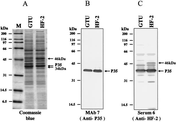
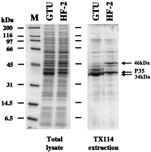
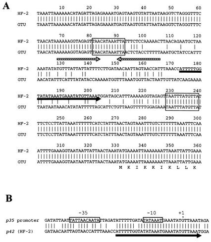
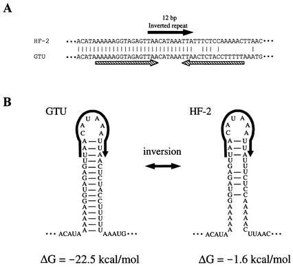
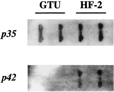
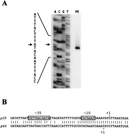
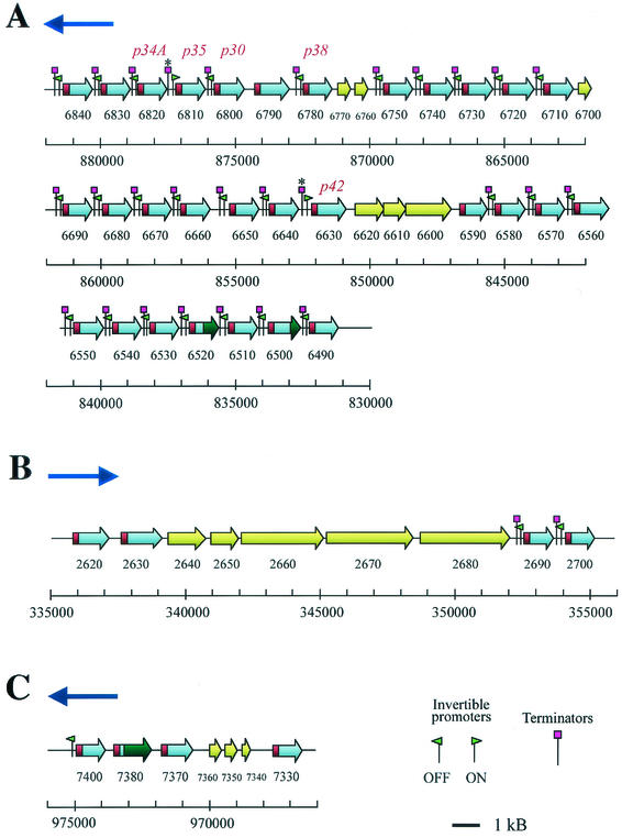
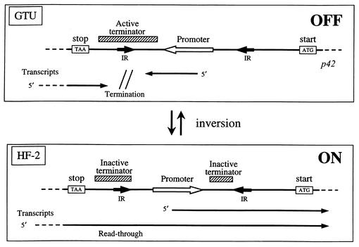
Mycoplasma penetrans is a newly identified species of the genus MYCOPLASMA: It was first isolated from a urine sample from a human immunodeficiency virus (HIV)-infected patient. M. penetrans changes its surface antigen profile with high frequency. The changes originate from ON<==>OFF phase variations of the P35 family of surface membrane lipoproteins. The P35 family lipoproteins are major antigens recognized by the human immune system during M. penetrans infection and are encoded by the mpl genes. Phase variations of P35 family lipoproteins occur at the transcriptional level of mpl genes; however, the precise genetic mechanisms are unknown. In this study, the molecular mechanisms of surface antigen profile change in M. penetrans were investigated. The focus was on the 46-kDa protein that is present in M. penetrans strain HF-2 but not in the type strain, GTU. The 46-kDa protein was the product of a previously reported mpl gene, pepIMP13, with an amino-terminal sequence identical to that of the P35 family lipoproteins. Nucleotide sequencing analysis of the pepIMP13 gene region revealed that the promoter-containing 135-bp DNA of this gene had the structure of an invertible element that functioned as a switch for gene expression. In addition, all of the mpl genes of M. penetrans HF-2 were identified using the whole-genome sequence data that has recently become available for this bacterium. There are at least 38 mpl genes in the M. penetrans HF-2 genome. Interestingly, most of these mpl genes possess invertible promoter-like sequences, similar to those of the pepIMP13 gene promoter. A model for the generation of surface antigenic variation by multiple promoter inversions is proposed.


Figures

FIG. 1.
FIG. 1.
Analysis of protein profiles of M. penetrans strains GTU and HF-2. (A) Coomassie blue-stained SDS-12% PAGE of total proteins of M. penetrans. A protein molecular mass marker (Bio-Rad, Hercules, Calif.) is in lane M, and the masses are shown on the left. The P35 and 34- and 46-kDa proteins are indicated. (B) Immunoblot analysis of M. penetrans proteins. Anti-P35 MAb (MAb 7) was used for detection. The P35 protein is indicated. (C) Immunoblot analysis with the polyclonal serum 6 (anti-HF-2). The positions of the P35 and 46-kDa proteins are indicated.
FIG. 2.
FIG. 2.
Analysis of LAMP profiles of M. penetrans. The total cell lysate and TX-114 phase-fractionated proteins of M. penetrans strains GTU and HF-2 were analyzed by SDS-12% PAGE. The proteins were stained with Coomassie blue. The positions of the P35 and 34- and 46-kDa proteins are indicated. A protein molecular mass marker is in lane M, and molecular masses are on the left.
FIG. 3.
FIG. 3.
Alignment of amino acid sequences of P35 and 46-kDa (pepIMP13) proteins. The 46-kDa protein was designated P42 based on the calculated molecular mass. The three partial amino acid sequences of P42 determined by Edman degradation of digested peptide are underlined. The signal sequence of the P35 family lipoprotein is shown in boldface characters. The cysteine residue marked with an asterisk is the potential binding site of fatty acid chains.
FIG. 4.
FIG. 4.
Nucleotide sequence of p42 gene upstream region. (A) Comparison of p42 gene upstream regions from strains GTU and HF-2. The 12-bp inverted-repeat sequences adjacent to the 135-bp inverted DNA region (see the text) are boxed. The solid arrow indicates the core of the promoter sequence in the HF-2 sequence (panel B). The hatched arrows indicate a 16-bp inverted repeat in the GTU sequence. The deduced amino acid sequence of the P42 protein (the first 10 aa) is shown. The nucleotides are counted from 330 bp upstream from the ATG start codon. (B) Comparison of p42 gene upstream sequence with promoter from p35 gene. The −10 and − 35 consensus sequences of the p35 promoter are boxed (24). The probable start site of p35 transcription is also marked +1. The highly conserved region between the p35 and p42 promoter sequences is underlined with an arrow.
FIG. 5.
FIG. 5.
Structure of 16-bp inverted-repeat sequence located upstream of p42 gene. (A) Nucleotide sequences of 16-bp inverted-repeat regions from strains HF-2 and GTU. The nucleotide sequence corresponds to nucleotides 63 to 110 in Fig. 4A. The solid arrow indicates a 12-bp inverted-repeat sequence adjacent to a 135-bp inverted DNA sequence. The hatched arrows indicate the 16-bp inverted repeat that is formed only in the GTU sequence. (B) Hairpin structures of the 16-bp inverted repeat. Formation of the hairpin structure is disrupted in the HF-2 sequence by DNA inversion between 12-bp inverted-repeat sequences. The hairpin structures are shown as RNA sequences. The calculated ΔGs of these hairpin structures are shown at the bottom.
FIG. 6.
FIG. 6.
RNA slot blot analysis of p35 and p42 gene transcription in M. penetrans strains GTU and HF-2. RNA samples from M. penetrans strains GTU and HF-2 were blotted in duplicate. The blots were hybridized with the oligonucleotide probes for p35 and p42 transcripts as indicated on the left.
FIG. 7.
FIG. 7.
Primer extension analysis of p42 gene transcription. (A) Autoradiogram of sequencing gel used to analyze the primer extension product. PE indicates the primer extension product obtained with M. penetrans HF-2 RNA as a template. The corresponding nucleotide sequence is shown on the left, and the probable start site is indicated by arrows. (B) Comparison of the transcriptional start sites in p35 and p42 promoters. Probable start sites determined by primer extension of the p35 promoter (24) and the p42 promoter (panel A) are indicated as +1. The putative −10 and −35 consensus regions are boxed.
FIG. 8.
FIG. 8.
Genomic organization of mpl genes in M. penetrans HF-2. Three mpl gene clusters are illustrated. The gray and red boxes in the arrows represent mpl genes. The red regions indicate signal sequence regions of mpl genes. The yellow arrows represent non-mpl genes. The numbers under the genes are MYPE serial numbers that are used to describe M. penetrans ORFs in the whole-genome sequencing analysis. The approximate nucleotide positions of mpl genes in the genome are also shown with the scale. The genes MYPE6500, -6520, and -7380 have frameshift mutations and are disrupted by internal stop codons. In these genes, the regions after the stop codons are shown as dark green. The small green arrowheads indicate invertible promoter-like sequences and their directions. The small purple squares represent 16-bp inverted-repeat terminator-like sequences. Two terminator-like sequences (upstream of the p35 and p42 genes that are marked with asterisks) are inactivated because of promoter inversions (see the text). The genes for P35, P42, and previously reported lipoproteins (P30, P38, and P34A) are labeled. The blue arrows above each cluster indicate relative directions in the genome.
FIG. 9.
FIG. 9.
Model for genetic switch of mpl genes by promoter inversion. The upstream intergenic regions of the p42 genes are illustrated. The open arrows represent promoter sequences and their directions. The short solid arrows represent 12-bp inverted repeats (IR). The hatched boxes represent terminator-like sequences (16-bp inverted repeats). When the promoter is in the OFF orientation (GTU), an active terminator is formed. The inversion between 12-bp inverted-repeat sequences changes the promoter direction from OFF to ON and splits the terminator sequence. RNA transcripts from the promoter and from upstream are represented by thin solid arrows.

References

    1. Abraham, J. M., C. S. Freitag, J. R. Clements, and B. I. Eisenstein. 1985. An invertible element of DNA controls phase variation of type 1 fimbriae of Escherichia coli. Proc. Natl. Acad. Sci. USA 82:5724-5727. - PMC - PubMed
    1. Behrens, A., M. Heller, H. Kirchhoff, D. Yogev, and R. Rosengarten. 1994. A family of phase- and size-variant membrane surface lipoprotein antigens (Vsps) of Mycoplasma bovis. Infect. Immun. 62:5075-5084. - PMC - PubMed
    1. Bhugra, B., L. L. Voelker, N. Zou, H. Yu, and K. Dybvig. 1995. Mechanism of antigenic variation in Mycoplasma pulmonis: interwoven, site-specific DNA inversions. Mol. Microbiol. 18:703-714. - PubMed
    1. Chambaud, I., R. Heilig, S. Ferris, V. Barbe, D. Samson, F. Galisson, I. Moszer, K. Dybvig, H. Wroblewski, A. Viari, E. P. Rocha, and A. Blanchard. 2001. The complete genome sequence of the murine respiratory pathogen Mycoplasma pulmonis. Nucleic Acids Res. 29:2145-2153. - PMC - PubMed
    1. Chambaud, I., H. Wroblewski, and A. Blanchard. 1999. Interactions between mycoplasma lipoproteins and the host immune system. Trends Microbiol. 7:493-499. - PubMed

MeSH terms