. 2003 Jan;185(1):254-61.
doi: 10.1128/JB.185.1.254-261.2003.

The crystal structure of the phosphorylation domain in PhoP reveals a functional tandem association mediated by an asymmetric interface

Affiliations

The crystal structure of the phosphorylation domain in PhoP reveals a functional tandem association mediated by an asymmetric interface

Catherine Birck et al. J Bacteriol. 2003 Jan.

Abstract

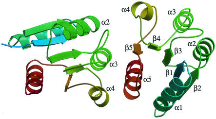
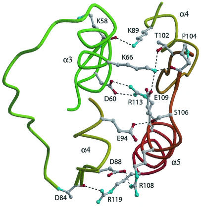
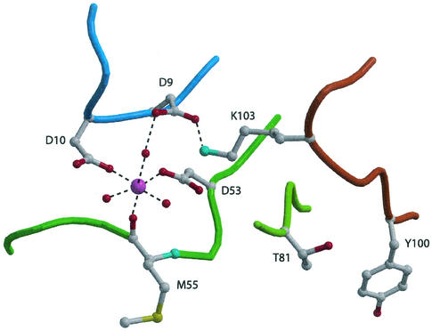
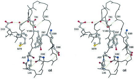
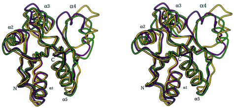
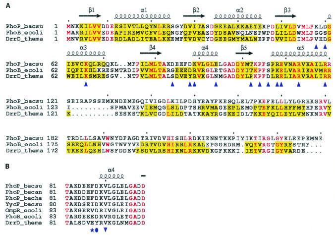
PhoP from Bacillus subtilis belongs to the OmpR subfamily of response regulators. It regulates the transcription of several operons and participates in a signal transduction network that controls adaptation of the bacteria to phosphate deficiency. The receiver domains of two members of this subfamily, PhoB from Escherichia coli and DrrD from Thermotoga maritima, have been structurally characterized. These modules have similar overall folds but display remarkable differences in the conformation of the beta4-alpha4 and alpha4 regions. The crystal structure of the receiver domain of PhoP (PhoPN) described in this paper illustrates yet another geometry in this region. Another major issue of the structure determination is the dimeric state of the protein and the novel mode of association between receiver domains. The protein-protein interface is provided by two different surfaces from each protomer, and the tandem unit formed through this asymmetric interface leaves free interaction surfaces. This design is well suited for further association of PhoP dimers to form oligomeric structures. The interprotein interface buries 970 A(2) from solvent and mostly involves interactions between charged residues. As described in the accompanying paper, mutations of a single residue in one salt bridge shielded from solvent prevented dimerization of the unphosphorylated and phosphorylated response regulator and had drastic functional consequences. The three structurally documented members of the OmpR family (PhoB, DrrD, and PhoP) provide a framework to consider possible relationships between structural features and sequence signatures in critical regions of the receiver domains.


Figures

FIG. 1.
FIG. 1.
Asymmetric interface between PhoPN protomers. Each protomer is represented by ribbons, and the color varies from light blue (N terminus) to red (C terminus). The two domains associate through surface A (α4, β5, loop β5-α5, and α5) from protomer 1 (right) and surface B (α3, loop β4-α4, and α4) from protomer 2 (left) (the protomers are arbitrarily numbered). This setting leaves surfaces B and A from protomers 1 and 2, respectively, free for association with other tandem units.
FIG. 2.
FIG. 2.
Network of polar interactions at the PhoPN dimer interface. The hydrogen bonds are represented by dashed lines. The salt bridge interaction between D60 and R113 is shielded from solvent.
FIG. 3.
FIG. 3.
Octahedral coordination of the manganese ion in the active site of PhoPN. The Mn2+ ion is represented by a large magenta sphere. Small red spheres and dashed lines indicate water molecules and polar interactions, respectively. Outside the active site, two conserved residues in receiver domains, T81 at the C-terminal edge of strand β4 and Y100 from strand β5, are also shown.
FIG. 4.
FIG. 4.
Stereo view of the β4-α4 and α4 areas of PhoPN. The protein backbone is indicated by black lines; oxygen atoms are red, carbon atoms are grey, nitrogen atoms are blue, and sulfur atoms are yellow. Residue F87 from the β4-α4 loop contributes to the hydrophobic pocket and anchors this region to the protein core. The hydrogen bond (green dashed lines) between D53 and the main chain carbonyl atom of E85 involves three water molecules (small red spheres) and may stabilize the conformation of the loop.
FIG. 5.
FIG. 5.
Stereo view of the superimposed receiver domains of PhoP from B. subtilis (purple), PhoB from E. coli (pdb 1B00) (green), and DrrD from T. maritima (pdb 1KGS) (yellow). The superimposition of the structures was based on the best fit of the central five-strand sheet and revealed the similarities and differences in the protein folds. The conformations of the β4-α4 loops and α4 helices are not related in these three members of the OmpR family.
FIG. 6.
FIG. 6.
Sequence alignment of PhoP from B. subtilis and other response regulators. (A) Structure-based sequence alignment of PhoP with PhoB from E. coli (pdb 1B00) and DrrD from T. maritima (pdb 1KGS). The secondary structure elements of PhoP are indicated by coils for α helices and arrows for β strands. The secondary structure elements in each receiver domain are indicated by a yellow background. Identical residues are indicated by red type. Residues that are involved in the PhoPN dimer interface are indicated by blue triangles. (B) Four response regulators displaying sequence signatures similar to that of PhoP in the region from position 81 to position 99: PhoP from B. anthracis (NP-658617), PhoP from B. halodurans (Q9K850), YycF from B. subtilis (P37478), and OmpR from E. coli (P03025). These proteins have a hydrophobic residue at position 87 (PhoP numbering). PhoB and DrrD have a glutamic acid residue at that position. Despite conservation of V90, substitution of D90 in PhoB by Y88 in DrrD induces a shift in register by two residues of the α4 anchor (7). PhoP_bacsu, B. subtilis PhoP; PhoB_ecoli, E. coli PhoB; DrrD_thema, T. maritima DrrD; PhoP_bacan, B. anthracis PhoP; PhoP_bacha, B. halodurans PhoP; YycF_bacsu, B. subtilis YycF; OmpR_ecoli, E. coli OmpR.

References

    1. Allen, M. P., K. B. Zumbrennen, and W. R. McCleary. 2001. Genetic evidence that the α5 helix of the receiver domain of PhoB is involved in interdomain interactions. J. Bacteriol. 183:2204-2211. - PMC - PubMed
    1. Baikalov, I., I. Schroder, M. Kaczor-Grzeskowiak, K. Grzeskowiak, R. P. Gunsalus, and R. E. Dickerson. 1996. Structure of the Escherichia coli response regulator NarL. Biochemistry 35:11053-11061. - PubMed
    1. Birck, C., L. Mourey, P. Gouet, B. Fabry, J. Schumacher, P. Rousseau, D. Kahn, and J. P. Samama. 1999. Conformational changes induced by phosphorylation of the FixJ receiver domain. Structure 7:1505-1515. - PubMed
    1. Blanco, A. G., M. Sola, F. X. Gomis-Rüth, and M. Coll. 2002. Tandem DNA recognition by PhoB, a two-component signal transduction transcriptional activator. Structure 10:701-713. - PubMed
    1. Bourret, R. B., N. W. Charon, A. M. Stock, and A. H. West. 2002. Bright lights, abundant operons—fluorescence and genomic technologies advance studies of bacterial locomotion and signal transduction: review of the BLAST meeting, Cuernavaca, Mexico, 14 to 19 January 2001 J. Bacteriol. 184:1-17. - PMC - PubMed

Publication types