. 2002 Dec 16;196(12):1575-84.
doi: 10.1084/jem.20021515.

CD1-mediated gamma/delta T cell maturation of dendritic cells

Affiliations

CD1-mediated gamma/delta T cell maturation of dendritic cells

David S Leslie et al. J Exp Med. .

Abstract

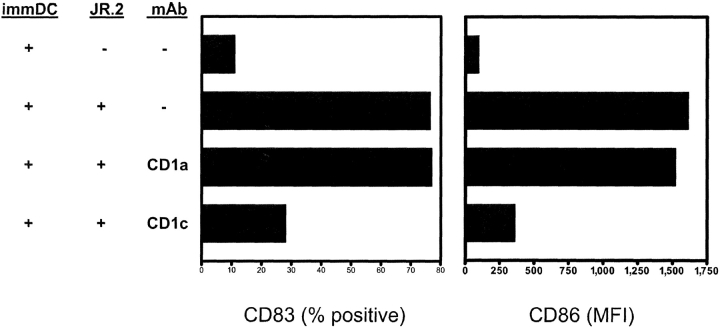
Immature myeloid dendritic cells (DCs) express only low levels of major histocompatibility complex (MHC) class II but express high levels of CD1 a, b, and c antigen-presenting molecules at the cell surface. As Vdelta1+ gamma/delta T cells are the main tissue subset of gamma/delta T cells and they are known to recognize CD1c in the absence of specific foreign antigen recognition, we examined the possible interaction of these T cells with immature DCs. We show that CD1-restricted gamma/delta T cells can mediate the maturation of DCs. DC maturation required cell-cell contact and could be blocked by antibodies against CD1c. The maturation process was partially mediated by tumor necrosis factor alpha. Importantly, immature DCs matured in the presence of lipopolysaccharide and CD1-restricted gamma/delta T cells produced bioactive interleukin-12p70. In addition, these DCs were able to efficiently present peptide antigens to naive CD4+ T cells. CD1-restricted gamma/delta T cell recognition of immature DCs provides the human immune system with the capacity to rapidly generate a pool of mature DCs early during microbial invasion. This may be an important source of critical host signals for T helper type 1 polarization of antigen-specific naive T cells and the subsequent adaptive immune response.


Figures

Figure 1.
Figure 1.
CD1-restricted γ/δ T cells induce phenotypic DC maturation. (A) Immature monocyte-derived DCs cultured with Vδ1+ CD1-restricted T cell clones JR.2 and XV.1 for 48 h expressed increased CD83 and CD86 cell surface molecules when compared with DCs cultured in medium alone as assessed by mAb staining and flow cytometry. (B) Immature DCs cultured with CD1c-restricted γ/δ clone JR.2 or TNF-α (50 ng/ml) for 48 h expressed increased levels of CD83 and CD86 cell surface molecules. Culture of DCs with Vδ1+ clones 10G4, 20H8, and a Vδ2+ γ/δ line (which are not CD1-restricted) all failed to promote maturation of DCs as assessed by CD83 and CD86 cell surface expression. Open squares, medium alone; open triangles, TNF-α, filled squares, JR.2; diamonds, XV.1; triangles, 10G4; inverted triangles, 20H8; circles, Vδ2 line. These results are representative of four independent experiments using different DC donors.
Figure 2.
Figure 2.
γ/δ T cell–induced DC maturation is mediated by CD1c. Immature monocyte-derived DCs were cultured with the CD1c-restricted γ/δ clone JR.2 at a T cell:DC ratio of 1:9 for 48 h in culture medium or in the presence of anti-CD1 mAb blocking. Treatment with anti-CD1c mAb F10/21A3 inhibited up-regulation of CD83 and CD86 by 64 and 78%, respectively. Blocking with mAb directed against CD1a (OKT6) did not inhibit maturation. These results are representative of three independent experiments using different DC donors.
Figure 3.
Figure 3.
CD1c-reactive T cells induce functional DC maturation. (A) Decreased uptake of soluble FITC dextran by mature DCs. Immature DCs were cultured in the presence of TNF-α (50 ng/ml), γ/δ clone JR.2 (T cell:DC ratio = 1:9), or medium alone for 48 h. These DCs were then washed and cultured in the presence of FITC–dextran during a 60 min time course and antigen uptake was assessed by flow cytometry. Note that DCs matured in the presence of clone JR.2 or TNFα had decreased uptake of FITC–dextran consistent with functional maturation. Open squares, medium; filled squares, JR.2; triangles, TNF-α. (B) Redistribution of MHC class II molecules in maturing DCs. Confocal microscopy was performed on DCs matured in the presence of TNF-α, JR.2 (T cell:DC ratio = 1:9) or medium alone. Cells were labeled with mAb against MHC class II (green staining) and the lysosomal marker LAMP 1 (red staining). Colocalization of these molecules is represented by the merged image (yellow). Note that DCs matured in the presence of JR.2 or TNF-α have increased staining of cell surface MHC class II and minimal colocalization with LAMP 1. Bar = 2 μm. (C) DC expression of cell surface CD83. DCs cultured in the presence of TNF-α or JR.2 expressed increased levels of the maturation marker CD83 as assessed by mAb staining and flow cytometry. These results are representative of three independent experiments using different DC donors.
Figure 3.
Figure 3.
CD1c-reactive T cells induce functional DC maturation. (A) Decreased uptake of soluble FITC dextran by mature DCs. Immature DCs were cultured in the presence of TNF-α (50 ng/ml), γ/δ clone JR.2 (T cell:DC ratio = 1:9), or medium alone for 48 h. These DCs were then washed and cultured in the presence of FITC–dextran during a 60 min time course and antigen uptake was assessed by flow cytometry. Note that DCs matured in the presence of clone JR.2 or TNFα had decreased uptake of FITC–dextran consistent with functional maturation. Open squares, medium; filled squares, JR.2; triangles, TNF-α. (B) Redistribution of MHC class II molecules in maturing DCs. Confocal microscopy was performed on DCs matured in the presence of TNF-α, JR.2 (T cell:DC ratio = 1:9) or medium alone. Cells were labeled with mAb against MHC class II (green staining) and the lysosomal marker LAMP 1 (red staining). Colocalization of these molecules is represented by the merged image (yellow). Note that DCs matured in the presence of JR.2 or TNF-α have increased staining of cell surface MHC class II and minimal colocalization with LAMP 1. Bar = 2 μm. (C) DC expression of cell surface CD83. DCs cultured in the presence of TNF-α or JR.2 expressed increased levels of the maturation marker CD83 as assessed by mAb staining and flow cytometry. These results are representative of three independent experiments using different DC donors.
Figure 4.
Figure 4.
Th1 cytokine production during JR.2:DC coculture is CD1c dependent. (A) JR.2 γ/δ T cells were incubated with immature DCs for 24 h in the presence of blocking mAbs against CD1a, CD1c, or an isotype-matched control antibody. The culture supernatants were collected and assayed for TNF-α, IFN-γ, IL-4, and IL-13. Note that production of both TNF-α and IFN-γ was inhibited by mAb against CD1c but not CD1a. IL-4 and IL-13 were not detected (data not shown). The sensitivity of the assay was 30 pg/ml. (B) Maturation of DCs by JR.2 is partially mediated by TNF-α. Immature DCs were cultured for 48 h in medium or with γ/δ clone JR.2 (T cell:DC ratio = 1:10) in the presence of blocking mAbs against TNF-α or IFN-γ. DC up-regulation of CD83 and CD86 was inhibited by the presence of mAbs against TNF-α but not IFN-γ. The combination of mAbs against both TNF-α and IFN-γ was similar to the effect seen with TNF-α mAbs alone. These results are representative of four independent experiments using different DC donors.
Figure 4.
Figure 4.
Th1 cytokine production during JR.2:DC coculture is CD1c dependent. (A) JR.2 γ/δ T cells were incubated with immature DCs for 24 h in the presence of blocking mAbs against CD1a, CD1c, or an isotype-matched control antibody. The culture supernatants were collected and assayed for TNF-α, IFN-γ, IL-4, and IL-13. Note that production of both TNF-α and IFN-γ was inhibited by mAb against CD1c but not CD1a. IL-4 and IL-13 were not detected (data not shown). The sensitivity of the assay was 30 pg/ml. (B) Maturation of DCs by JR.2 is partially mediated by TNF-α. Immature DCs were cultured for 48 h in medium or with γ/δ clone JR.2 (T cell:DC ratio = 1:10) in the presence of blocking mAbs against TNF-α or IFN-γ. DC up-regulation of CD83 and CD86 was inhibited by the presence of mAbs against TNF-α but not IFN-γ. The combination of mAbs against both TNF-α and IFN-γ was similar to the effect seen with TNF-α mAbs alone. These results are representative of four independent experiments using different DC donors.
Figure 5.
Figure 5.
Role of γ/δ T cell surface molecules and soluble mediators in DC maturation. (A) CD40 ligand cell surface expression by JR.2. γ/δ T cells. CD40 ligand expression on the surface of γ/δ T cell clone JR.2 increased approximately fourfold during a 24 h coculture with immature DCs as assessed by mAb staining and flow cytometry. Triangles, culture with DC; squares, medium control. (B) Inhibition of DC maturation by blocking of γ/δ T cell surface molecules and the cytokines TNF-α and IFN-γ. Immature DCs were cocultured with LPS (10 ng/ml) or JR.2 for 48 h. In the JR.2:DC cocultures the presence of mAb against TNF-α inhibited up-regulation of DCs expressing CD83 by ∼50%, while culture in the presence of mAbs against IFN-γ, CD40L, FasL, or the RANKL antagonist OPG had no effect. These results are representative of four independent experiments using different DC donors.
Figure 5.
Figure 5.
Role of γ/δ T cell surface molecules and soluble mediators in DC maturation. (A) CD40 ligand cell surface expression by JR.2. γ/δ T cells. CD40 ligand expression on the surface of γ/δ T cell clone JR.2 increased approximately fourfold during a 24 h coculture with immature DCs as assessed by mAb staining and flow cytometry. Triangles, culture with DC; squares, medium control. (B) Inhibition of DC maturation by blocking of γ/δ T cell surface molecules and the cytokines TNF-α and IFN-γ. Immature DCs were cocultured with LPS (10 ng/ml) or JR.2 for 48 h. In the JR.2:DC cocultures the presence of mAb against TNF-α inhibited up-regulation of DCs expressing CD83 by ∼50%, while culture in the presence of mAbs against IFN-γ, CD40L, FasL, or the RANKL antagonist OPG had no effect. These results are representative of four independent experiments using different DC donors.
Figure 6.
Figure 6.
IL-12p70 production by DCs requires both CD1-restricted γ/δ T cells and LPS. Immature DCs were incubated for 24 h with the JR.2 γ/δ T cell clone, LPS (10 ng/ml), or both JR.2 and LPS in the presence of blocking mAbs against IFN-γ, CD1c, or CD1a. The culture supernatants were collected and assayed for IL-12p70. Note that production of IL-12p70 occurred only in the presence of both JR.2 and LPS and was inhibited by mAb against IFN-γ and CD1c but not CD1a. The sensitivity of the assay was 0.3 ng/ml. These results are representative of three independent experiments using different DC donors.
Figure 7.
Figure 7.
γ/δ clone JR.2 promotes DC antigen presentation to CD4+ T cells. (A) Allogeneic MLR. Immature monocyte-derived DCs were cultured for 72 h in the presence of TNF-α, γ/δ clone JR.2, or medium alone. Irradiated DC were then cultured at DC:T cell ratios from 1:1,000 to 1:10 with allogeneic CD4+ T cells for 7 d and proliferation was measured. DCs that had been matured in the presence of TNF-α or γ/δ clone JR.2 resulted in increased proliferation of allogeneic CD4+ T cells when compared with immature DCs. Squares, medium alone; inverted triangles, TNF-α; diamonds, JR.2. (B) KLH antigen presentation. Immature monocyte-derived DCs were cultured for 72 h in the presence of TNF-α, γ/δ clone JR.2, or medium alone in the presence or absence of 25 μg/ml Keyhole Limpet Hemacyanin (KLH). Irradiated DCs were then cultured with 5 × 10⁴ autologous CD4+CD45RA+ naive T cells/well at a DC:T cell ratio of 1:10 for 5 d and proliferation was measured. Only DCs pulsed with KLH and matured in the presence of TNFα or γ/δ clone JR.2 resulted in proliferation by naive responder T cells. Results are expressed as relative stimulation index. Results are representative of two independent experiments using different DC donors.
Figure 7.
Figure 7.
γ/δ clone JR.2 promotes DC antigen presentation to CD4+ T cells. (A) Allogeneic MLR. Immature monocyte-derived DCs were cultured for 72 h in the presence of TNF-α, γ/δ clone JR.2, or medium alone. Irradiated DC were then cultured at DC:T cell ratios from 1:1,000 to 1:10 with allogeneic CD4+ T cells for 7 d and proliferation was measured. DCs that had been matured in the presence of TNF-α or γ/δ clone JR.2 resulted in increased proliferation of allogeneic CD4+ T cells when compared with immature DCs. Squares, medium alone; inverted triangles, TNF-α; diamonds, JR.2. (B) KLH antigen presentation. Immature monocyte-derived DCs were cultured for 72 h in the presence of TNF-α, γ/δ clone JR.2, or medium alone in the presence or absence of 25 μg/ml Keyhole Limpet Hemacyanin (KLH). Irradiated DCs were then cultured with 5 × 10⁴ autologous CD4+CD45RA+ naive T cells/well at a DC:T cell ratio of 1:10 for 5 d and proliferation was measured. Only DCs pulsed with KLH and matured in the presence of TNFα or γ/δ clone JR.2 resulted in proliferation by naive responder T cells. Results are expressed as relative stimulation index. Results are representative of two independent experiments using different DC donors.

Similar articles

Cited by

References

    1. Mellman, I., and R.M. Steinman. 2001. Dendritic cells: specialized and regulated antigen processing machines. Cell. 106:255–258. - PubMed
    1. Steinman, R.M., K. Inaba, S. Turley, P. Pierre, and I. Mellman. 1999. Antigen capture, processing, and presentation by dendritic cells: recent cell biological studies. Hum. Immunol. 60:562–567. - PubMed
    1. Lanzavecchia, A. 1999. Dendritic cell maturation and generation of immune responses. Haematologica 84 Suppl EHA-4:23-25. - PubMed
    1. Langenkamp, A., M. Messi, A. Lanzavecchia, and F. Sallusto. 2000. Kinetics of dendritic cell activation: impact on priming of TH1, TH2 and nonpolarized T cells. Nat. Immunol. 1:311–316. - PubMed
    1. Shreedhar, V., A.M. Moodycliffe, S.E. Ullrich, C. Bucana, M.L. Kripke, and L. Flores-Romo. 1999. Dendritic cells require T cells for functional maturation in vivo. Immunity. 11:625–636. - PubMed

Publication types

MeSH terms