. 2002 Dec 23;159(6):1071-86.
doi: 10.1083/jcb.200205014. Epub 2002 Dec 16.

The fibronectin-binding integrins alpha5beta1 and alphavbeta3 differentially modulate RhoA-GTP loading, organization of cell matrix adhesions, and fibronectin fibrillogenesis

Affiliations

The fibronectin-binding integrins alpha5beta1 and alphavbeta3 differentially modulate RhoA-GTP loading, organization of cell matrix adhesions, and fibronectin fibrillogenesis

Erik H J Danen et al. J Cell Biol. .

Abstract

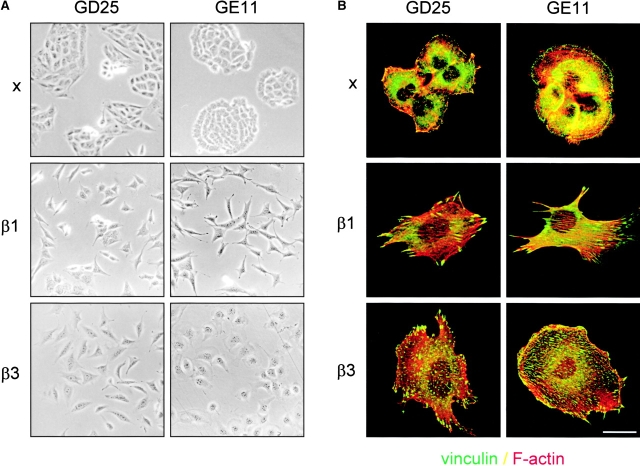
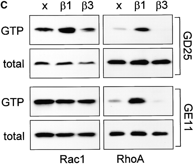
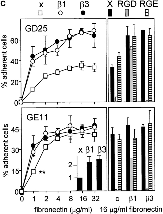
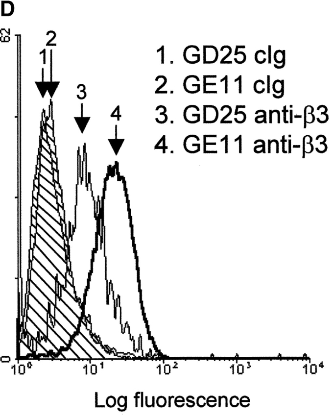
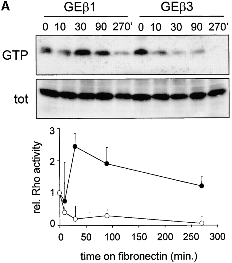
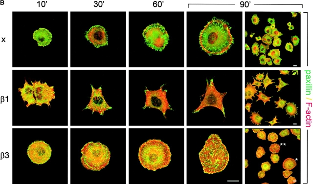
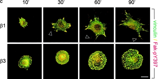
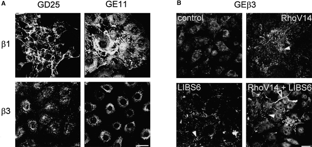
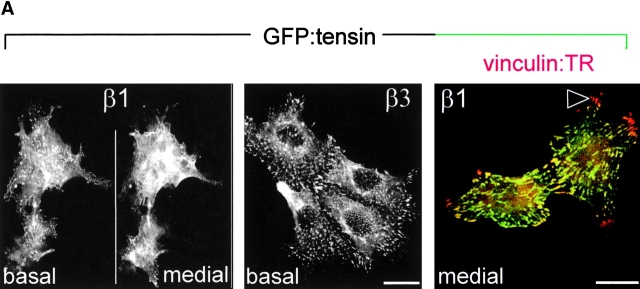
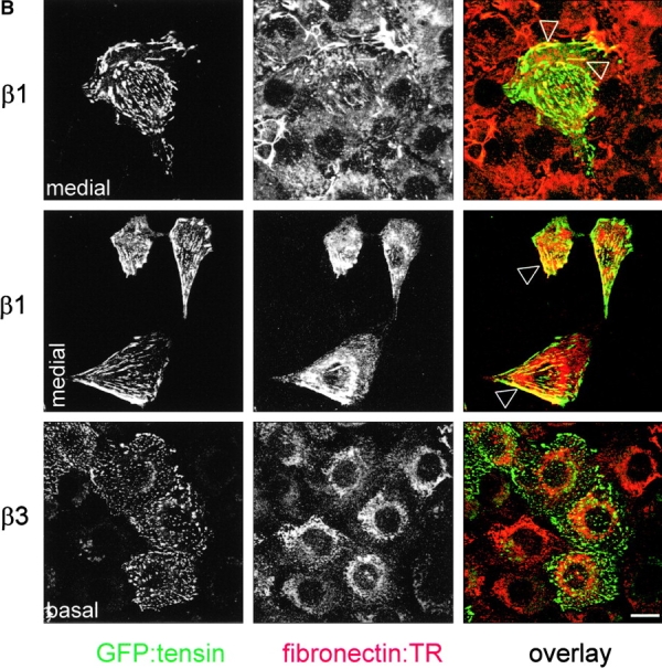
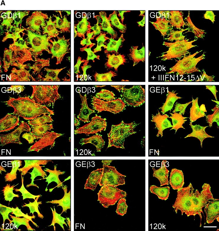
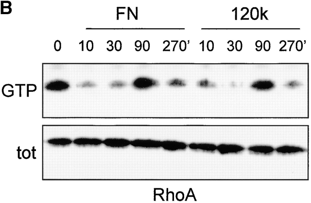
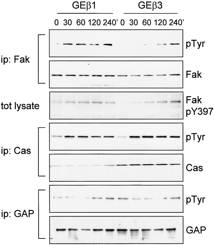
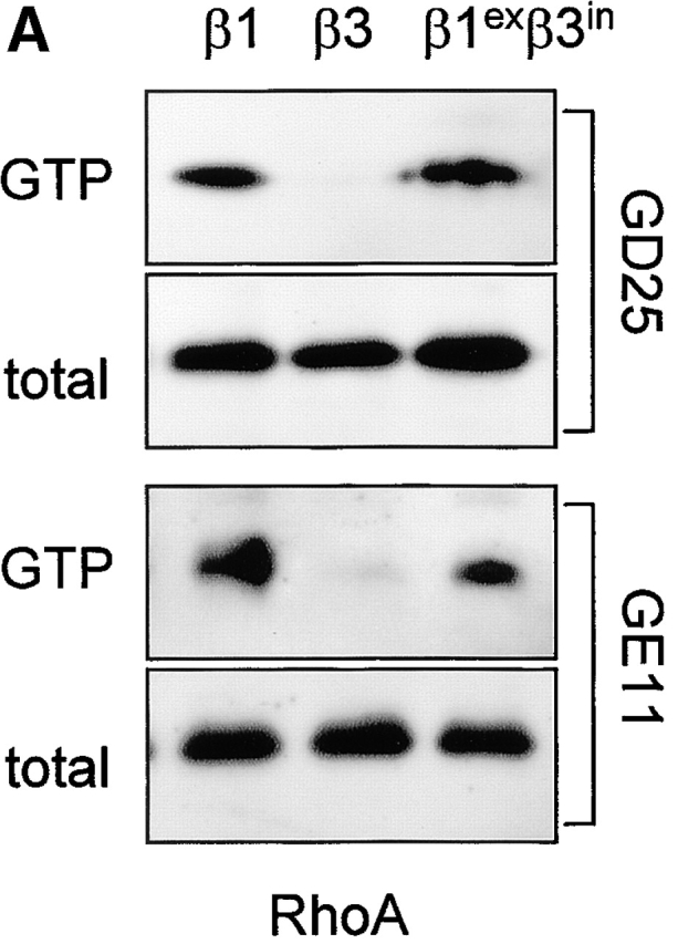
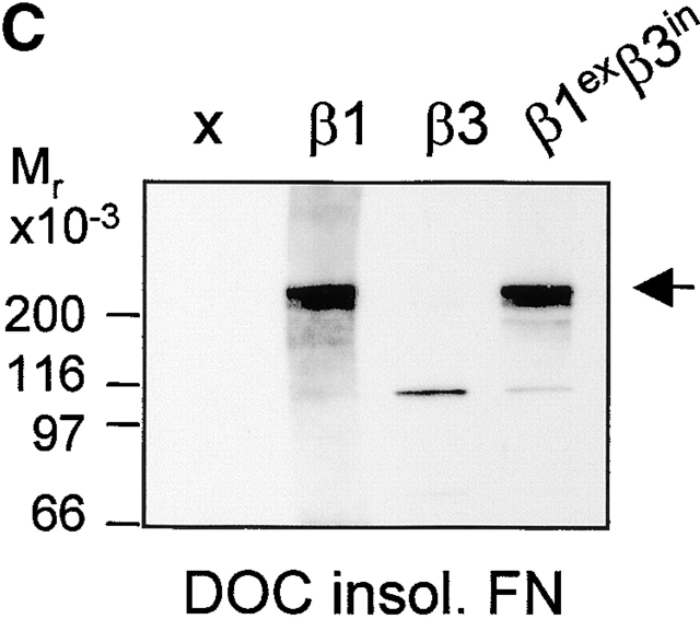
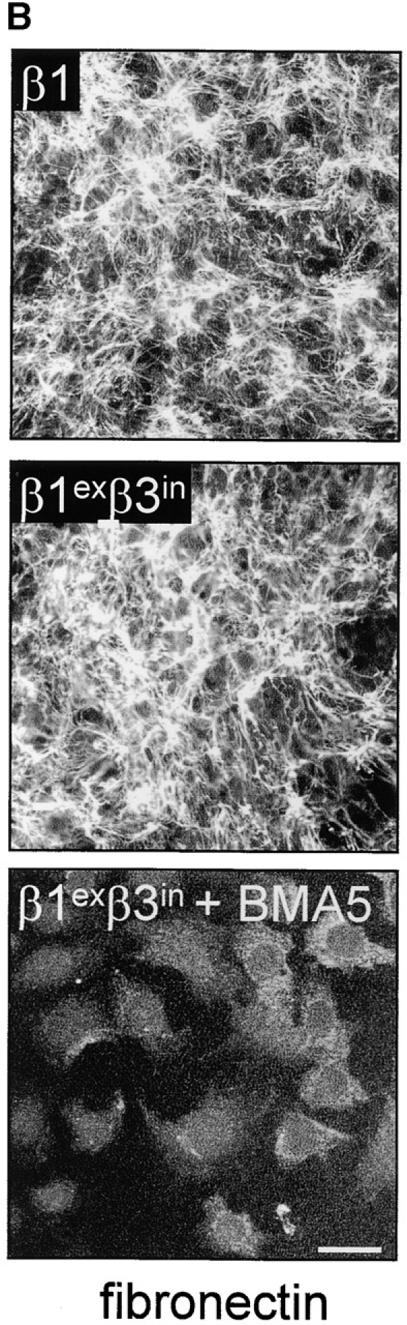
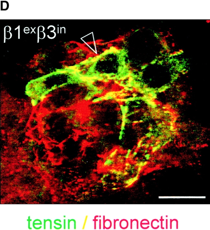
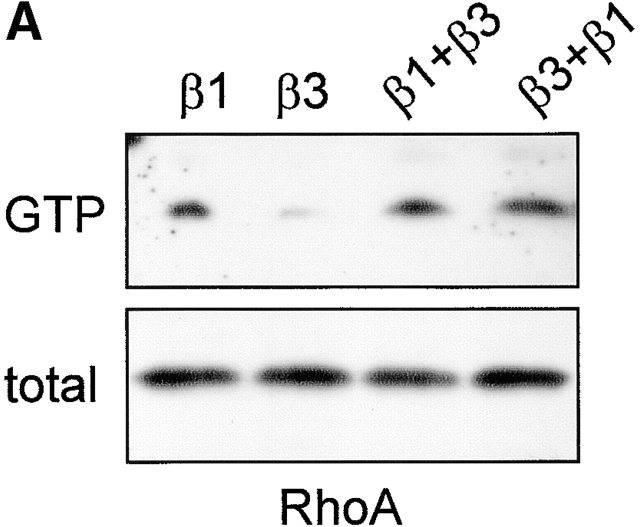
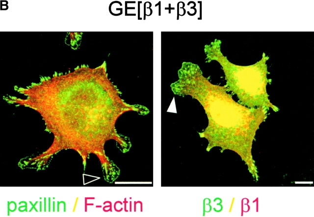
We have studied the formation of different types of cell matrix adhesions in cells that bind to fibronectin via either alpha5beta1 or alphavbeta3. In both cases, cell adhesion to fibronectin leads to a rapid decrease in RhoA activity. However, alpha5beta1 but not alphavbeta3 supports high levels of RhoA activity at later stages of cell spreading, which are associated with a translocation of focal contacts to peripheral cell protrusions, recruitment of tensin into fibrillar adhesions, and fibronectin fibrillogenesis. Expression of an activated mutant of RhoA stimulates alphavbeta3-mediated fibrillogenesis. Despite the fact that alpha5beta1-mediated adhesion to the central cell-binding domain of fibronectin supports activation of RhoA, other regions of fibronectin are required for the development of alpha5beta1-mediated but not alphavbeta3-mediated focal contacts. Using chimeras of beta1 and beta3 subunits, we find that the extracellular domain of beta1 controls RhoA activity. By expressing both beta1 and beta3 at high levels, we show that beta1-mediated control of the levels of beta3 is important for the distribution of focal contacts. Our findings demonstrate that the pattern of fibronectin receptors expressed on a cell dictates the ability of fibronectin to stimulate RhoA-mediated organization of cell matrix adhesions.


Figures

Figure 1.
Figure 1.
Ectopic expression of β3 in β1 integrin-deficient cells stimulates cell scattering and formation of cell matrix adhesions but not RhoA–GTP loading. (A) Microphotographs of GD25 fibroblastoid and GE11 epithelioid β1-deficient cells ectopically expressing the integrin β1 or β3 subunit. (B) GD25 and GE11 cells expressing the indicated integrins were grown in complete medium for 1 d on glass coverslips, fixed, permeabilized, stained for vinculin (FITC) and F-actin (phalloidin:TR), and analyzed by fluorescence microscopy. Bar, 5 μm. (C) GD25 or GE11 cells transduced with integrin β subunits as indicated were grown in standard culture medium, lysed, and processed for RhoA and Rac1 activity assays as described in the Materials and methods. One representative experiment of three is shown.
Figure 1.
Figure 1.
Ectopic expression of β3 in β1 integrin-deficient cells stimulates cell scattering and formation of cell matrix adhesions but not RhoA–GTP loading. (A) Microphotographs of GD25 fibroblastoid and GE11 epithelioid β1-deficient cells ectopically expressing the integrin β1 or β3 subunit. (B) GD25 and GE11 cells expressing the indicated integrins were grown in complete medium for 1 d on glass coverslips, fixed, permeabilized, stained for vinculin (FITC) and F-actin (phalloidin:TR), and analyzed by fluorescence microscopy. Bar, 5 μm. (C) GD25 or GE11 cells transduced with integrin β subunits as indicated were grown in standard culture medium, lysed, and processed for RhoA and Rac1 activity assays as described in the Materials and methods. One representative experiment of three is shown.
Figure 2.
Figure 2.
Integrin expression profiles and adhesion to fibronectin. (A) FACS® analysis showing surface expression of the indicated human (hu) and mouse (mo) integrin subunits on GE11, GEβ1, and GEβ3 cells (cIg, control Ig). Numbers indicate mean fluorescence units. (B) Immunoprecipitations of the indicated biotinylated integrin subunits from cell lysates of GE11, GEβ1, and GEβ3 cells were separated on 4–20% (left) or 5% SDS PAGE (right) and subjected to Western blotting using HRPO-labeled streptavidin. (C) The line graphs show adhesion of control (□) or β1- (○) or β3-transduced (•) GD25 or GE11 cells to wells coated with the indicated concentrations of fibronectin. Mean ± SD of one out of three experiments performed in triplicate is shown. **For GE11, GEβ1, and GEβ3 cells, the relative adhesion to wells coated with 1 μg/ml of fibronectin calculated from three individual experiments is shown in the inset. The column graphs show the adhesion to wells coated with 16 μg/ml of fibronectin in the absence (black bars) or presence of 0.5 mg/ml GRGDSP peptide (white bars) or control GRGESP peptide (hatched bars). (D) FACS® analysis showing expression of endogenous mouse β3 on GD25 (profile 3) and GE11 cells (profile 4).
Figure 2.
Figure 2.
Integrin expression profiles and adhesion to fibronectin. (A) FACS® analysis showing surface expression of the indicated human (hu) and mouse (mo) integrin subunits on GE11, GEβ1, and GEβ3 cells (cIg, control Ig). Numbers indicate mean fluorescence units. (B) Immunoprecipitations of the indicated biotinylated integrin subunits from cell lysates of GE11, GEβ1, and GEβ3 cells were separated on 4–20% (left) or 5% SDS PAGE (right) and subjected to Western blotting using HRPO-labeled streptavidin. (C) The line graphs show adhesion of control (□) or β1- (○) or β3-transduced (•) GD25 or GE11 cells to wells coated with the indicated concentrations of fibronectin. Mean ± SD of one out of three experiments performed in triplicate is shown. **For GE11, GEβ1, and GEβ3 cells, the relative adhesion to wells coated with 1 μg/ml of fibronectin calculated from three individual experiments is shown in the inset. The column graphs show the adhesion to wells coated with 16 μg/ml of fibronectin in the absence (black bars) or presence of 0.5 mg/ml GRGDSP peptide (white bars) or control GRGESP peptide (hatched bars). (D) FACS® analysis showing expression of endogenous mouse β3 on GD25 (profile 3) and GE11 cells (profile 4).
Figure 3.
Figure 3.
Regulation of GTP–RhoA levels and organization of cell matrix adhesions during cell spreading on fibronectin. (A) Cells were serum starved overnight, maintained in suspension for 2 h, and subsequently plated in the absence of serum on dishes coated with 10 μg/ml fibronectin and processed for RhoA activity assays. The Western blot of one representative experiment is shown, and the graph indicates the mean ± SD of three experiments in which the amount of GTP-bound RhoA is shown relative to that in suspended cells (•, GEβ1; ○, GEβ3). (B and C) GE11 cells expressing the indicated integrins were plated in the absence of serum on fibronectin-coated coverslips for the indicated times, fixed, permeabilized, stained for paxillin (FITC) and F-actin (phalloidin:TR) (B) or vinculin (FITC) and phospho-specific pY397-FAK (TR) (C), and analyzed by fluorescence microscopy. Bars, 5 μm.
Figure 3.
Figure 3.
Regulation of GTP–RhoA levels and organization of cell matrix adhesions during cell spreading on fibronectin. (A) Cells were serum starved overnight, maintained in suspension for 2 h, and subsequently plated in the absence of serum on dishes coated with 10 μg/ml fibronectin and processed for RhoA activity assays. The Western blot of one representative experiment is shown, and the graph indicates the mean ± SD of three experiments in which the amount of GTP-bound RhoA is shown relative to that in suspended cells (•, GEβ1; ○, GEβ3). (B and C) GE11 cells expressing the indicated integrins were plated in the absence of serum on fibronectin-coated coverslips for the indicated times, fixed, permeabilized, stained for paxillin (FITC) and F-actin (phalloidin:TR) (B) or vinculin (FITC) and phospho-specific pY397-FAK (TR) (C), and analyzed by fluorescence microscopy. Bars, 5 μm.
Figure 4.
Figure 4.
Absence of fibronectin matrix assembly in β1-deficient cells ectopically expressing β3 and partial rescue by V14RhoA. (A) GD25 or GE11 cells expressing the indicated integrins were grown on glass coverslips for 36 h in standard culture medium, fixed, stained with antifibronectin antibodies, and analyzed by immunofluorescence microscopy. Bar, 20 μm. (B) GEβ3 cells, either untransfected or transiently expressing V14RhoA, were seeded for 4 h on fibronectin-coated glass coverslips, washed, and subsequently incubated for 20 h in medium containing fibronectin-depleted serum supplemented with 10 μg/ml biotinylated fibronectin in the absence or presence of 5 μM LIBS6 antibody as indicated. Cells were fixed, stained with TR-conjugated streptavidin, and analyzed by fluorescence microscopy. Filled arrowheads indicate short (< 5 μm) and open arrowheads indicate longer fibrils (10–20 μm). Bar, 20 μm.
Figure 5.
Figure 5.
Absence of tensin-containing fibrillar adhesions in β1-deficient cells ectopically expressing β3. (A) GD25 cells expressing the indicated integrins were transiently transfected with GFP-tensin, seeded in standard culture medium on glass coverslips for 24 h, fixed, and permeabilized for analysis by confocal fluorescence microscopy. Shown is the localization of GFP-tensin alone (black and white pictures) or in combination with vinculin (TR; color picture). Note the absence of tensin in many of the peripheral focal contacts that stain for vinculin in GDβ1 cells (arrowhead). (B) GE11 cells expressing the indicated integrins and transiently expressing GFP-tensin were seeded for 4 h on fibronectin-coated glass coverslips, washed, and subsequently incubated for 20 h in medium containing fibronectin-depleted serum supplemented with 10 μg/ml biotinylated fibronectin. Cells were fixed, stained with TR-conjugated streptavidin, and analyzed by fluorescence microscopy. Note that coalignment of fibronectin and tensin can be observed in confluent (top) and subconfluent cultures (middle) of GEβ1 (arrowheads) but not GEβ3 cells. Focal planes are as indicated. Bars, 10 μm.
Figure 5.
Figure 5.
Absence of tensin-containing fibrillar adhesions in β1-deficient cells ectopically expressing β3. (A) GD25 cells expressing the indicated integrins were transiently transfected with GFP-tensin, seeded in standard culture medium on glass coverslips for 24 h, fixed, and permeabilized for analysis by confocal fluorescence microscopy. Shown is the localization of GFP-tensin alone (black and white pictures) or in combination with vinculin (TR; color picture). Note the absence of tensin in many of the peripheral focal contacts that stain for vinculin in GDβ1 cells (arrowhead). (B) GE11 cells expressing the indicated integrins and transiently expressing GFP-tensin were seeded for 4 h on fibronectin-coated glass coverslips, washed, and subsequently incubated for 20 h in medium containing fibronectin-depleted serum supplemented with 10 μg/ml biotinylated fibronectin. Cells were fixed, stained with TR-conjugated streptavidin, and analyzed by fluorescence microscopy. Note that coalignment of fibronectin and tensin can be observed in confluent (top) and subconfluent cultures (middle) of GEβ1 (arrowheads) but not GEβ3 cells. Focal planes are as indicated. Bars, 10 μm.
Figure 6.
Figure 6.
Roles of fibronectin regions other than the CCBD in β1- and β3-mediated regulation of cell matrix adhesions and RhoA–GTP loading. (A) GD25 and GE11 cells transduced with the indicated integrin subunits were serum starved overnight and plated for 120 min in the absence or presence of 0.5 μg/ml GST–IIIFN12-15ΔV on glass coverslips coated with 10 μg/ml fibronectin or 5 μg/ml fibronectin 120-kD chymotryptic fragment as indicated. Cells were fixed, permeabilized, stained for paxillin (FITC) and F-actin (phalloidin:TR), and analyzed by fluorescence microscopy. Bar, 10 μm. (B) GDβ1 cells were serum starved overnight, maintained in suspension for 2 h in the presence of cyclohexamide, plated for the indicated times on dishes coated with 10 μg/ml fibronectin or 5 μg/ml 120-kD chymotryptic fragment, and processed for RhoA activity assays (note that GTP–RhoA levels are high in suspended cells and in cells plated 90 min on FN or the 120-kD fragment).
Figure 6.
Figure 6.
Roles of fibronectin regions other than the CCBD in β1- and β3-mediated regulation of cell matrix adhesions and RhoA–GTP loading. (A) GD25 and GE11 cells transduced with the indicated integrin subunits were serum starved overnight and plated for 120 min in the absence or presence of 0.5 μg/ml GST–IIIFN12-15ΔV on glass coverslips coated with 10 μg/ml fibronectin or 5 μg/ml fibronectin 120-kD chymotryptic fragment as indicated. Cells were fixed, permeabilized, stained for paxillin (FITC) and F-actin (phalloidin:TR), and analyzed by fluorescence microscopy. Bar, 10 μm. (B) GDβ1 cells were serum starved overnight, maintained in suspension for 2 h in the presence of cyclohexamide, plated for the indicated times on dishes coated with 10 μg/ml fibronectin or 5 μg/ml 120-kD chymotryptic fragment, and processed for RhoA activity assays (note that GTP–RhoA levels are high in suspended cells and in cells plated 90 min on FN or the 120-kD fragment).
Figure 7.
Figure 7.
Fibronectin-stimulated tyrosine phosphorylation of FAK, p130Cas, and p190RhoGAP. GEβ1 and GEβ3 cells were serum starved overnight and replated on dishes coated with 10 μg/ml fibronectin in serum-free medium. Cells were lysed in modified RIPA buffer at the indicated time points for immunoprecipitation with antibodies indicated on the left. Immunoprecipitates or total lysates were separated on 8% SDS-PAGE followed by Western blotting with antibodies indicated on the right.
Figure 8.
Figure 8.
The role of the cytoplasmic domain in RhoA activation, fibronectin matrix assembly, and tensin recruitment. (A) GD25 and GE11 cells expressing the indicated wild-type or chimeric integrin subunits were grown in standard culture medium, lysed, and processed for RhoA activity assays. (B) GEβ1 and GEβ1exβ3in cells were serum starved overnight, seeded for 4 h on fibronectin-coated glass coverslips in serum-free medium, washed, and subsequently incubated in the absence or presence of 0.5 μg/ml BMA5 anti-α5 antibody for 20 h in medium containing fibronectin-depleted serum supplemented with 10 μg/ml biotinylated fibronectin. Cells were fixed, stained with TR-conjugated streptavidin, and analyzed by fluorescence microscopy. Bars, 20 μm. (C) GE11 cells expressing the indicated wild-type or chimeric integrin subunits were serum starved, seeded for 4 h on fibronectin-coated dishes in serum-free medium, washed, and subsequently incubated for 20 h in medium containing fibronectin-depleted serum supplemented with 10 μg/ml biotinylated fibronectin. Cells were lysed in DOC buffer, and insoluble material was separated by SDS-PAGE and subjected to Western blotting using HRPO-conjugated streptavidin. (D) GDβ1exβ3in cells were transiently transfected with GFP-tensin, serum starved, seeded for 4 h on fibronectin-coated glass coverslips, washed, and subsequently incubated for 20 h in medium containing fibronectin-depleted serum supplemented with 10 μg/ml biotinylated fibronectin. Cells were fixed, stained with TR-conjugated streptavidin, and analyzed by fluorescence microscopy. A cluster of ∼10 cells is shown; the arrowhead points to localization of tensin along fibronectin fibrils. Bar, 20 μm.
Figure 8.
Figure 8.
The role of the cytoplasmic domain in RhoA activation, fibronectin matrix assembly, and tensin recruitment. (A) GD25 and GE11 cells expressing the indicated wild-type or chimeric integrin subunits were grown in standard culture medium, lysed, and processed for RhoA activity assays. (B) GEβ1 and GEβ1exβ3in cells were serum starved overnight, seeded for 4 h on fibronectin-coated glass coverslips in serum-free medium, washed, and subsequently incubated in the absence or presence of 0.5 μg/ml BMA5 anti-α5 antibody for 20 h in medium containing fibronectin-depleted serum supplemented with 10 μg/ml biotinylated fibronectin. Cells were fixed, stained with TR-conjugated streptavidin, and analyzed by fluorescence microscopy. Bars, 20 μm. (C) GE11 cells expressing the indicated wild-type or chimeric integrin subunits were serum starved, seeded for 4 h on fibronectin-coated dishes in serum-free medium, washed, and subsequently incubated for 20 h in medium containing fibronectin-depleted serum supplemented with 10 μg/ml biotinylated fibronectin. Cells were lysed in DOC buffer, and insoluble material was separated by SDS-PAGE and subjected to Western blotting using HRPO-conjugated streptavidin. (D) GDβ1exβ3in cells were transiently transfected with GFP-tensin, serum starved, seeded for 4 h on fibronectin-coated glass coverslips, washed, and subsequently incubated for 20 h in medium containing fibronectin-depleted serum supplemented with 10 μg/ml biotinylated fibronectin. Cells were fixed, stained with TR-conjugated streptavidin, and analyzed by fluorescence microscopy. A cluster of ∼10 cells is shown; the arrowhead points to localization of tensin along fibronectin fibrils. Bar, 20 μm.
Figure 9.
Figure 9.
Regulation of αvβ3 levels by β1 affects focal contact maturation not RhoA–GTP loading. (A) GE11 cells expressing the indicated integrin subunits were grown in standard culture medium, lysed, and processed for RhoA activity assays. (B) GE[β1+β3] cells were plated in the absence of serum on fibronectin-coated coverslips for 90 min, fixed, permeabilized, and stained for paxillin (FITC) and F-actin (phalloidin:TR) or with polyclonal anti-β1 (TR) and FITC-conjugated monoclonal anti-β3 as indicated. Arrowheads indicate membrane blebs containing many small matrix adhesions that stain for paxillin (open arrowhead) and β3 integrin (filled arrowhead). Bars, 5 μm.
Figure 9.
Figure 9.
Regulation of αvβ3 levels by β1 affects focal contact maturation not RhoA–GTP loading. (A) GE11 cells expressing the indicated integrin subunits were grown in standard culture medium, lysed, and processed for RhoA activity assays. (B) GE[β1+β3] cells were plated in the absence of serum on fibronectin-coated coverslips for 90 min, fixed, permeabilized, and stained for paxillin (FITC) and F-actin (phalloidin:TR) or with polyclonal anti-β1 (TR) and FITC-conjugated monoclonal anti-β3 as indicated. Arrowheads indicate membrane blebs containing many small matrix adhesions that stain for paxillin (open arrowhead) and β3 integrin (filled arrowhead). Bars, 5 μm.

Similar articles

Cited by

References

    1. Aota, S., M. Nomizu, and K. Yamada. 1994. The short amino acid sequence Pro-His-Ser-Arg-Asn in human fibronectin enhances cell-adhesive function. J. Biol. Chem. 269:24756–24761. - PubMed
    1. Arthur, W., L. Petch, and K. Burridge. 2000. Integrin engagement suppresses RhoA activity via a c-Src-dependent mechanism. Curr. Biol. 10:719–722. - PubMed
    1. Baneyx, G., L. Baugh, and V. Vogel. 2001. Coexisting conformations of fibronectin in cell culture imaged using fluorescence resonance energy transfer. Proc. Natl. Acad. Sci. USA. 98:14464–14468. - PMC - PubMed
    1. Barry, S., H. Flinn, M. Humphries, D. Critchley, and A. Ridley. 1997. Requirement for Rho in integrin signalling. Cell Adhes. Commun. 4:387–398. - PubMed
    1. Berrier, A.L., R. Martinez, G.M. Bokoch, and S.L. LaFlamme. 2002. Integrin β tails activate Rac1. J. Cell Sci. 115:4285–4291 - PubMed

Publication types

MeSH terms

Substances