. 2002 Dec 23;159(6):1029-37.
doi: 10.1083/jcb.200207165. Epub 2002 Dec 16.

Ghrelin and des-acyl ghrelin inhibit cell death in cardiomyocytes and endothelial cells through ERK1/2 and PI 3-kinase/AKT

Affiliations

Ghrelin and des-acyl ghrelin inhibit cell death in cardiomyocytes and endothelial cells through ERK1/2 and PI 3-kinase/AKT

Gianluca Baldanzi et al. J Cell Biol. .

Abstract

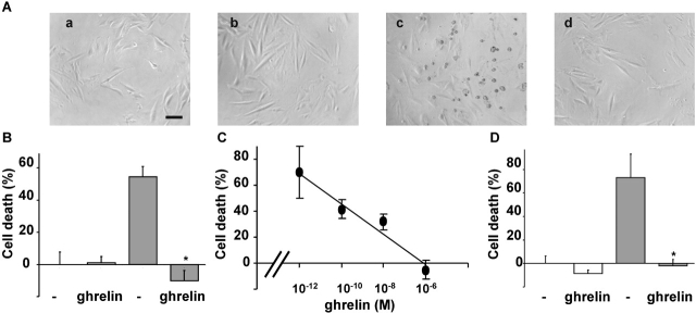
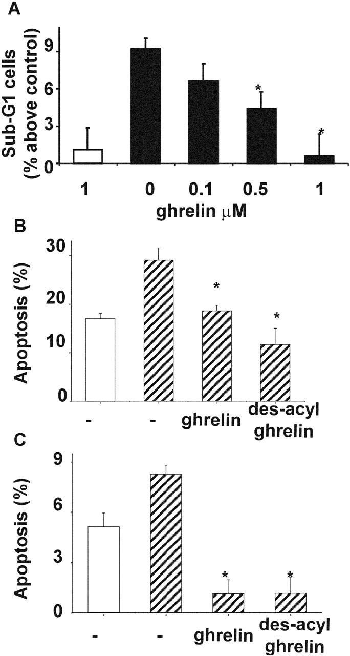
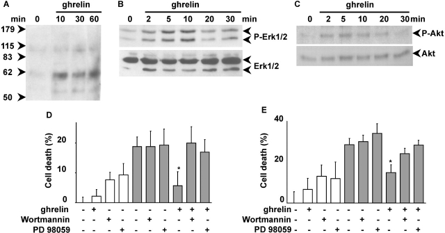
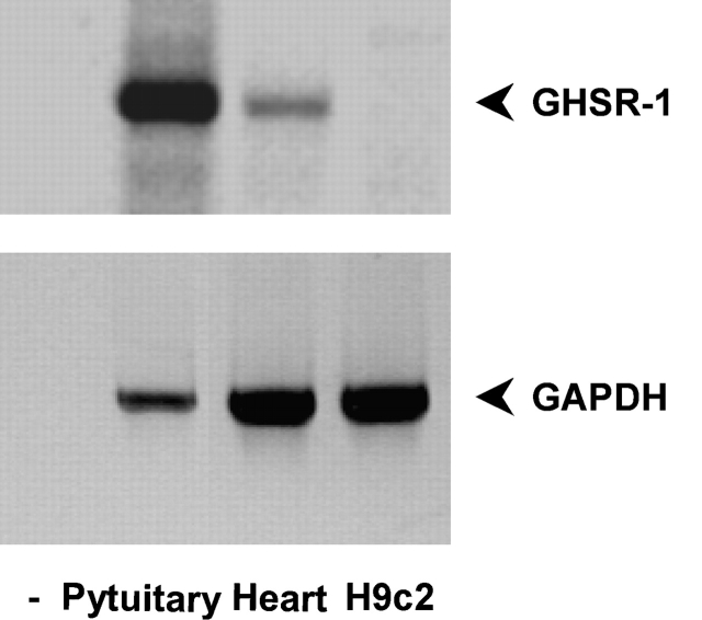
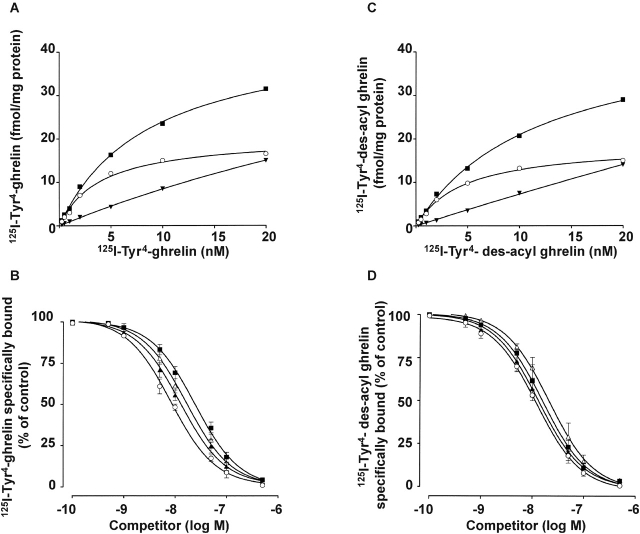
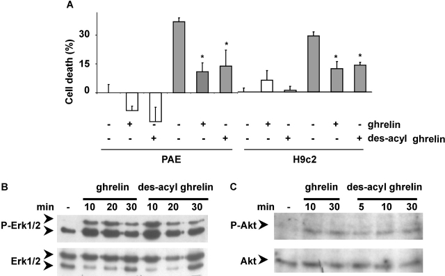
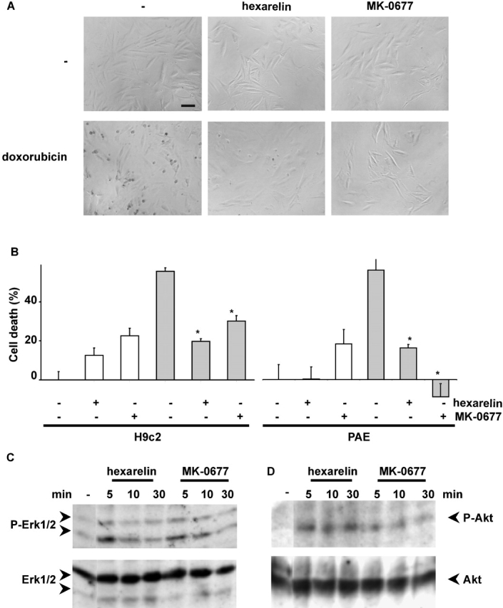
Ghrelin is an acyl-peptide gastric hormone acting on the pituitary and hypothalamus to stimulate growth hormone (GH) release, adiposity, and appetite. Ghrelin endocrine activities are entirely dependent on its acylation and are mediated by GH secretagogue (GHS) receptor (GHSR)-1a, a G protein-coupled receptor mostly expressed in the pituitary and hypothalamus, previously identified as the receptor for a group of synthetic molecules featuring GH secretagogue (GHS) activity. Des-acyl ghrelin, which is far more abundant than ghrelin, does not bind GHSR-1a, is devoid of any endocrine activity, and its function is currently unknown. Ghrelin, which is expressed in heart, albeit at a much lower level than in the stomach, also exerts a cardio protective effect through an unknown mechanism, independent of GH release. Here we show that both ghrelin and des-acyl ghrelin inhibit apoptosis of primary adult and H9c2 cardiomyocytes and endothelial cells in vitro through activation of extracellular signal-regulated kinase-1/2 and Akt serine kinases. In addition, ghrelin and des-acyl ghrelin recognize common high affinity binding sites on H9c2 cardiomyocytes, which do not express GHSR-1a. Finally, both MK-0677 and hexarelin, a nonpeptidyl and a peptidyl synthetic GHS, respectively, recognize the common ghrelin and des-acyl ghrelin binding sites, inhibit cell death, and activate MAPK and Akt.These findings provide the first evidence that, independent of its acylation, ghrelin gene product may act as a survival factor directly on the cardiovascular system through binding to a novel, yet to be identified receptor, which is distinct from GHSR-1a.


Figures

Figure 1.
Figure 1.
Ghrelin protects H9c2 cardiomyocytes from doxorubicin-induced cell death. H9c2 cardiomyocytes were treated with 1 μM doxorubicin (grey bar) in the presence or absence of 1 μM ghrelin. (A) Phase–contrast images (200×; bar, 0.1 mm) after 24 h of treatment: untreated (a), ghrelin (b), doxorubicin (c), and doxorubicin + ghrelin (d). (B) Cell death measured by MTT assay after 24 h of treatment is expressed as the percentage of cell death. Values are the mean ± SE of eight samples (*t test, P < 0.01). (C) Dose response of ghrelin in MTT cell death assay after 24 h of treatment. Values are the mean ± SE of eight samples. (D) Cell death measured by LDH release after 48 h of treatment. Values are the mean ± SE of eight samples (*t test, P < 0.01).
Figure 2.
Figure 2.
Ghrelin and des-acyl ghrelin inhibit apoptosis. (A) Primary cultures of adult cardiomyocytes were pretreated with either 0.1, 0.5, or 1 μM ghrelin for 16 h. Then FAS was stimulated (black bars), and sub-G1 cells were measured by FACS® analysis after 6 h. Data were calculated as the percentage of apoptotic nuclei induced by FAS stimulation above untreated control. Values are the mean ± SE of three independent experiments (*t test, P < 0.01). H9c2 cardiomyocytes (B) and PAE cells (C) cultured in either 10% FCS (white bar) or 0% FCS (hatched bars) were pretreated with 1 μM ghrelin or des-acyl ghrelin for 48 h. Apoptosis was measured by FACS® analysis with fluorescent annexin V; values are mean ± SE of three samples (*t test, P < 0.02).
Figure 3.
Figure 3.
Ghrelin stimulates protein-tyrosine phosphorylation and activates ERK1/2 and Akt. Overnight serum-starved H9c2 cells were stimulated with 1 μM ghrelin and lysed at different times. Total lysates were analyzed by Western blot with antiphosphotyrosine antibodies (A), specific anti-phosphoERK1/2 antibodies (B, top) and anti-ERK1/2 antibodies (B, bottom), and specific anti-phosphoAkt antibodies (C, top) and anti-Akt antibodies (C, bottom). H9c2 cardiomyocytes (D) and PAE cells (E) were treated with 1 μM doxorubicin for 20 h (black bars) in the presence or absence of 1 μM ghrelin and in the presence or absence of 100 nM wortmannin or 30 μM PD98059. Cell death was measured by MTT assay. Values are the mean ± SE of eight samples (*t test, P < 0.01).
Figure 4.
Figure 4.
GHSr-1a is expressed in the whole heart but not in H9c2 cardiomyocytes. RT-PCR was performed on total RNA extracted from H9c2 cardiomyocytes and rat heart with primers specific for GHSR-1a (top) or GAPDH (bottom).
Figure 5.
Figure 5.
H9c2 cardiomyocytes express a ghrelin receptor distinct from GHSR-1a. (A and C) Specific binding was determined by incubation of crude membranes with increasing concentrations (0.25–20 nM) of either radio-labeled ghrelin (A) or radio-labeled des-acyl ghrelin (C) in the presence or absence of 1 μM unlabeled ghrelin (A) or unlabeled des-acyl ghrelin (C), respectively: total binding (▪), specific binding (▾), and nonspecific binding (○). Data are the average of duplicate assay determinants. Similar results were obtained in at least two other independent experiments. (B and D) Displacement curves of radio-labeled ghrelin (B) or radio-labeled des-acyl ghrelin (D) binding by unlabeled ghrelin (○), des-acyl ghrelin (▪), hexarelin (▴), and MK-0677 (▵). Displacement binding values are the mean ± SE of four separate experiments.
Figure 6.
Figure 6.
Des-acyl ghrelin inhibits cell death and activates ERK1/2 and Akt in H9c2 cardiomyocytes and PAE. (A) H9c2 cardiomyocytes and PAE cells were treated with 1 μM doxorubicin (grey bar) for 24 h in the presence or absence of either 1 μM ghrelin or des-acyl ghrelin. Cell death was assessed by the MTT assay. Values are mean ± SE of eight samples (*t test, P < 0.01). (B and C) H9c2 cardiomyocytes were serum starved overnight, stimulated with 1 μM des-acyl ghrelin, and lysed at different times. Total lysates were analyzed by Western blot with specific anti-phosphoERK1/2 antibodies (B, top) and anti-ERK1/2 antibodies (B, bottom) and specific anti-phosphoAkt antibodies (C, top) and anti-Akt antibodies (C, bottom).
Figure 7.
Figure 7.
Hexarelin and MK-0677 inhibit cell death and activate ERK1/2 and Akt in H9c2 cardiomyocytes and PAE. (A) H9c2 cardiomyocytes were treated with 1 μM of either doxorubicin, hexarelin, and/or MK-0677 as indicated. Phase–contrast images (200×; bar, 0.1 mm) were captured after 24 h of treatment. Representative fields are shown. (B) H9c2 cardiomyocytes and PAE cells were treated with 1 μM doxorubicin (grey bar) for 24 h in the presence or absence of either 1 μM hexarelin or MK-0677. Cell death was assessed by the MTT assay. Values are mean ± SE of eight samples (*t test, P < 0.01). (C and D) H9c2 cardiomyocytes were serum starved overnight, stimulated with either 1 μM hexarelin or MK-0677, and lysed at different times. Total lysates were analyzed by Western blot with specific anti-phosphoERK1/2 antibodies (C, top) and anti-ERK1/2 antibodies (C, bottom) and specific anti-phosphoAkt antibodies (D, top) and anti-Akt antibodies (D, bottom).

Similar articles

Cited by

References

    1. Alessi, D.R., M. Andjelkovic, B. Caudwell, P. Cron, N. Morrice, P. Cohen, and B.A. Hemmings. 1996. Mechanism of activation of protein kinase B by insulin and IGF-1. EMBO J. 15:6541–6551. - PMC - PubMed
    1. Arola, O.J., A. Saraste, K. Pulkki, M. Kallajoki, M. Parvinen, and L.M. Voipio-Pulkki. 2000. Acute doxorubicin cardiotoxicity involves cardiomyocyte apoptosis. Cancer Res. 60:1789–1792. - PubMed
    1. Bisi, G., V. Podio, M.R. Valetto, F. Broglio, G. Bertuccio, G. Aimaretti, E. Pelosi, G. Del Rio, G. Muccioli, H. Ong, et al. 1999. Cardiac effects of hexarelin in hypopituitary adults. Eur. J. Pharmacol. 381:31–38. - PubMed
    1. Bodart, V., M. Febbraio, A. Demers, N. McNicoll, P. Pohankova, A. Perreault, T. Sejlitz, E. Escher, R.L. Silverstein, D. Lamontagne, and H. Ong. 2002. CD36 mediates the cardiovascular action of growth hormone-releasing peptides in the heart. Circ. Res. 90:844–849. - PubMed
    1. Bowers, C.Y. 2001. Unnatural growth hormone-releasing peptide begets natural ghrelin. J. Clin. Endocrinol. Metab. 86:1464–1469. - PubMed

Publication types

MeSH terms

Substances