. 2002 Dec 23;159(6):1087-96.
doi: 10.1083/jcb.200208050. Epub 2002 Dec 16.

Cell migration requires both ion translocation and cytoskeletal anchoring by the Na-H exchanger NHE1

Affiliations

Cell migration requires both ion translocation and cytoskeletal anchoring by the Na-H exchanger NHE1

Sheryl P Denker et al. J Cell Biol. .

Abstract

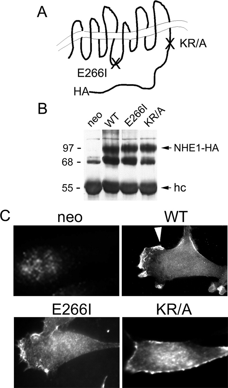
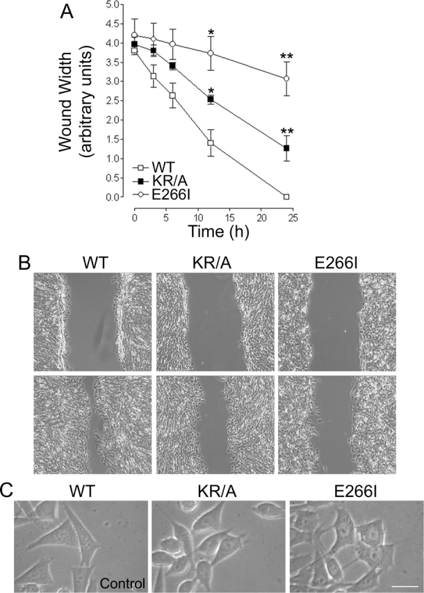
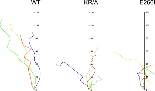
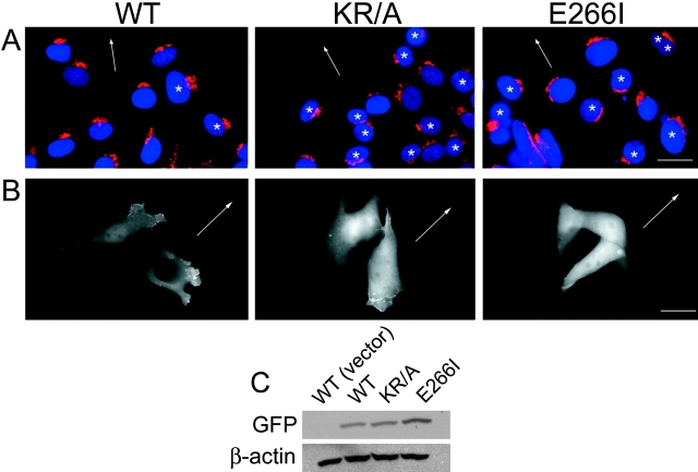
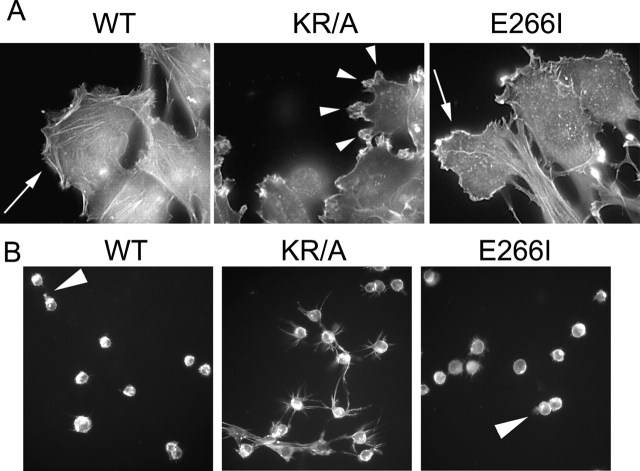
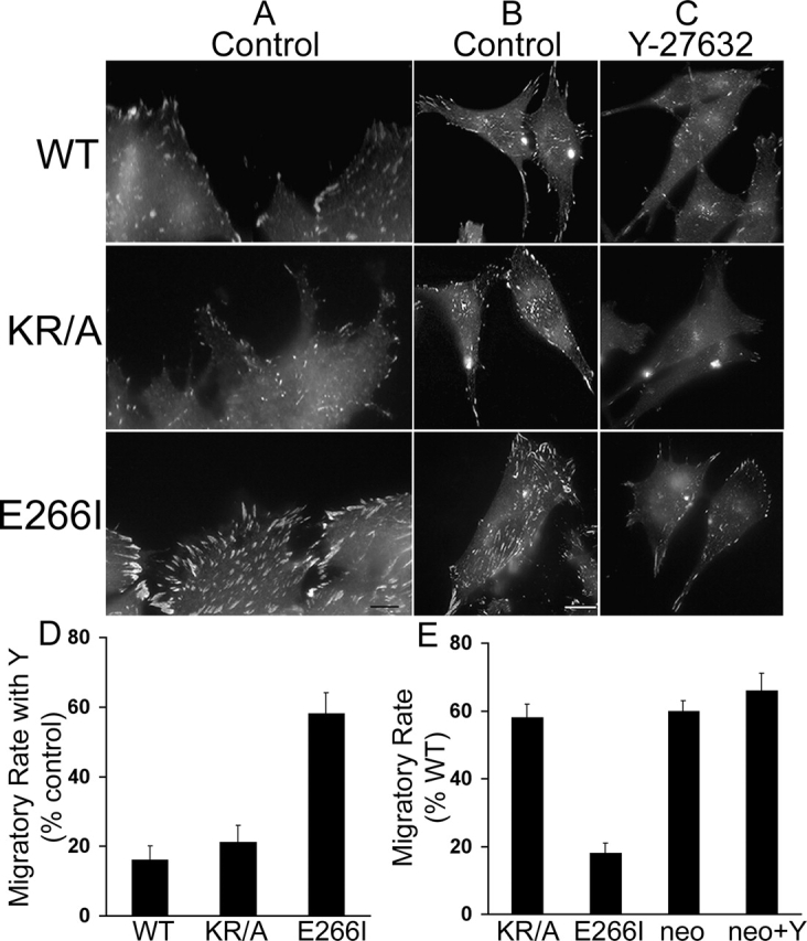
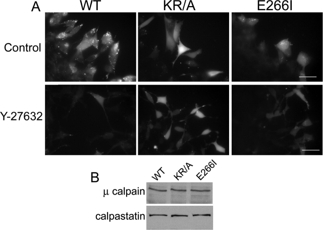
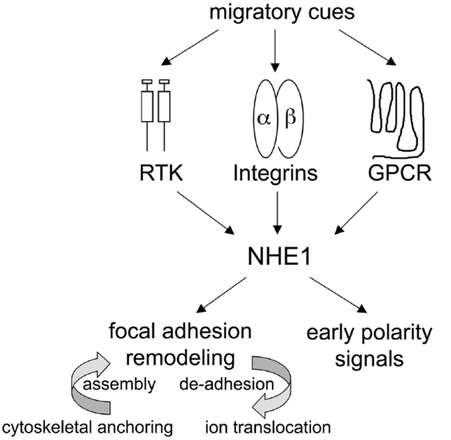
Directed cell movement is a multi-step process requiring an initial spatial polarization that is established by asymmetric stimulation of Rho GTPases, phosphoinositides (PIs), and actin polymerization. We report that the Na-H exchanger isoform 1 (NHE1), a ubiquitously expressed plasma membrane ion exchanger, is necessary for establishing polarity in migrating fibroblasts. In fibroblasts, NHE1 is predominantly localized in lamellipodia, where it functions as a plasma membrane anchor for actin filaments by its direct binding of ezrin/radixin/moesin (ERM) proteins. Migration in a wounding assay was impaired in fibroblasts expressing NHE1 with mutations that independently disrupt ERM binding and cytoskeletal anchoring or ion transport. Disrupting either function of NHE1 impaired polarity, as indicated by loss of directionality, mislocalization of the Golgi apparatus away from the orientation of the wound edge, and inhibition of PI signaling. Both functions of NHE1 were also required for remodeling of focal adhesions. Most notably, lack of ion transport inhibited de-adhesion, resulting in trailing edges that failed to retract. These findings indicate that by regulating asymmetric signals that establish polarity and by coordinating focal adhesion remodeling at the cell front and rear, cytoskeletal anchoring by NHE1 and its localized activity in lamellipodia act cooperatively to integrate cues for directed migration.


Figures

Figure 1.
Figure 1.
Characterization of cell lines. (A) Schematic diagram of NHE1 indicating positions of the HA-epitope tag, the KR/A mutation (amino acids 553–564 of rat sequence, alanines substituted for lysine and arginine residues) that abolishes ERM binding, and the E266I mutation that abolishes ion translocation. (B) Expression and (C) localization of NHE1 wild-type (WT; arrowhead) and mutant proteins in PS120 fibroblasts. Expression of the selection plasmid alone (neo) served as a control in initial studies. hc, IgG heavy chain.
Figure 2.
Figure 2.
Wounding assays demonstrate that mutations in NHE1 impair cell migration. (A) The migratory rate of fibroblasts in a wounding assay was determined by measuring wound width as a function of time for cells plated on gridded coverslips. Wound closure at 24 h was 100% for WT cells, 60–70% for KR/A cells, and 20–25% for E266I cells. Data are expressed as the mean ± SEM of three experiments. Asterisks indicate a significant difference from WT cells. *, 0.05%; **, 0.01%. (B) Images from time-lapse videos acquired at 0 and 12 h. WT cells migrated together as a sheet, whereas both KR/A and E266I cells displayed asynchronous movement. Videos 1–3 were taken with a 10× phase-contrast objective. (C) High magnification images (63×) of cells at the wound edge demonstrate the fusiform shape of KR/A cells and extended tails in E266I cells. Bar, 10 μm. Videos available at http://www.jcb.org/cgi/content/full/jcb.200208050/DC1.
Figure 3.
Figure 3.
Cell tracking demonstrates that both ion translocation and cytoskeletal anchoring by NHE1 are required for directionality. The paths and distance traveled by representative cells at the wound edge were plotted as a function of time over 16 h. WT cells moved farthest with few turns and no reversals. KR/A and E266I cells turned frequently and reversed direction.
Figure 4.
Figure 4.
Both ion translocation and cytoskeletal anchoring by NHE1 regulate cell polarity. (A) Cells migrating in a wounding assay were fixed after 10 h and processed for immunolocalization of the Golgi complex (anti-Giantin antibody) and nuclei (DAPI). Representative fields are shown, with arrows indicating the direction of migration and asterisks indicating cells with misoriented Golgi apparatus. The large cluster of nuclei in the bottom left panel of E266I cells was not included in determining the percentage of cells with correct Golgi apparatus orientation. (B) GFP-PH-Akt was detected in the leading-edge membrane of WT, but not KR/A or E266I cells moving at the front of a migrating monolayer. GFP-PH-Akt was primarily cytosolic in both mutant cell lines. Images are representative of 90% of cells at the wound edge expressing GFP-PH-Akt in three separate cell preparations. Arrows indicate the direction of migration. (C) Immunoblotting with antibodies to GFP indicated that the relative expression level of GFP-PH-Akt was similar in WT and KR/A cells, but higher in E266I cells. There was no GFP signal in WT cells transfected with a vector control. Immunoblotting for β-actin confirmed equivalent loading of protein in all samples. Bars: (A) 5 μm; (B) 2 μm.
Figure 5.
Figure 5.
ERM binding and cytoskeletal anchoring by NHE1 are required for development of a primary lamellipod. (A) Migrating WT and E266I cells exhibit one broad lamellipod (arrows), whereas KR/A cells have multiple, small protrusions (arrowheads). Bar, 5 μm. (B) When plated on Matrigel™ for 4 h, WT and E266I cells develop one primary protrusion (arrowhead), whereas KR/A cells develop multiple protrusions. (A and B) TRITC-phalloidin staining.
Figure 6.
Figure 6.
Inhibition of focal adhesion turnover in the absence of NHE1 activity is rate-limiting for migration speed. Cells migrating in the absence (Control) or presence of 10 μM Y-27632 for 15 h were fixed and labeled for paxillin. (A–C). Paxillin labeling of control cells at the wound edge (A) and of representative cells that had migrated into the wound (B) to visualize focal contacts at the front and rear of the cell. Compared with WT cells, KR/A cells have reduced paxillin labeling, whereas E266I cells have increased paxillin labeling. (C) The abundance and intensity of paxillin labeling is decreased in cells treated with 10 μM Y-27632. (D) Migratory rate of cells in the absence or presence of Y-27632 was determined between 0 and 12 h and expressed as a percentage of increase induced by Y-27632 over control rate. (E) Migratory rates of KR/A cells, E266I cells, and NHE1-null PS120neo cells in the absence or presence of Y-27632 were determined between 0 and 12 h and expressed as percentage of the migratory rate of WT cells. Data represent the mean ± SEM of three separate wounding preparations. Bars: (A) 1 μm; (B) 5 μm.
Figure 7.
Figure 7.
NHE1 and ROCK regulate calpain activity. (A) Top, calpain activity in control cells, indicated by bright punctate and cytosolic fluorescence, was detected along the front of migrating WT (but not E266I) cells. In KR/A cells, the diffuse, but not the punctate, fluorescence pattern was observed. Bottom, calpain activity in cells treated with Y-27632. (B) Immunoblotting indicated equal abundance of μ-calpain and calpastatin in the three cell lines. Images are representative of cells at the wound edge in four separate wounding assays for each cell type. Bars, 20 μm.
Figure 8.
Figure 8.
NHE1 integrates migratory cues to spatially amplify early polarity signals and to regulate focal adhesion remodeling. Membrane receptors, including receptor tyrosine kinases (RTK), integrins, and G protein–coupled receptors (GPCR), receive migratory cues. All three classes of receptors activate NHE1, which is spatially restricted to the leading-edge membrane of lamellipodia by cytoskeletal anchoring through the binding of ERM proteins. Localized NHE1 activity integrates signals from diverse membrane receptors to coordinate early polarity signals and promote focal adhesion remodeling. The two functions of NHE1 have distinct effects on focal adhesion remodeling. Cytoskeletal anchoring is necessary for focal adhesion assembly and ion translocation is necessary for de-adhesion.

Similar articles

Cited by

References

    1. Alblas, J., L. Ulfman, P. Hordijk, and L. Koenderman. 2001. Activation of RhoA and ROCK are essential for detachment of migrating leukocytes. Mol. Biol. Cell. 12:2137–2145. - PMC - PubMed
    1. Bailly, M., J. Wyckoff, B. Bouzahzah, R. Hammerman, V. Sylvestre, M. Cammer, R. Pestell, and J.E. Segall. 2000. Epidermal growth factor receptor distribution during chemotactic responses. Mol. Biol. Cell. 11:3873–3883. - PMC - PubMed
    1. Bernstein, B.W., W.B. Painter, H. Chen, L.S. Minamide, H. Abe, and J.R. Bamburg. 2000. Intracellular pH modulation of ADF/cofilin proteins. Cell Motil. Cytoskeleton. 47:319–336. - PubMed
    1. Bertrand, B., S. Wakabayashi, T. Ikeda, J. Pouyssegur, and M. Shigekawa. 1994. The Na+/H+ exchanger isoform 1 (NHE1) is a novel member of the calmodulin-binding proteins. Identification and characterization of calmodulin-binding sites. J. Biol. Chem. 269:13703–13709. - PubMed
    1. Bialkowska, K., S. Kulkarni, X. Du, D.E. Goll, T.C. Saido, and J.E. Fox. 2000. Evidence that beta3 integrin-induced Rac activation involves the calpain-dependent formation of integrin clusters that are distinct from the focal complexes and focal adhesions that form as Rac and RhoA become active. J. Cell Biol. 151:685–696. - PMC - PubMed

Publication types

MeSH terms