. 2002 Dec 15;22(24):10509-13.
doi: 10.1523/JNEUROSCI.22-24-10509.2002.

Starburst cells nondirectionally facilitate the responses of direction-selective retinal ganglion cells

Affiliations

Starburst cells nondirectionally facilitate the responses of direction-selective retinal ganglion cells

Chuan-Chin Chiao et al. J Neurosci. .

Abstract

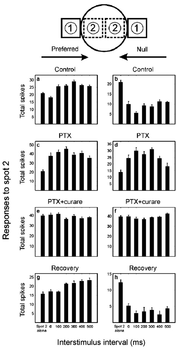
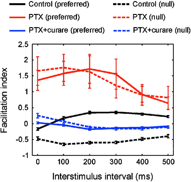
The mechanism of direction selectivity in retinal ganglion cells remains controversial. An important issue is how the starburst amacrine cells, which are known to provide a major synaptic input to the direction-selective ganglion cells, participate in the directional discrimination. Here, we present evidence that the cholinergic outputs of the starburst cells affect the responses of the ganglion cells symmetrically; they provide a feedforward excitation that facilitates the response of the ganglion cells to movement in both the preferred and null directions. This seems to place a constraint on models of the directional discrimination in which the starburst cells participate, namely, that their cholinergic synapses be nondirectional in their effects on the ganglion cells.


Figures

Fig. 1.
Fig. 1.
A, An ON–OFF DS cell injected with Lucifer yellow immediately after recording. The ON layer is in focus, with the OFF layer out of focus in the image. The characteristic morphology confirms the identity of the recorded unit as a classic ON–OFF DS cell. Scale bar, 100 μm. B, Two-spot apparent motion under control condition and in the presence of 50 μm curare. The index of facilitation was calculated as (RtR0)/R0, where Rt is response of the second spot when preceded by the first spot, and R0 is the response of the second spot alone. The experimental paradigm is diagrammed in Figure 2. A negative facilitation index indicates inhibition. Data points show mean ± SEM for three cells.
Fig. 2.
Fig. 2.
Two-spot apparent motion experiment for an ON–OFF DS cell of the rabbit retina. The first spot was placed just outside of the excitatory center of receptive field for both preferred and null directions (the first spot alone caused no response). The second spot was placed 30 μm inward from the first spot. Responses to the second spot are shown, either alone or preceded by the first spot, with various delays (0–500 msec). Ten trials were averaged for each stimulus sequence. Data shown are mean ± SEM, and the ON and OFF components of the response are combined. Note that different ordinates are used under the various conditions (this was done to make the facilitation more easily compared). Pooled results for a population of cells are shown in Figure 3.
Fig. 3.
Fig. 3.
Pooled results (n = 27) for the two-spot apparent motion experiment described in Figure 2. The index of facilitation was calculated as in Figure 1. Data points show mean ± SEM.
Fig. 4.
Fig. 4.
Schematic diagrams interpreting the present results. a, A two-stage model for the generation of direction selectivity. The first stage creates motion sensitivity by symmetric facilitation (red lines). The interaction is shown (conventionally) as multiplicative, but a nondirectional facilitation could be additive without fundamentally changing the overall concept. The second stage creates direction selectivity by asymmetric inhibition (green lines). Both stages require delay components (ε). The two stages could occur in sequence or in parallel and could in principle be mediated by the starburst cell plus another amacrine or by the starburst cell alone. b, If the starburst cells mediate both the facilitation and the inhibition, their excitatory synapses (red dots) would need to contact the DS cell nonselectively to account for symmetrical facilitation (present results), but their inhibitory, GABAergic synapses (green dots) would contact them asymmetrically (only on the left sides of the starburst cells). Excitatory and inhibitory synapses are shown in a restricted set of dendritic crossings because the outputs of the starburst cells are restricted to the distal third of their dendrites (Famiglietti, 1991). This diagram shows only four starburst cells, a small subset of the total that cover each DS cell (Tauchi and Masland, 1984); in actuality, red and green dots would cover the entire dendritic arbor of the DS cell uniformly (Jeon et al., 2002).

References

    1. Amthor FR, Grzywacz NM, Merwine DK. Extra-receptive-field motion facilitation in on-off directionally selective ganglion cells of the rabbit retina. Vis Neurosci. 1996;13:303–309. - PubMed
    1. Ariel M, Daw NW. Pharmacological analysis of directionally selective rabbit retinal ganglion cells. J Physiol (Lond) 1982;324:161–185. - PMC - PubMed
    1. Barlow HB, Levick WR. The mechanism of directionally selective units in rabbit's retina. J Physiol (Lond) 1965;178:477–504. - PMC - PubMed
    1. Barlow HB, Hill RM, Levick WR. Retinal ganglion cells responding selectively to direction and speed of image motion in the rabbit. J Physiol (Lond) 1964;173:377–407. - PMC - PubMed
    1. Borg-Graham LJ. The computation of directional selectivity in the retina occurs presynaptic to the ganglion cell. Nat Neurosci. 2001;4:176–183. - PubMed

Publication types

LinkOut - more resources