Comparative Study
. 2002 Dec 15;22(24):10539-48.
doi: 10.1523/JNEUROSCI.22-24-10539.2002.

Modulation of Alzheimer-like synaptic and cholinergic deficits in transgenic mice by human apolipoprotein E depends on isoform, aging, and overexpression of amyloid beta peptides but not on plaque formation

Affiliations
Comparative Study

Modulation of Alzheimer-like synaptic and cholinergic deficits in transgenic mice by human apolipoprotein E depends on isoform, aging, and overexpression of amyloid beta peptides but not on plaque formation

Manuel Buttini et al. J Neurosci. .

Abstract

The most frequent human apolipoprotein (apo) E isoforms, E3 and E4, differentially affect Alzheimer's disease (AD) risk (E4 > E3) and age of onset (E4 < E3). Compared with apoE3, apoE4 promotes the cerebral deposition of amyloid beta (Abeta) peptides, which are derived from the amyloid precursor protein (APP) and play a central role in AD. However, it is uncertain whether Abeta deposition into plaques is the main mechanism by which apoE isoforms affect AD. We analyzed murine apoE-deficient transgenic mice expressing in their brains human APP (hAPP) and Abeta together with apoE3 or apoE4. Because cognitive decline in AD correlates better with decreases in synaptophysin-immunoreactive presynaptic terminals, choline acetyltransferase (ChAT) activity, and ChAT-positive fibers than with plaque load, we compared these parameters in hAPP/apoE3 and hAPP/apoE4 mice and singly transgenic controls at 6-7, 12-15, and 19-24 months of age. Brain aging in the context of high levels of nondeposited human Abeta resulted in progressive synaptic/cholinergic deficits. ApoE3 delayed the synaptic deficits until old age, whereas apoE4 was not protective at any of the ages analyzed. Old hAPP/apoE4 mice had more plaques than old hAPP/apoE3 mice, but synaptic/cholinergic deficits preceded plaque formation in hAPP/apoE4 mice. Moreover, despite their different plaque loads, old hAPP/apoE4 and hAPP/apoE3 mice had comparable synaptic/cholinergic deficits, and these deficits were found not only in the hippocampus but also in the neocortex, which in most mice contained no plaques. Thus, apoE3, but not apoE4, delays age- and Abeta-dependent synaptic deficits through a plaque-independent mechanism. This difference could contribute to the differential effects of apoE isoforms on the risk and onset of AD.


Figures

Fig. 1.
Fig. 1.
Comparable levels of human Aβ and apoE in brain tissues of hAPP/apoE3 and hAPP/apoE4 mice. A–E, Snap-frozen neocortex from 12- to 15-month-old hAPP/apoE3 mice and hAPP/apoE4 mice (n = 4–7 per genotype) was homogenized and analyzed for human Aβ1–x or Aβ1–42 by ELISA and for human apoE by quantitative Western blot analysis. hAPP/apoE3 (open bars) and hAPP/apoE4 (filled bars) mice had comparable Aβ1–x levels (A), Aβ1–42 levels (B), Aβ1–42/Aβ1–x ratios (C), and apoE levels (D). Values in A–D represent group means ± SEM. E depicts a representative Western blot demonstrating similar apoE levels in hAPP/apoE3 mice (lanes 1–6) and hAPP/apoE4 mice (lanes 7–10).F–H, The distribution of human apoE in the hippocampus of 12- to 15-month-old mice was determined by anti-apoE immunoperoxidase staining of paraformaldehyde-fixed vibratome sections in a hAPP/apoE3 mouse (F) and a hAPP/apoE4 mouse (G). AnApoe−/− mouse served as a control (H). Scale bar, 400 μm (applies toF–H).
Fig. 2.
Fig. 2.
Age-related changes in presynaptic terminals of apoE singly transgenic (left) and hAPP/apoE doubly transgenic (right) mice lacking murine apoE. Littermates expressing neither human nor murine apoE served as additional controls. The density of SYN-IR presynaptic terminals in the strata radiatum and lacunosum of the hippocampus and in the neocortex was determined by confocal microscopy and computer-assisted image analysis. ApoE4 mice and hAPP/apoE4 mice showed a significant loss of SYN-IR presynaptic terminals at 12–15 and 19–24 months in all three brain regions. Like nontransgenic, wild-type (Apoe+/+) mice (Buttini et al., 1999, 2000) (data not shown), apoE3 mice had no significant age-dependent loss of SYN-IR presynaptic terminals. In hAPP/apoE3 mice, the loss of SYN-IR presynaptic terminals was delayed until 19–24 months of age, when it was comparable to that in age-matched hAPP/apoE4 mice. ApoE-deficient mice with or without hAPP/Aβ expression developed an age-related loss of SYN-IR presynaptic terminals similar to that of apoE4 or hAPP/apoE4 mice. Results represent means ± SEM; n = 4–7 mice per genotype and age range; *p < 0.05, age-matched mice expressing apoE3 versus apoE4 (Tukey–Kramer test).
Fig. 3.
Fig. 3.
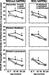
Age-related changes in ChAT activity (top row) and ChAT-IR fibers (bottom rows) in apoE singly transgenic (left) and hAPP/apoE doubly transgenic (right) mice lacking murine apoE. Littermates expressing neither human nor murine apoE served as additional controls. ChAT activity was measured in the medial septum. Levels of ChAT-IR fibers in the strata radiatum and lacunosum of the hippocampus and in the neocortex were expressed as percentage of corresponding levels in nontransgenic, wild-type (Apoe+/+) control mice. Compared with apoE3 mice, apoE4 mice showed a loss of ChAT activity and ChAT-IR fibers at 12–15 and 19–24 months of age but not at 6–7 months of age. hAPP/apoE mice showed an age-related loss of ChAT regardless of whether they expressed apoE3 or apoE4. Age-related losses of ChAT in apoE-deficient mice with or without hAPP expression were similar to those in apoE4 mice. Results represent means ± SEM; n = 3–10 (ChAT activity) andn = 4–7 (ChAT-IR fibers) mice per genotype and age range; *p < 0.05, age-matched mice expressing apoE3 versus apoE4 (Tukey–Kramer test). ChAT activity measurements for one 19-month-old apoE3 mouse and one 12-month-old apoE4 mouse were excluded from the statistical analysis because they were well outside of the range of the other values.
Fig. 4.
Fig. 4.
Age-dependent progression of neuropathological alterations in hAPP/apoE3 and hAPP/apoE4 mice. SYN-IR presynaptic terminals (stratum lacunosum) and ChAT-IR fibers (strata lacunosum and radiatum) of defined signal intensity were measured in the hippocampus. Age-dependent decreases in SYN-IR presynaptic terminals developed later in hAPP/apoE3 mice than in hAPP/apoE4 mice. Thio-S-positive plaques and Aβ-IR deposits in the hippocampus were detected only in the oldest age group, with more deposits found in hAPP/apoE4 mice than in hAPP/apoE3 mice. Note that 12- to 15-month-old hAPP/apoE4 mice had decreased levels of SYN-IR presynaptic terminals and ChAT-IR fibers but no amyloid deposits, and that 19- to 24-month-old hAPP/apoE4 mice had a larger amyloid burden than hAPP/apoE3 mice (Fig. 5), although at this age both groups had comparable decreases in SYN-IR presynaptic terminals and ChAT-IR fibers (Figs. 2, 3). Scale bars: top row, 30 μm; second row, 65 μm; third row, 250 μm; fourth row, 400 μm.
Fig. 5.
Fig. 5.
Age-dependent formation of thio-S-positive plaques in the hippocampus of hAPP/apoE mice. Brain sections were stained with thio-S and imaged by confocal microscopy (FITC filter setting). The hippocampal area occupied by thio-S-positive plaques was determined (y-axis on left, solid symbols and lines). Thio-S-positive amyloid plaques were detected only in the 19–24 month age group. hAPP/apoE4 mice had a significantly higher plaque load than hAPP/apoE3 mice. Results represent means ± SEM; n = 4–7 mice per genotype and age range; *p < 0.05 by Mann–Whitney U test. Synaptophysin data (stratum lacunosum) from Figure 2 were superimposed (y-axis on right, open symbols and dashed lines) to highlight that the differential effects of apoE isoforms on plaques and SYN-IR presynaptic terminals occur at different ages.

Similar articles

Cited by

References

    1. Allen SJ, MacGowan SH, Tyler S, Wilcock GK, Robertson AGS, Holden PH, Smith SKF, Dawbarn D. Reduced cholinergic function in normal and Alzheimer's disease brain is associated with apolipoprotein E4 genotype. Neurosci Lett. 1997;239:33–36. - PubMed
    1. Arendt T, Schindler C, Brückner MK, Eschrich K, Bigl V, Zedlick D, Marcova L. Plastic neuronal remodeling is impaired in patients with Alzheimer's disease carrying apolipoprotein ε4 allele. J Neurosci. 1997;17:516–529. - PMC - PubMed
    1. Berr C, Hauw J-J, Delaäre P, Duyckaerts C, Amouyel P. Apolipoprotein E allele 4 is linked to increased deposition of the amyloid β-peptide (A-β) in cases with or without Alzheimer's disease. Neurosci Lett. 1994;178:221–224. - PubMed
    1. Bierer L, Haroutunian V, Gabriel S, Knott PJ, Carlin LS, Purohit DP, Perl DP, Schmeidler J, Kanof P, Davis KL. Neurochemical correlates of dementia severity in Alzheimer's disease: relative importance of the cholinergic deficits. J Neurochem. 1995;64:749–760. - PubMed
    1. Boncristiano S, Calhoun ME, Kelly PH, Pfeifer M, Bondolfi L, Stalder M, Phinney AL, Abramowski D, Sturchler-Pierrat C, Enz A, Sommer B, Staufenbiel M, Jucker M. Cholinergic changes in the APP23 transgenic mouse model of cerebral amyloidosis. J Neurosci. 2002;22:3234–3243. - PMC - PubMed

Publication types

MeSH terms