Comparative Study
. 2002 Dec 15;22(24):10580-92.
doi: 10.1523/JNEUROSCI.22-24-10580.2002.

Bursting in leech heart interneurons: cell-autonomous and network-based mechanisms

Affiliations
Comparative Study

Bursting in leech heart interneurons: cell-autonomous and network-based mechanisms

Gennady S Cymbalyuk et al. J Neurosci. .

Abstract

Rhythmic activity within the heartbeat pattern generator of the medicinal leech is based on the alternating bursting of mutually inhibitory pairs of oscillator heart interneurons (half-center oscillators). Bicuculline methiodide has been shown to block mutual inhibition between these interneurons and to cause them to spike tonically while recorded intracellularly (Schmidt and Calabrese, 1992). Using extracellular recording techniques, we show here that oscillator and premotor heart interneurons continue to burst when pharmacologically isolated with bicuculline, although the bursting is not robust in some preparations. We propose that a nonspecific leak current introduced by the intracellular microelectrode suppresses endogenous bursting activity to account for the discrepancy with results using intracellular recording. A two-parameter bifurcation diagram (E(leak) vs g(leak)) of a mathematical model of a single heart interneuron shows a narrow stripe of parameter values where bursting occurs, separating large zones of tonic spiking and silence. A similar analysis performed for a half-center oscillator model outlined a much larger area of bursting. Bursting in the half-center oscillator model is also less sensitive to variation in the maximal conductances of voltage-gated currents than in the single-neuron model. Thus, in addition to ensuring appropriate bursting characteristics such as period, phase, and duty cycles, the half-center configuration enhances oscillation robustness, making them less susceptible to random or imposed changes in membrane parameters. Endogenous bursting, in turn, ensures appropriate bursting if the strength of mutual inhibition is weakened and limits the minimum period of the half-center oscillator to a period near that of the single neuron.


Figures

Fig. 1.
Fig. 1.
A, Connectivity diagram of the heartbeat neuronal network. Large open circles represent neurons. The neurons playing similar functional roles in the heartbeat network and with similar input and output connections are lumped together. Numbers identify ganglia where somata are located. Small filled ellipses represent inhibitory chemical synapses. Two pairs of reciprocally inhibitory heart interneurons located in ganglia 3 and 4 form half-center oscillators. These oscillators are coupled by coordinating interneurons whose somata are located in ganglia 1 and 2. Other premotor interneurons located in ganglia 6 and 7, along with the oscillator interneurons, inhibit heart motor neurons. Switch interneurons located in ganglion 5 interface between oscillator interneurons and premotor interneurons to produce two alternating coordination states, peristaltic and synchronous, of the motor neurons. B-D, Pharmacologically isolated oscillator heart interneurons fire tonically when recorded with sharp microelectrodes but burst rhythmically when recorded extracellularly. The left oscillator interneuron from a mutually inhibitory pair is recorded intracellularly, and the right one is recorded extracellularly. B, Under control conditions, the two neurons produce rhythmic alternating bursts. C, Addition of bicuculline results in tonic spiking in the intracellularly recorded cell and continued bursting in the other. D, The effects of bicuculline were reversible with washout. In this and all subsequent figures, voltage traces recorded from heart interneurons are labeledHN and indexed by body side (R, L) and ganglion number.
Fig. 2.
Fig. 2.
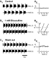
Oscillator heart interneurons recorded extracellularly burst independently when pharmacologically isolated with bicuculline. A1, Oscillator interneurons burst rhythmically in alternation in normal saline.A2, Instantaneous phase between the activity of the neurons plotted against burst number stays close to 0.5.B1, The oscillator interneurons burst independently in bicuculline methiodide (1 mm).B2, The instantaneous phase drifts gradually from 0 to 1, demonstrating independent bursting with different cycle periods. C1, The oscillator interneurons burst in alternation after washout with normal saline.C2, The instantaneous phase stays near 0.5, although with larger deviations. The instantaneous phase was defined as the delay of the HN(R,3) burst median spike relative to the HN(L,3) burst median spike divided by the currentHN(L,3) cycle period.
Fig. 3.
Fig. 3.
In some preparations, pharmacologically isolated oscillator interneurons show bursting interspersed with bouts of tonic spiking. A, Normal pattern of alternating bursting in saline. B, In a solution containing bicuculline methiodide (1 mm), bursting is sporadic. Trains of bursts are interrupted by long intervals of tonic spiking. One heart interneuron can be bursting while the other is spiking tonically.C, The normal alternating bursting pattern was restored after washout with normal saline (exposure to bicuculline <10 min).
Fig. 4.
Fig. 4.
Seizure-like synchronous bursting observed in oscillator interneurons and other neurons in bicuculline (1 mm). A, Electrical activity of a Retzius cell (top trace) and left and right oscillator interneurons from ganglion 4 (bottom 2 traces) showing a synchronous burst two to three times longer than the asynchronous bursts in the oscillator interneurons. The Retzius cell was recorded with a sharp microelectrode. In bicuculline, the Retzius cell was silent most of time. A strong depolarization underlies the seizure-like burst. B, Graph of the cycle period plotted against time for the heart interneurons HN(L,4) and HN(R,4) (open circles, asterisks, respectively) shows that the burst period shortens right after the synchronous burst occurred and returns to baseline after ∼25 sec. The cycle period was defined as the interval between the median spikes of two consecutive bursts.
Fig. 5.
Fig. 5.
Paired extracellular recordings of a coordinating interneuron (ganglion 2) and an ipsilateral oscillator interneuron (ganglion 3). A, In normal saline (Control), the coordinating interneuron and the oscillator interneuron produced alternating bursts. B, In bicuculline (1 mm, after 8 min of exposure), both interneurons burst independently. C, On washout with normal saline, normal alternating bursting returned.
Fig. 6.
Fig. 6.
Bifurcation diagram of the single-cell oscillator interneuron model activities (A) and bifurcation diagram of the bursting activity in a half-center oscillator model (B). A, Pink, white, and yellow areas mark the parameter regimes in which tonic spiking, bursting, and silence are stable, respectively. Green areas mark parameter regimes of multistability in which more than one activity is stable.Multistability(A) points to the area where bursting coexists with silence; multistability(B) points to the area where bursting coexists with tonic spiking;multistability(C) points to the area where tonic spiking coexists with silence. The asterisk corresponds to thegleak and Eleakparameter values (g leakel = 0) used in the models illustrated in Figure 9. Plus sign, open circle, filled circle, asterisk, and open diamond correspond to the gleak andEleak parameter values for oscillator interneuron model activity illustrated in Figure 7 and fall along a line generated when the introducedg leakel changes from −0.35 to 1 nS about model parameters of E leako andg leako (asterisk) as used in Figure 9, A and B. Open triangles mark the parameter values corresponding to Figure7E1–E3. B, The blue areacorresponds to the parameter region where stationary bursting activity was observed, and the white area corresponds to the region where stationary bursting was not observed. The borders separating the major activity regimes of the single-cell model taken from A are plotted for comparison. The pink patch delimits the single-cell model parameter values that produced bursting characteristics within the ranges measured experimentally. The green line was generated by varyingg leakel from 0 to 1 nS about model parameters of E leako andg leako (asterisk). The marked points in A (except foropen triangles) fall along this line. The pink line was generated similarly by varyingg leakel from 0 to 1 nS starting withE leako andg leako at the lowest point of the experimentally constrained patch. A family of such lines originating in the pink patch defines by their end points the upper delimited area.
Fig. 7.
Fig. 7.
Characteristic activities of the single-oscillator interneuron model. A, Tonic spiking (Fig.6A, plus sign). B, Two-spike bursting (Fig. 6A, open circle). C, Multispike bursting near the border of bursting and tonic spiking (Fig.6A, filled circle). D, Multispike bursting near the border of bursting and silence (Fig.6A, open diamond). E, Three coexisting oscillatory activities of the single oscillator interneuron model at a point (gleak = 12.703 nS; Eleak = −61 mV) that is marked in Figure 6A by open triangles.E1, Bursting with three spikes. E2, Bursting with two spikes. E3, Tonic spiking. Initial conditions leading to each of these states are as follows:E1, V = −2.1 mV;mCaF = 0.9940;hCaF = 0.0123;mCaS = 0.7722; hCaS = 0.1255;mK1 = 0.7880;hK1 = 0.8775;mK2 = 0.1947;mKA = 0.8911;hKA = 0.0211;mh = 0.3611;mP = 0.7529;mNa = 0.9834;hNa = 0.2891; E2, V = −7.0 mV; mCaF = 0.9940;hCaF = 0.0090;mCaS = 0.8325;hCaS = 0.1023;mK1 = 0.2914;hK1 = 0.8527;mK2 = 0.1683;mKA = 0.8098;hKA = 0.0216;mh = 0.3201;mP = 0.6739;mNa = 0.9589;hNa = 0.6024; E3, V = −46.0 mV; mCaF = 0.5485;hCaF = 0.0456;mCaS = 0.5403;hCaS = 0.1080;mK1 = 0.0267;hK1 = 0.9009;mK2 = 0.0976;mKA = 0.4296;hKA = 0.0654;mh = 0.3667;mP = 0.2979;mNa = 0.0726;hNa = 0.9997.
Fig. 8.
Fig. 8.
Major bursting characteristics for the single-oscillator interneuron model: the burst period (A), duty cycle (B), and spike frequency (C) observed in the model when the values of (gleak,Eleak) are varied. The minimal and maximal values experimentally measured for oscillator interneurons are marked by the pairs of green lines on the axes, except for the spike frequency (C), for which the maximum value (upper boundary) was never exceeded by the model; thus only the lower boundary was marked. Projection of the surfaces of characteristics values on the plane (gleak,Eleak) describes the area where bursting occurs. It includes the green area that defines the model values conforming to the experimentally measured boundaries.
Fig. 9.
Fig. 9.
Voltage and current traces produced by the single-cell (A) and half-center oscillator (B) models. ParametersEleak = −63.5 mV andgleak = 9.9 nS (Fig.6A,B, asterisks) were chosen to comply with the experimental data, and all other parameters were canonical. Major inward and outward currents contributing to the slow wave of oscillation in each model are illustrated. Dotted linesmark 0 nA.
Fig. 10.
Fig. 10.
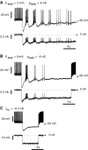
Subtraction of a leak current using a dynamic clamp elicits a few bursts in intracellular recordings (sharp microelectrodes) from oscillator heart interneurons isolated pharmacologically with bicuculline (1 mm).A, Leak current subtraction (Eleak = 0 mV;gleak = −6 nS) caused five bursts followed by irregular tonic spiking. B, Leak current subtraction (Eleak = 0 mV;gleak = −5 nS) caused three bursts followed by silence. C, Injection of a steady hyperpolarizing current (−0.4 nA) did not lead to bursting.

Similar articles

Cited by

References

    1. Angstadt JD, Friesen WO. Synchronized oscillatory activity in leech neurons induced by calcium channel blockers. J Neurophysiol. 1991;66:1858–1873. - PubMed
    1. Arbas EA, Calabrese RL. Rate modification in the heartbeat central pattern generator of the medicinal leech. J Comp Physiol [A] 1984;155:783–794.
    1. Arbas EA, Calabrese RL. Leydig neuron activity modulates heartbeat in the medicinal leech. J Comp Physiol [A] 1990;167:665–671. - PubMed
    1. Arshavsky YI, Deliagina TG, Orlovsky GN, Panchin YV, Pavlova GA, Popova LB. Control of locomotion in marine mollusc Clione limacina. VI. Activity of isolated neurons of pedal ganglia. Exp Brain Res. 1986;63:106–112. - PubMed
    1. Arshavsky YI, Orlovsky GN, Panchin YV, Roberts A, Soffe SR. Neuronal control of swimming locomotion: analysis of the pteropod mollusc Clione and embryos of the amphibian Xenopus. Trends Neurosci. 1993;16:227–233. - PubMed

Publication types