. 2002 Dec 15;22(24):10653-61.
doi: 10.1523/JNEUROSCI.22-24-10653.2002.

Correlated calcium uptake and release by mitochondria and endoplasmic reticulum of CA3 hippocampal dendrites after afferent synaptic stimulation

Affiliations

Correlated calcium uptake and release by mitochondria and endoplasmic reticulum of CA3 hippocampal dendrites after afferent synaptic stimulation

Natalia B Pivovarova et al. J Neurosci. .

Abstract

Mitochondria and endoplasmic reticulum (ER) are important modulators of intracellular calcium signaling pathways, but the role of these organelles in shaping synaptic calcium transients in dendrites of pyramidal neurons remains speculative. We have measured directly the concentrations of total Ca (bound plus free) within intracellular compartments of proximal dendrites of CA3 hippocampal neurons at times after synaptic stimulation corresponding to the peak of the cytoplasmic free Ca2+ transient (1 sec), to just after its decay (30 sec), and to well after its return to prestimulus levels (180 sec). Electron probe microanalysis of cryosections from rapidly frozen slice cultures has revealed that afferent mossy fiber stimulation evokes large, rapid elevations in the concentration of total mitochondrial Ca ([Ca](mito)) in depolarized dendrites. A single tetanus (50 Hz/1 sec) elevated [Ca](mito) more than fivefold above characteristically low basal levels within 1 sec of stimulation and >10-fold by 30 sec after stimulation. This strong Ca accumulation was reversible, because [Ca](mito) had recovered by 180 sec after the tetanus. Ca sequestered within mitochondria was localized to small inclusions that were distributed heterogeneously within, and probably among, individual mitochondria. By 30 sec after stimulation an active subpopulation of ER cisterns had accumulated more Ca than had mitochondria despite a approximately 1 sec delay before the onset of accumulation. Active ER cisterns retained their Ca load much longer (>3 min) than mitochondria. The complementary time courses of mitochondrial versus ER Ca2+ uptake and release suggest that these organelles participate in a choreographed interplay, each shaping dendritic Ca2+ signals within characteristic regimes of cytosolic Ca2+ concentration and time.


Figures
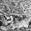
Fig. 1.
Fig. 1.
Ultrastructural organization of proximal dendrites in CA3 hippocampal neurons. Shown are representative digital transmission electron micrographs of freeze-dried cryosections prepared from rapidly frozen hippocampal slice cultures, illustrating the subcellular structure of proximal dendritic compartments and demonstrating that the three compartments targeted for EPMA (ER, mitochondria, and cytoplasm) are readily identifiable. Images were recorded using standard low-dose techniques in a LEO 912 Omega cryoanalytical electron microscope at −170°C by means of a ProScan slow-scan CCD camera (1024 × 1024). Survey view (A) shows the general appearance of the proximal dendritic field of CA3 neuropil in unstained cryosections from slice cultures. Three primary dendrites (asterisks) course across the field; cytoplasm, characterized by longitudinal bundles of microtubules, and elongated mitochondria (adjacent toasterisks) are evident. At higher magnification (B) both mitochondria and ER [essentially the only intracellular organelles present in proximal apical dendrites at the distance that was sampled (50–100 μm from the soma)] are apparent. The latter is seen as a network of smooth-surfaced cisterns, often in clusters (arrowheads). Together, the ER, mitochondria, and cytosol comprise essentially all of the dendritic volume while also including all important components of the Ca2+ regulatory system. Scale bars, 1 μm.
Fig. 2.
Fig. 2.
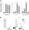
Calcium accumulation in dendritic compartments of CA3 hippocampal neurons. Comparison of calcium concentrations at different time points after synaptic stimulation in three dendritic compartments. In certain instances data were not distributed normally; therefore, all results are given as medians. In A, data are in primary units of mmol/kg dry weight. In B, Ca concentrations have been converted to mmol/kg wet weight (as described in Materials and Methods) to facilitate quantitative comparisons of Ca concentrations between compartments. Uncertainties introduced by this conversion are discussed in Results. Statistical significance (p values) relative to corresponding resting Ca concentrations is given above the bars; other significant differences are indicated by bracketed pairs. Statistical tests are described in Materials and Methods. A, At 1 sec after afferent synaptic stimulation [Ca]mito and [Ca]cyto, but not [Ca]ER, were elevated compared with resting levels. By 30 sec after stimulation Ca concentrations were elevated in all three compartments. (For ER, only data from the subpopulation that actively sequesters Ca in responsive dendrites, as presented in Table 1and discussed in Results, are included.) By 180 sec after stimulation only [Ca]mito had recovered. Note the differenty-axes. B, At rest, ER Ca levels are significantly higher than those in mitochondria and cytosol (compareblack bars). The ER also contains by far the highest Ca concentrations at 30 and 180 sec (compare light gray andwhite bars), but not at 1 sec (dark gray bars).
Fig. 3.
Fig. 3.
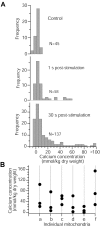
Heterogeneous distribution of [Ca]mito within individual mitochondria after synaptic stimulation. A, Mean [Ca]mito in resting dendrites is low, and the distribution of individual measurements is normal (top panel), as indicated by a good fit (R2 = 0.99) to a single Gaussian with a width (ς = 2.5 mmol/kg dry weight) expected because of statistical uncertainties in EDX analysis. At 1 sec (middle panel) and 30 sec (bottom panel) the distribution becomes progressively skewed toward higher [Ca]mito as a result of a sequestration mechanism that concentrates Ca in small inclusions within the mitochondrial matrix.B, Distribution of single [Ca]mitomeasurements from six representative mitochondria with elevated Ca levels that were analyzed at three separate locations using a 100 nm probe. Large differences in [Ca]mito within each of these mitochondria reflect the heterogeneous distribution of calcium within individual mitochondria.
Fig. 4.
Fig. 4.
Calcium dynamics in dendritic compartments after synaptic stimulation. Time courses for [Ca]cyto(opencircles), [Ca]mito(filled circles), and [Ca]ER(filled triangles; responsive subset only at 30 and 180 sec) illustrate complementary temporal relationship for activity-dependent Ca2+ uptake and release by dendritic mitochondria and ER. These organelles appear to transport Ca2+ within distinct, possibly cooperative, time domains. For example, [Ca]mito increases earlier and declines faster than [Ca]ER, which remained elevated for >10 min. Data are given as medians. Note the log scale on the abscissa.

Similar articles

Cited by

References

    1. Albrecht MA, Colegrove SL, Hongpaisan J, Pivovarova NB, Andrews SB, Friel DD. Multiple modes of calcium-induced calcium release in sympathetic neurons I: attenuation of endoplasmic reticulum Ca2+ accumulation at low [Ca2+]i during weak depolarization. J Gen Physiol. 2001;118:83–100. - PMC - PubMed
    1. Andrews SB, Leapman RD, Landis DM, Reese TS. Activity-dependent accumulation of calcium in Purkinje cell dendritic spines. Proc Natl Acad Sci USA. 1988;85:1682–1685. - PMC - PubMed
    1. Babcock DF, Hille B. Mitochondrial oversight of cellular Ca2+ signaling. Curr Opin Neurobiol. 1998;8:398–404. - PubMed
    1. Babcock DF, Herrington J, Goodwin PC, Park YB, Hille B. Mitochondrial participation in the intracellular Ca2+ network. J Cell Biol. 1997;136:833–844. - PMC - PubMed
    1. Berridge MJ. Neuronal calcium signaling. Neuron. 1998;21:13–26. - PubMed

Publication types

LinkOut - more resources