. 2002 Dec 15;22(24):10662-70.
doi: 10.1523/JNEUROSCI.22-24-10662.2002.

Proinflammatory mediators, stimulators of sensory neuron excitability via the expression of acid-sensing ion channels

Affiliations

Proinflammatory mediators, stimulators of sensory neuron excitability via the expression of acid-sensing ion channels

Julien Mamet et al. J Neurosci. .

Abstract

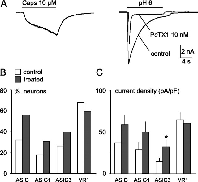
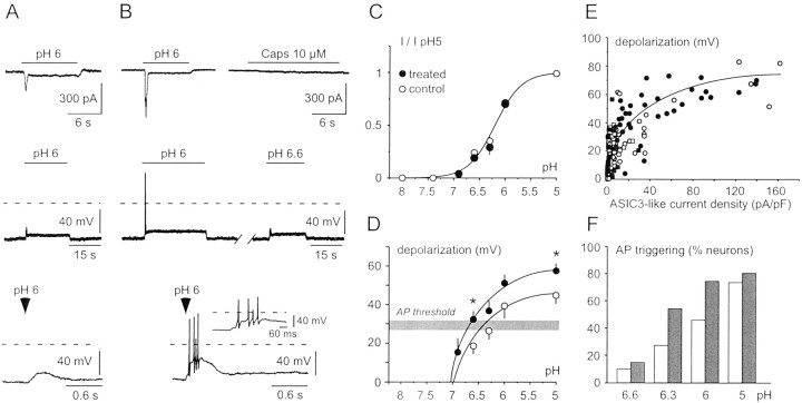
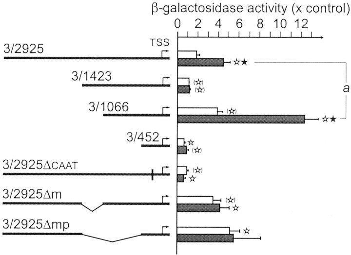
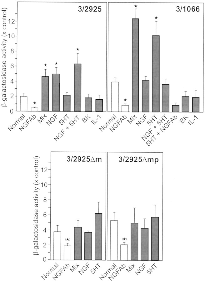
Tissue acidosis is an important feature of inflammation. It is a direct cause of pain and hyperalgesia. Protons activate sensory neurons mainly through acid-sensing ion channels (ASICs) and the subsequent membrane depolarization that leads to action potential generation. We had previously shown that ASIC transcript levels were increased in inflammatory conditions in vivo. We have now found that this increase is caused by the proinflammatory mediators NGF, serotonin, interleukin-1, and bradykinin. A mixture of these mediators increases ASIC-like current amplitude on sensory neurons as well as the number of ASIC-expressing neurons and leads to a higher sensory neuron excitability. An analysis of the promoter region of the ASIC3 encoding gene, an ASIC specifically expressed in sensory neurons and associated with chest pain that accompanies cardiac ischemia, reveals that gene transcription is controlled by NGF and serotonin.


Figures

Fig. 1.
Fig. 1.
Effect of proinflammatory mediators on proton-gated currents in rat DRG neurons. A, Original current traces from a DRG neuron expressing capsaicin-activated VR1 current (left) and PcTX1-sensitive ASIC1a current and ASIC3-like current (right). This neuron was treated overnight with the proinflammatory mix. Holding potential was −50 mV.B, Percentage of recorded neurons expressing an ASIC-like current (ASIC), a PcTX1-sensitive ASIC1a current (ASIC1), an ASIC3-like current (ASIC3), and a capsaicin-activated VR1 current (VR1), in control neurons (white bars) and in neurons treated overnight with the proinflammatory mix (dark gray bars). Some neurons can express a mixture of these different current types because each current type (VR1, ASIC1a, or ASIC3-like) could be found alone or associated with one or two of the others. The proportion of neurons expressing each current type, whether alone or associated with one or two other types, was determined. The total number of recorded neurons was 34 for each condition, from 5 different cell cultures in which similar numbers of control and treated neurons were recorded. C, Mean current density (picoamperes per picoFarads) of ASIC-like current (ASIC), PcTX1-sensitive ASIC1a current (ASIC1), ASIC3-like current (ASIC3), and capsaicin-activated VR1 current (VR1), in control neurons (white bars) and in neurons treated overnight with the proinflammatory mix (dark gray bars). Holding potential was −50 mV; ASIC current amplitudes were measured at pH 5. Mean ± SEM is shown; n ranges from 8 to 23 from 5 different cell cultures. *p < 0.05; significantly different from the control value.
Fig. 2.
Fig. 2.
Effect of the proinflammatory mediators on the excitability of DRG neurons containing an ASIC3-like current.A, Original current and potential recordings from a control DRG neuron. Top, Voltage-clamp recording of ASIC3-like current induced by a pH drop from 7.4 to 6, whereas capsaicin induced no VR1 current (data not shown). Holding potential was −50 mV. Middle, Current-clamp recording (I = 0 pA) of the depolarization induced by a pH drop from 7.4 to 6. No action potential was triggered by the membrane depolarization. The dashed line represents the 0 mV level. Bottom, Enlargement of the initial depolarization induced by the pH drop. B, Original current and potential recordings from a proinflammatory mix-treated DRG neuron.Top, Voltage-clamp recording of ASIC3-like current induced by pH drop from 7.4 to 6, whereas capsaicin induced no VR1 current (right). Holding potential was −50 mV.Middle, Current-clamp recording (I = 0 pA) of the depolarization induced by a pH drop from 7.4 to 6 and 6.6. Action potentials were triggered by the membrane depolarization induced by pH 6 but not by pH 6.6. Bottom, Enlargement of the initial depolarization induced by the pH drop, showing action potential bursts. C, pH-dependent activation of the peak ASIC3-like current in control (○) and treated (●) neurons. Holding potential was −50 mV. The current amplitude was expressed as a fraction of the current induced by pH 5 (I/I pH 5) and plotted as mean ± SEM; n ranges from 5 to 19. The data could be fitted by a sigmoid with a pH0.5 = 6.2 and a Hill slope factor of 1.7. D, Mean peak membrane depolarization as a function of extracellular pH in control (○) and treated (●) neurons, measured from current-clamp recordings (I= 0 pA). The resting potential was −57.4 ± 2.6 mV (n = 15) and −59.8 ± 3.0 mV (n = 9) in control and treated neurons, respectively. The depolarization values during APs were excluded from measurements. The membrane potential area corresponding to the AP threshold is indicated by a gray bar (see Results for value). Mean ± SEM values are shown; n ranges from 4 to 19. *p < 0.05; significantly different from the control value. E, Membrane depolarization as a function of ASIC3-like current density activated by the same pH drop on the same neuron. The membrane potential was measured by current-clamp (I = 0 pA), and the depolarization values during APs were excluded from measurements. ASIC3-like current density (picoamperes per picoFarads) was subsequently measured on the same neuron, at a holding potential similar to the resting potential measured in current clamp (I = 0). Membrane potential and ASIC3-like current density were measured during the transient peak (○, control; ●, treated neurons) and during the sustained plateau phase (■, control; ▪, treated neurons) for the same pH value. F, Percentage of recorded neurons in which APs were triggered by various pH drops (white bars, control; dark gray bars, treated neurons).
Fig. 3.
Fig. 3.
ASIC3 gene promoter region study by reporter gene assays. The activities of different constructs of the promoter region of the ASIC3 gene were tested using β-galactosidase as a reporter gene. The clones were transfected into DRG neurons in primary culture, and the cells were either maintained in the regular medium (white bars) or treated with the proinflammatory mix (NGF, bradykinin, IL-1, and serotonin) (dark gray bars) shown to mimic inflammation action on ASIC encoding gene transcription. NGF antibody corresponds to incubation with the anti-NGF antibody. Results are normalized on SEAP activity (which takes into account the transfection yield) and the amount of proteins. They are expressed as the ratio of a treated condition with the background level (given by the β-galactosidase vector basal activity; i.e., without any promoter region insert added) and are given as mean ± SEM. The following constructs were studied: the full-length sequence of ASIC3 encoding gene promoter region (the 2925-base long fragment, named3/2925, from −249 to −3174), three shorter fragments (3/1423, from −249 to −1672; 3/1066, from −249 to −1315; and 3/452, from −249 to −701), and three deletion fragments (3/2925ΔCAAT, lacking the putative CCAAT box from −405 to −416;3/2925Δm, lacking from −1315 to −1672; 3/2925Δmp, lacking from −701 to −1672). n ≥ 3; ★ corresponds to a significant difference (p < 0.05) between the control and the treated conditions for the same clone, ⋆ corresponds to a significant difference (p< 0.05), and (⋆) corresponds to a difference withp < 0.1, between the control of clone 3/2925 and any other condition.
Fig. 4.
Fig. 4.
Effects of the different active proinflammatory mediators on ASIC3 encoding gene transcription. The same constructs as in Figure 3 are tested in the same system with the mediators used individually. Results are given as in Figure 3 (mean ± SEM) withn ≥ 3. ★ corresponds to a significant difference (p < 0.05), and (★) corresponds to a difference with p < 0.1, between the control and the treated conditions for the same clone. NGFAb, NGF antibody; BK, bradykinin; 5HT, serotonin.

References

    1. Babinski K, Catarsi S, Biagini G, Seguela P. Mammalian ASIC2a and ASIC3 subunits co-assemble into heteromeric proton-gated channels sensitive to Gd3+. J Biol Chem. 2000;275:28519–28525. - PubMed
    1. Baker MD, Wood JN. Involvement of Na+ channels in pain pathways. Trends Pharmacol Sci. 2001;22:27–31. - PubMed
    1. Baron A, Waldmann R, Lazdunski M. ASIC-like, proton-activated currents in rat hippocampal neurons. J Physiol (Lond) 2002;539:485–494. - PMC - PubMed
    1. Bennett DL, Koltzenburg M, Priestley JV, Shelton DL, McMahon SB. Endogenous nerve growth factor regulates the sensitivity of nociceptors in the adult rat. Eur J Neurosci. 1998;10:1282–1291. - PubMed
    1. Benson CJ, Xie J, Wemmie JA, Price MP, Henss JM, Welsh MJ, Snyder PM. Heteromultimers of DEG/ENaC subunits form H+-gated channels in mouse sensory neurons. Proc Natl Acad Sci USA. 2002;99:2338–2343. - PMC - PubMed

Publication types

MeSH terms