. 2002 Dec 15;22(24):10671-9.
doi: 10.1523/JNEUROSCI.22-24-10671.2002.

Modulation of the readily releasable pool of transmitter and of excitation-secretion coupling by activity and by serotonin at Aplysia sensorimotor synapses in culture

Affiliations

Modulation of the readily releasable pool of transmitter and of excitation-secretion coupling by activity and by serotonin at Aplysia sensorimotor synapses in culture

Yali Zhao et al. J Neurosci. .

Abstract

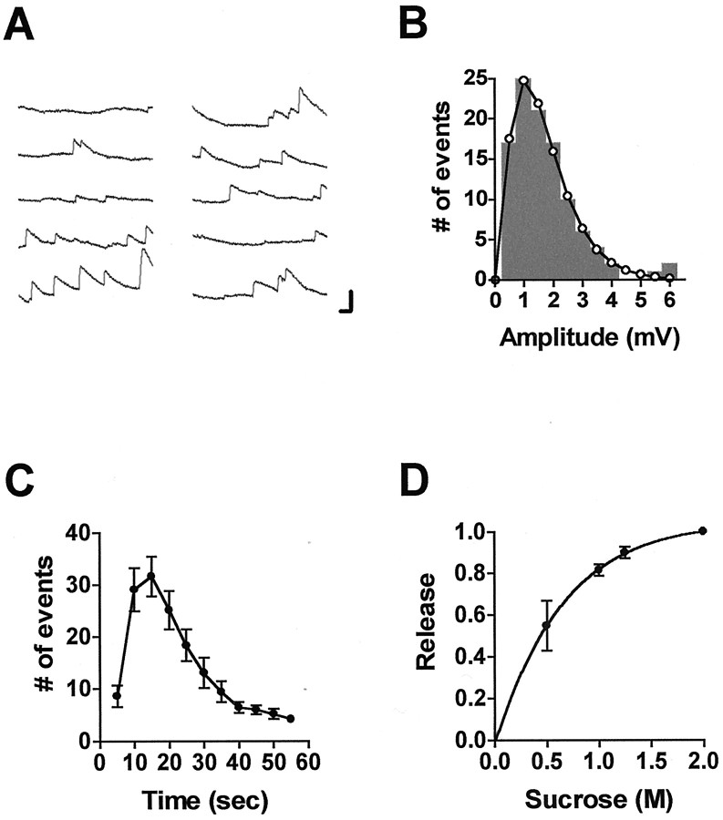
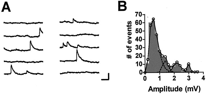
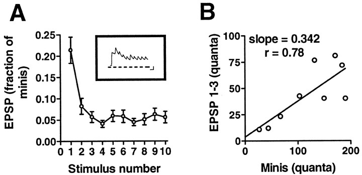
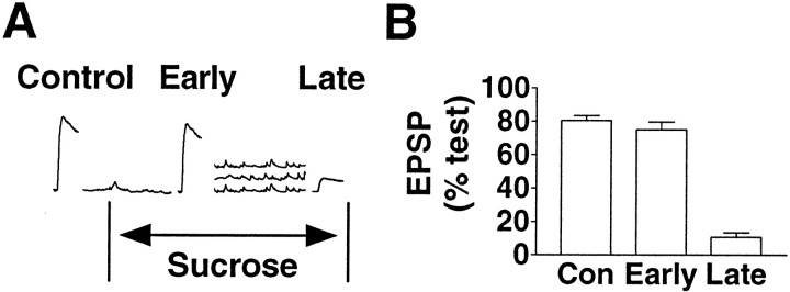
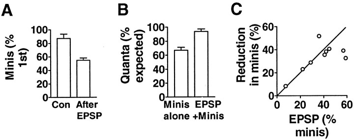
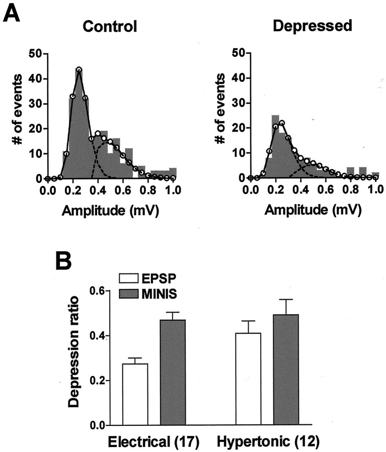
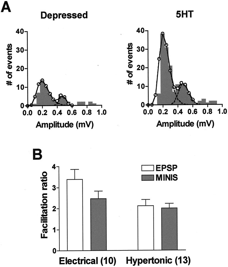
Short-term homosynaptic depression and heterosynaptic facilitation of transmitter release from mechanoreceptor sensory neurons of Aplysia are involved in habituation and sensitization, respectively, of defensive withdrawal reflexes. We investigated whether synaptic transmission is regulated in these forms of plasticity by means of changes in the size of the pool of transmitter available for immediate release [the readily releasable pool (RRP)] or in the efficacy of release from an unchanging pool. Using sensorimotor synapses formed in cell culture, we estimated the number of transmitter quanta in the RRP from the asynchronous release of neurotransmitter caused by application of a hypertonic bathing solution. Our experiments indicate that the transmitter released by action potentials and by hypertonic solution comes from the same pool. The RRP was reduced after homosynaptic depression of the EPSP by low-frequency stimulation and increased after facilitation of the EPSP by application of the endogenous facilitatory transmitter serotonin (5-HT) after homosynaptic depression. However, although the fractional changes in the RRP and in the EPSP were similar for both synaptic depression and facilitation when depression was induced by repeated hypertonic stimulation, the changes in the EPSP were significantly greater than the changes in the RRP when depression was induced by repeated electrical stimulation. These observations indicate that homosynaptic depression and restoration of depressed transmission by 5-HT are caused by changes in both the amount of transmitter available for immediate release and in processes involved in the coupling of the action potential to transmitter release.


Figures

Fig. 1.
Fig. 1.
Asynchronous transmitter release triggered by hypertonic solution at Aplysia sensorimotor synapses in culture. A, Ten consecutive 1 sec records of miniature synaptic potentials elicited in a postsynaptic neuron ∼3 sec after application of bathing solution with 1 m added sucrose. Calibration: 2 mV, 100 msec. B, Amplitude histogram of all the miniature potentials within 1 min after application of hypertonic solution; same experiment as inA. The open circles connected by theline represent the best fit to the experimental points (mean amplitude, 1.67 mV) using the equation of Bekkers et al. (1990).C, Kinetics of asynchronous transmitter release elicited with hypertonic solution containing 1 m sucrose. Error bars represent SEM for 20 experiments. D, Dependence of the asynchronous response on the concentration of added sucrose (n = 6; ±SEM). The total number of minis detected in the first 55 sec after application of solutions containing different concentrations of sucrose is plotted against sucrose concentration and normalized to the response at 2 m.
Fig. 2.
Fig. 2.
Apparent multiquantal miniature synaptic potentials in response to application of hypertonic solution.A, Consecutive 1 sec records from a postsynaptic neuron taken ∼40 sec after application of solution containing 1m sucrose. The relatively low frequency of minis at this time indicates that the large events are not likely to be coincidences of release from different sites. Calibration: 2 mV, 200 msec.B, Amplitude histogram of all the events recorded within 1 min of sucrose application in the same experiment.Peaks appear at approximately evenly spaced intervals.Circles connected by the solid linerepresent the fit to the data assuming four peaks with means of 0.67, 1.40, 2.21, and 2.97 mV, respectively. Broken lines represent fits of the individual peaks. See Figures 6 and7 for other examples of amplitude histograms with multiple peaks.
Fig. 3.
Fig. 3.
Transmitter release elicited with high-frequency electrical stimulation and with hypertonic solution. A 25 Hz train of action potentials was triggered with the aim of depleting the RRP, and the RRP was then estimated from the total number of quanta released before the EPSP reached a steady state. A, The number of quanta (see Materials and Methods) released in response to each action potential of the train (see inset for sample record; calibration: 5 mV, 50 msec) was divided by the total number of quanta released in response to an earlier application of 1 mhypertonic solution (n = 7). B, The total number of quanta released in response to the first three action potentials of each train is plotted against the total number of quanta released in response to the hypertonic solution (n= 9). Although the first three action potentials released only approximately one-third of the amount released with the hypertonic solution, the correlation between the number of quanta released by the action potentials and by the hypertonic stimulus is statistically significant (one-tailed p < 0.01).
Fig. 4.
Fig. 4.
Transmitter release by hypertonic solution reduces the release evoked with an action potential. A, Illustration of the experimental protocol. After a single test EPSP was elicited (data not shown), a second EPSP was evoked in one of the following three conditions: (1) without any intervening treatment (Control), (2) in the presence of the hypertonic solution before asynchronous transmitter release had occurred (Early); or (3) in the presence of hypertonic solution immediately after the decay of the barrage of miniature synaptic potentials (Late). B, Summary of experimental results. The EPSP was reduced only if it was triggered after the asynchronous response to the hypertonic stimulus. (Control, n = 8;Early, n = 12; Late,n = 8).
Fig. 5.
Fig. 5.
An EPSP reduces the subsequent asynchronous response to hypertonic solution. A, A second application of a hypertonic solution 10 min after a first test application gave an asynchronous response that was on average ∼87% of the test response [control (Con); n = 22]. However, if the second response was immediately preceded by an EPSP, the second response was reduced to ∼55% of the first (After EPSP; n = 6). B, Although the number of quanta in the asynchronous response was reduced by a preceding EPSP (Minis alone), the sum of the number of quanta in the EPSP and in the asynchronous response was equal to the total number expected for a second application of hypertonic solution (EPSP+Minis; n = 9).C, In nine experiments, the fractional reduction in the asynchronous response caused by a preceding EPSP was similar to the fraction of the asynchronous response estimated to be released in the EPSP (p = 0.28; paired ttest), indicating a common pool of quanta for the EPSP and the asynchronous response. The slope of the line is 1.
Fig. 6.
Fig. 6.
The RRP is reduced in homosynaptic depression.A, In the first part of the experiment, a test EPSP was elicited, and the number of minis released in response to a hypertonic stimulus immediately afterward was recorded (Control). After a 10 min wash and rest, homosynaptic depression was induced by repeatedly eliciting EPSPs at a low frequency. Steady-state homosynaptic depression was tested with an EPSP 2 min after the train, and the response to the hypertonic solution was measured again (Depressed). B, After repeated low-frequency stimulation, the EPSP was reduced more than the number of minis in the asynchronous response relative to the test (Electrical; 0.273 ± 0.027 and 0.469 ± 0.034, respectively; p < 0.0001; pairedt test). However, when depression was induced by repeated applications of hypertonic solution, the reductions in the EPSP and in the asynchronous response were comparable (Hypertonic; 0.409 ± 0.055 and 0.490 ± 0.068, respectively; p > 0.2).Numbers in parentheses denote numbers of independent experiments. The depression ratio is the ratio of the EPSP or number of minis in the asynchronous response after depression to their respective test values.
Fig. 7.
Fig. 7.
The RRP is increased by 5-HT applied after homosynaptic depression. A, Facilitation of the EPSP by 5-HT after homosynaptic depression induced by electrical stimulation was accompanied by an increase in the response to the hypertonic solution. B, Facilitation of the EPSP was greater than the increase in the RRP after action potential-induced depression (Electrical; 3.387 ± 0.474 and 2.462 ± 0.368, respectively; p < 0.02), whereas the facilitation of the EPSP and the increase in the RRP were comparable after depression induced by repeated exposure to the hypertonic solution (Hypertonic; 2.125 ± 0.287 and 2.010 ± 0.213, respectively; p > 0.6). Facilitation is expressed as the EPSP or number of minis after application of 5-HT relative to the depressed EPSP or number of minis, respectively.

Similar articles

Cited by

References

    1. Angers A, Fioravante D, Chin J, Cleary LJ, Bean AJ, Byrne JH. Serotonin stimulates phosphorylation of Aplysia synapsin and alters its subcellular distribution in sensory neurons. J Neurosci. 2002;22:5412–5422. - PMC - PubMed
    1. Armitage BA, Siegelbaum SA. Presynaptic induction and expression of homosynaptic depression at Aplysia sensorimotor neuron synapses. J Neurosci. 1998;18:8770–8779. - PMC - PubMed
    1. Ashery U, Varoqueaux F, Voets T, Betz A, Thakur P, Koch H, Neher E, Brose N, Rettig J. Munc13-1 acts as a priming factor for large dense-core vesicles in bovine chromaffin cells. EMBO J. 2000;19:3586–3596. - PMC - PubMed
    1. Auger C, Kondo S, Marty A. Multivesicular release at single functional synaptic sites in cerebellar stellate and basket cells. J Neurosci. 1998;18:4532–4547. - PMC - PubMed
    1. Bailey CH, Chen M. Morphological basis of short-term habituation in Aplysia. J Neurosci. 1988;8:2452–2459. - PMC - PubMed

Publication types

MeSH terms

LinkOut - more resources