. 2002 Dec 15;22(24):10690-8.
doi: 10.1523/JNEUROSCI.22-24-10690.2002.

Endoplasmic reticulum stress and the unfolded protein response in cellular models of Parkinson's disease

Affiliations

Endoplasmic reticulum stress and the unfolded protein response in cellular models of Parkinson's disease

Elizabeth J Ryu et al. J Neurosci. .

Abstract

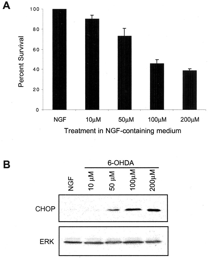
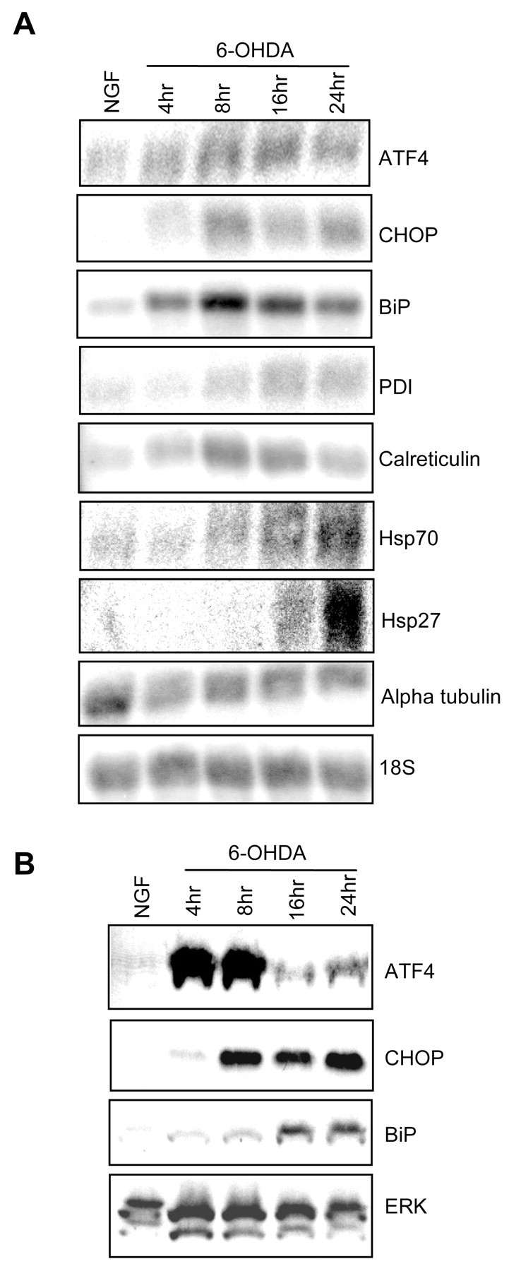
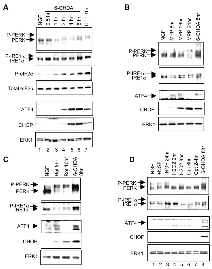
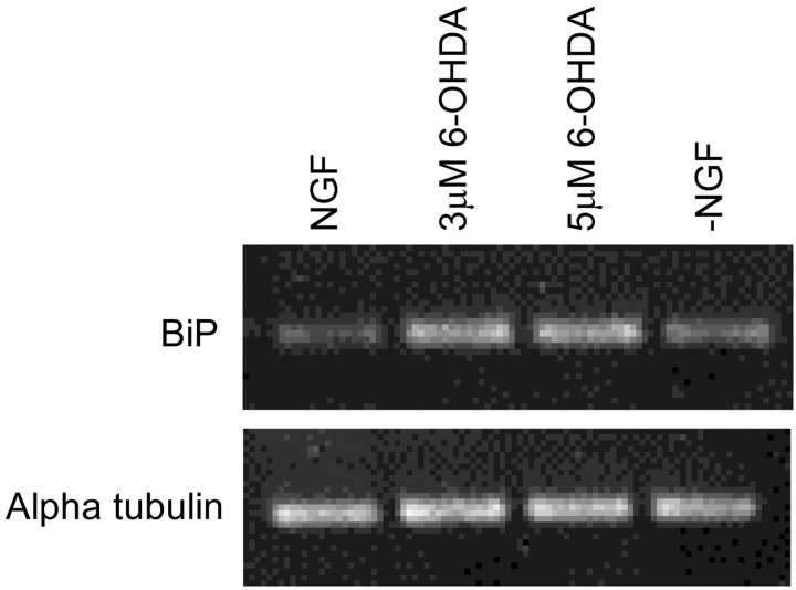
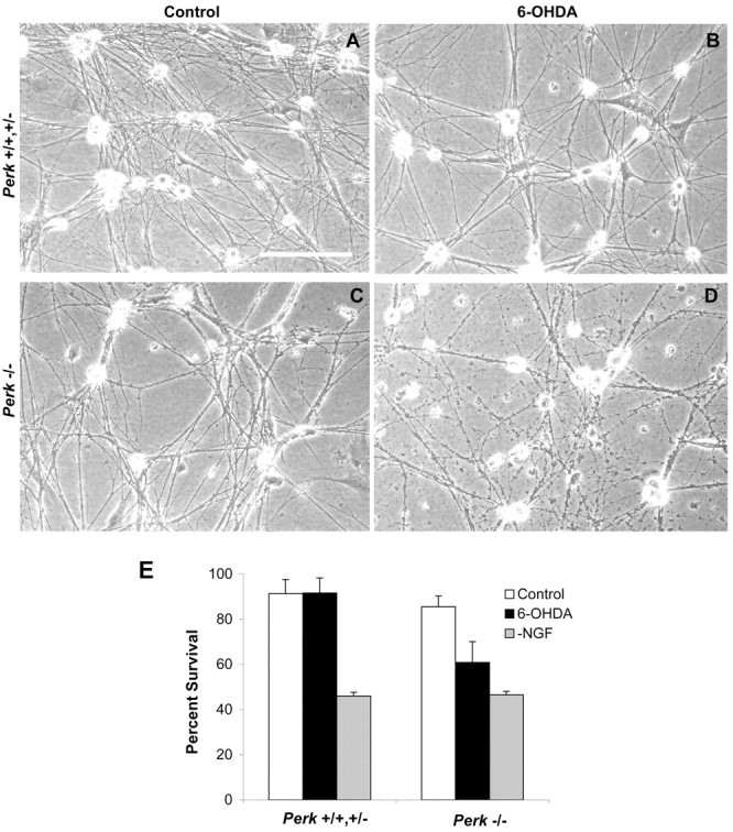
6-hydroxydopamine, 1-methyl-4-phenyl-pyridinium (MPP+), and rotenone cause the death of dopaminergic neurons in vitro and in vivo and are widely used to model Parkinson's disease. To identify regulated genes in such models, we performed serial analysis of gene expression on neuronal PC12 cells exposed to 6-hydroxydopamine. This revealed a striking increase in transcripts associated with the unfolded protein response. Immunoblotting confirmed phosphorylation of the key endoplasmic reticulum stress kinases IRE1alpha and PERK (PKR-like ER kinase) and induction of their downstream targets. There was a similar response to MPP+ and rotenone, but not to other apoptotic initiators. As evidence that endoplasmic reticulum stress contributes to neuronal death, sympathetic neurons from PERK null mice in which the capacity to respond to endoplasmic reticulum stress is compromised were more sensitive to 6-hydroxydopamine. Our findings, coupled with evidence from familial forms of Parkinson's disease, raise the possibility of widespread involvement of endoplasmic reticulum stress and the unfolded protein response in the pathophysiology of this disease.


Figures

Fig. 1.
Fig. 1.
Dose–response for survival and induction of CHOP in neuronal PC12 cell cultures exposed to various concentrations of 6-OHDA. A, Quantitation of PC12 cell survival at 24 hr after exposure to the indicated concentrations of 6-OHDA. Data are presented as the percentages of surviving cells in treated cultures compared with control cultures and are expressed as the means ± SEM (n = 3 replicate cultures). Similar results were achieved in an independent experiment. B, Western immunoblot of CHOP induction in neuronal PC12 cells after exposure to the indicated concentrations of 6-OHDA for 16 hr. Nuclear extracts were prepared and analyzed by immunoblotting with an anti-CHOP antibody. The blot was stripped and reprobed with anti-ERK1 to indicate equality of loading. Similar results were achieved in an independent experiment.
Fig. 2.
Fig. 2.
Confirmation of SAGE data by Northern and Western blotting. A, Total RNA from neuronal PC12 cells treated without or with 100 μm 6-OHDA in NGF-containing medium was subjected to Northern blot analysis with the indicated radiolabeled probes. Blots were stripped and reprobed for 18S RNA to assess equality of loading. The 18S blot that is pictured was preprobed for CHOP and calreticulin and is representative of the results obtained with the other blots in the figure. Similar results were achieved in an additional one to three independent experiments. B, Whole-cell (ATF4, BiP) or nuclear (CHOP) extracts from neuronal PC12 cells treated with or without 100 μm 6-OHDA in NGF-containing medium were subjected to Western blotting and probed with antibodies against the indicated proteins. Blots were stripped and reprobed with ERK-1 antiserum to assess equality of loading. The ERK blot that is shown also was probed for BiP and is representative of the results obtained with the other blots in the figure. Similar results were achieved in an additional one to three independent experiments.
Fig. 3.
Fig. 3.
Activation of ER stress response proteins by 6-OHDA, MPP+, and rotenone, but not by other inducers of apoptosis. A, Immunoblot analysis of ER stress proteins in neuronal PC12 cells after various times of 6-OHDA treatment. Neuronal PC12 cells were treated without or with 100 μm 6-OHDA in NGF-containing medium or with 2 μm DTT and were used to prepare cytoplasmic (PERK, IRE1α, and eIF2α) or nuclear (ATF4 and CHOP) extracts. PERK and IRE1α were immunoprecipitated and analyzed by Western immunoblotting with antisera that recognize both the phospho and non-phospho forms of these proteins; other proteins were analyzed by Western immunoblotting of extracts. The eIF2α blot was probed with an antiserum specific for phospho-eIF2α and then with an antibody that recognizes total eIF2α. The CHOP blot was reprobed with anti-ERK1 to indicate equality of loading; results were similar with the ATF4 blot (data not shown). Similar results were achieved in an additional one to three independent experiments. MPP+ (B; 200 μm) and rotenone (C; 1 μm) activate ER stress proteins. Experimental details are as inA. D, NGF withdrawal (−NGF), H2O2 (300 μm), and campthothecin (Cpt; 10 μm), under conditions that promote the death of neuronal PC12 cells, show little or no activation of ER stress proteins. Experimental details are as inA.
Fig. 4.
Fig. 4.
Induction of BiP in sympathetic neurons exposed to 6-OHDA. Cultured sympathetic neurons from newborn mice were exposed to 3 or 5 μm 6-OHDA or subjected to NGF withdrawal, all for 8 hr. BiP mRNA expression was analyzed by semiquantitative RT-PCR (35 cycles) and agarose gel electrophoresis. The samples were analyzed for the expression of α-tubulin (35 cycles) to ensure the use of an equal amount of template in each case. There was no signal when water was substituted for template. Comparable results were obtained in an independent experiment.
Fig. 5.
Fig. 5.
Perk−/−sympathetic neurons exhibit increased sensitivity to 6-OHDA treatment.A–D, Photomicrographs of sympathetic neurons cultured from newborn Perk+/+,+/− (A, B) and Perk−/− (C, D) mice without (A, C) or with (B, D) 8 hr of exposure to 4 μm 6-OHDA. Scale bar, 30 μm. E, Quantitation of effects of 6-OHDA (2 μm for 40 hr; black bars) and NGF deprivation (32 hr; gray bars) on survival of sympathetic neurons cultured from newbornPerk+/+,+/− andPerk−/− mice. Strip counts of phase-bright live cells were performed on each culture just before and after the various treatments. Values are reported as the percentage of neurons present at the end of treatment compared with the numbers present before treatment and are represented as the means ± SEM (n = 3 cultures; p = 0.02 null control vs null 6-OHDA). Comparable results were achieved in an independent experiment.

Similar articles

Cited by

References

    1. Angelastro JM, Klimaschewski L, Tang S, Vitolo OV, Weissman TA, Donlin LT, Shelanski ML, Greene LA. Identification of diverse nerve growth factor-regulated genes by serial analysis of gene expression (SAGE) profiling. Proc Natl Acad Sci USA. 2000;97:10424–10429. - PMC - PubMed
    1. Bence NF, Sampat RM, Kopito RR. Impairment of the ubiquitin-proteasome system by protein aggregation. Science. 2001;292:1552–1555. - PubMed
    1. Bertolotti A, Zhang Y, Hendershot LM, Harding HP, Ron D. Dynamic interaction of BiP and ER stress transducers in the unfolded-protein response. Nat Cell Biol. 2000;2:326–332. - PubMed
    1. Betarbet R, Sherer TB, MacKenzie G, Garcia-Osuna M, Panov AV, Greenamyre JT. Chronic systemic pesticide exposure reproduces features of Parkinson's disease. Nat Neurosci. 2000;3:1301–1306. - PubMed
    1. DeGracia DJ, Sullivan JM, Neumar RW, Alousi SS, Hikade KR, Pittman JE, White BC, Rafols JA, Krause GS. Effect of brain ischemia and reperfusion on the localization of phosphorylated eukaryotic initiation factor 2α. J Cereb Blood Flow Metab. 1997;17:1291–1302. - PubMed

Publication types

MeSH terms