. 2002 Dec 15;22(24):10699-709.
doi: 10.1523/JNEUROSCI.22-24-10699.2002.

Functional and biochemical analysis of a sodium channel beta1 subunit mutation responsible for generalized epilepsy with febrile seizures plus type 1

Affiliations

Functional and biochemical analysis of a sodium channel beta1 subunit mutation responsible for generalized epilepsy with febrile seizures plus type 1

Laurence S Meadows et al. J Neurosci. .

Abstract

Generalized epilepsy with febrile seizures plus type 1 is an inherited human epileptic syndrome, associated with a cysteine-to-tryptophan (C121W) mutation in the extracellular immunoglobin domain of the auxiliary beta1 subunit of the voltage-gated sodium channel. The mutation disrupts beta1 function, but how this leads to epilepsy is not understood. In this study, we make several observations that may be relevant for understanding why this beta1 mutation results in seizures. First, using electrophysiological recordings from mammalian cell lines, coexpressing sodium channel alpha subunits and either wild-type beta1 or C121Wbeta1, we show that loss of beta1 functional modulation, caused by the C121W mutation, leads to increased sodium channel availability at hyperpolarized membrane potentials and reduced sodium channel rundown during high-frequency channel activity, compared with channels coexpressed with wild-type beta1. In contrast, neither wild-type beta1 nor C121Wbeta1 significantly affected sodium current time course or the voltage dependence of channel activation. We also show, using a Drosophila S2 cell adhesion assay, that the C121W mutation disrupts beta1-beta1 homophilic cell adhesion, suggesting that the mutation may alter the ability of beta1 to mediate protein-protein interactions critical for sodium channel localization. Finally, we demonstrate that neither functional modulation nor cell adhesion mediated by wild-type beta1 is occluded by coexpression of C121Wbeta1, arguing against the idea that the mutant beta1 acts as a dominant-negative subunit. Together, these data suggest that C121Wbeta1 causes subtle effects on channel function and subcellular distribution that bias neurons toward hyperexcitabity and epileptogenesis.


Figures

Fig. 1.
Fig. 1.
The C121W mutation reduces the efficacy of β1-mediated modulation of brain sodium channels expressed inXenopus oocytes. A, Typical whole-cell sodium currents in oocytes expressing the rat Nav1.2a subtype of the sodium channel α subunit, either alone or with different concentrations of wild-type β1 (left-hand traces) or C121Wβ1 (right-hand traces). The values given for each trace correspond to moles of β1 RNA per moles of α RNA injected into each oocyte. The currents were evoked by depolarization to 0 mV, from a holding voltage of −90 mV. The traces were normalized with respect to the peak currents to enable comparison of inactivation time course. B, The proportion of fast decay, plotted as a function of moles of wild-type β1 (○,n = 6–8) or C121Wβ1 (●, n= 6–8) per mole of α. The proportion of fast decay for each experiment was determined by fitting inactivation of whole-cell currents elicited at 0 mV with the sum of two exponentials and then assessing the fraction of inactivation described by the faster of the two time constants. The fast and slow time constants were fairly constant over a range of β1 concentrations (τfast, ∼1 msec; τslow, ∼5–10 msec), whereas the proportion of the fast and slow decay varied as a function of β1 concentration. In this and subsequent figures, the data points correspond to means ± SEM. Data for α alone (▴, n = 7) are shown for comparison.
Fig. 2.
Fig. 2.
Wild-type β1 and C121Wβ1 associate with human Nav1.3 α subunits in CNahIII-12 cells. Coimmunoprecipitation experiments in two different CNahIII-12-derived cell lines, one stably coexpressing the human Nav1.3 α subunit and the human β1 subunit (right-hand blot) and the other stably coexpressing Nav1.3 and C121Wβ1 (left-hand blot). In each experiment, sodium channels were immunoprecipitated from solubilized membranes using an anti-Nav1.3 antibody and then probed using an anti-β1 polyclonal antiserum.
Fig. 3.
Fig. 3.
Neither wild-type β1 nor C121Wβ1 affect sodium current time course in CNahIII-12 cells. A, Mean time course of currents evoked at 0 mV in cells stably expressing hNav1.3 alone (solid line,n = 5), hNav1.3 plus β1 (dashed line, n = 6), or hNav1.3 plus C121Wβ1 (dotted line,n = 7). For each cell, current elicited by a 90 msec pulse to 0 mV was normalized, and then the normalized traces for each cell type were averaged together. Vertical linesindicate SEM determined at 0.2 msec intervals. B, Averaged traces over the entire 90-msec-long pulse duration, rescaled to show the persistent currents. In this case, the error bars are not shown. C, Current decay for each cell was fit according toAfastexp−t/τfast +Aslowexp−t/τslow +c, in which τfast and τsloware fast and slow time constants andAfast andAslow are scaling factors, respectively. The graph shows fast (filled symbols) and slow (open symbols) time constants for hNav1.3 alone (squares), hNav1.3 plus β1 (diamonds), and hNav1.3 plus C121Wβ1 (triangles), determined over a range of test potentials. D, The proportion of current decay corresponding to the slow time constant.Symbols are the same as in C. For all experiments in this figure, we used TTX subtraction to eliminate capacitive and leak currents.
Fig. 4.
Fig. 4.
The voltage dependence of sodium channel availability is more positive in cells expressing C121Wβ1 than in cells expressing wild-type β1. A, Activation curves for CNahIII-12 cells, expressing human Nav1.3 alone (■,n = 19) or transiently coexpressing wild-type β1 (⋄, n = 13) or C121Wβ1 (▵,n = 10). Current–voltage relationships were converted to activation curves as described in Materials and Methods. The smooth lines are according to the Boltzmann equation (see Materials and Methods), using the following mean values forV1/2 and k determined from fits of individual experiments: hNav1.3:V1/2 = −12.1 ± 1.6,k = −5.3 ± 0.3; hNav1.3β1: −14.7 ± 1.6, −5.3 ± 0.5; hNav1.3C121Wβ1: −9.2 ± 3, −5.5 ± 0.6. B, Availability curves from the same cells as in A. The data were generated as described in Materials and Methods and fit with the Boltzmann equation as in A, using the following mean values for V1/2 and k: hNav1.3: V1/2 = −47.5 ± 1.2, k = 7 ± 0.2; hNav1.3β1: −55.9 ± 1.7, 7.4 ± 0.4; hNav1.3C121Wβ1: −44.1 ± 2, 7.1 ± 0.4. β1 shifted inactivation significantly negative compared with Nav1.3 alone (p < 0.001) or Nav1.3 with C121Wβ1 (p < 0.001).
Fig. 5.
Fig. 5.
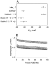
Sodium currents in CNahIII-12 cells expressing C121Wβ1 show reduced frequency-dependent rundown and faster recovery from inactivation, compared with currents in cells expressing wild-type β1. A, Mean amplitudes of currents elicited by 80 Hz pulse trains in CNahIII-12 cells expressing hNav1.3 alone (■, n = 5) and for cells stably coexpressing wild-type β1 (⋄, n = 5) or C121W β1 (▵,n = 6). The pulse trains consisted of 100 pulses, each 5 msec long, to +10 mV, from a holding voltage of −80 mV. Current amplitudes in each experiment were normalized with respect to the current evoked by the first pulse. B, Mean time course of recovery from inactivation for the same cells as inA. Recovery time course was assessed as described in Materials and Methods. The smooth lines are means of exponential fits of the data, with time constants of 3.7 ± 0.5, 4.9 ± 0.5, and 10.5 ± 0.3 msec, for hNav1.3 alone, hNav1.3 plus C121Wβ1, and hNav 1.3 plus β1, respectively. The vertical dashed line shows the extent of recovery after 7.5 msec, the duration between pulses in the 80 Hz trains. Both rundown and recovery time course were significantly different in cells coexpressing β1 than in cells expressing hNav1.3 alone or with C121Wβ1 (p < 0.0001).
Fig. 6.
Fig. 6.
C121Wβ1 does not act as a dominant-negative subunit. A, Mean V1/2 values of availability curves for CNahIII-12 cells expressing Nav1.3 alone (V1/2 = −47.5 ± 1.2; k = 7 ± 0.2;n = 18), for lines stably coexpressing β1 (−60.7 ± 0.9; 7 ± 0.3; n = 6), or for C121Wβ1 (−47.3 ± 1.5; 7.1 ± 0.2; n = 8), for the stable β1 line transiently coexpressing C121W β1 (−62.1 ± 1.8; 6.9 ± 0.6; n = 6), and for the stable C121Wβ1 line transiently coexpressing β1 (−60.4 ± 2; 7.1 ± 0.6; n = 8). β1 caused significant negative shifts in V1/2(p < 0.00001), even when coexpressed with C121Wβ1. B, Frequency-dependent rundown for CNahIII-12 cells expressing hNav1.3 alone (■) or hNav1.3 plus β1 (⋄; same data as in Fig. 5) and for the stable β1 line transiently coexpressing C121Wβ1 (○, n = 4).
Fig. 7.
Fig. 7.
Ecdysone-inducible expression of β1 or C121Wβ1 subunits and association of β1 or C121Wβ1 with rat Nav1.2a α subunits. A, SNaIIA-pIND.β1 or SNaIIA-pIND.C121Wβ1 cells were treated with vehicle (0 ponasterone) or hormone (20 μm ponasterone) for 48 hr in culture, solubilized in 5% SDS, and boiled in SDS-PAGE sample buffer containing 5% β-mercaptoethanol. Samples were separated by 10% acrylamide SDS-PAGE and transferred to nitrocellulose. Western blots were probed with anti-β1EX antibody (1:500 dilution) and then with horseradish peroxidase-conjugated goat anti-rabbit antibody (1:100,000). Immunoreactive bands were visualized with Westdura chemiluminescent substrate. Arrow indicates position of β1 immunoreactive band. B, Equal aliquots of SNaIIA-pIND.β1 or SNaIIA-pIND.C121Wβ1 cells were treated with 20 μm ponasterone for 48 hr in culture, and then equal aliquots of cells were immunoprecipitated with anti-Nav1.2a antibody as described in Materials and Methods. The samples were then separated by SDS-PAGE, transferred to nitrocellulose, and probed with anti-β1EX antibody (1:500), followed by horseradish peroxidase-conjugated goat anti-rabbit antibody (1:100,000). The blot was detected with Westdura chemiluminescent substrate and exposed to ECL Hyperfilm. Arrow indicates migration of immunoreactive β1 subunits.
Fig. 8.
Fig. 8.
The C121W mutation causes loss of β1-mediated functional modulation of rat Nav1.2a sodium channels expressed in SNaIIA cells. A, Mean current time courses at 0 mV for uninduced SNaIIA-pIND.β1 and SNaIIA-pIND.C121Wβ1 cells, which express rat Nav1.2a alone (solid line,n = 8), and for induced cells expressing Nav1.2a plus β1 (dashed line,n = 6) or Nav1.2a plus C121Wβ1 (dotted line, n = 4). Neither wild-type nor mutant β1 significantly altered current time course.B, Mean V1/2 values for activation (open symbols) and availability (solid symbols) for uninduced SNaIIA cells (SNaIIA-pIND.β1: activation: V1/2 = −16.3 ± 1.1 mV, k = −6.1 ± 0.2; availability: −49.4 ± 1.4, 5.4 ± 0.3, n = 4; SNaIIA-pIND.C121Wβ1: −17.1 ± 1.4, −6.3 ± 0.4, −48.2 ± 2, 5.7 ± 0.4; n = 4) and for induced cells coexpressing β1 (−19 ± 1.4, −6.3 ± 0.3, −59.8 ± 0.8, 5.3 ± 0.2, n = 6) or C121Wβ1 (−18.1 ± 1.5, −6 ± 0.2, −50.1 ± 1.5, 5.6 ± 0.5, n = 4). Induction of wild-type β1 shifted the midpoint of availability significantly negative compared with cells expressing Nav1.2a alone or Nav1.2a with C121Wβ1 (p < 0.001). C, Mean frequency-dependent rundown in uninduced SNaIIA cells (■, n = 8) and in induced cells coexpressing β1 (⋄, n = 6) or C121Wβ1 (▵,n = 4). Wild-type β1 significantly increased frequency-dependent rundown compared with uninduced cells or cells coexpressing C121Wβ1 (p < 0.01).
Fig. 9.
Fig. 9.
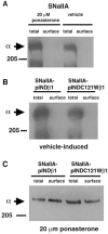
β1 and C121Wβ1 subunits promote translocation of Nav1.2a α subunits to the plasma membrane. SNaIIA, SNaIIA-pIND.β1, or SNaIIA-pIND.C121Wβ1 cells were treated with vehicle or 20 μm ponasterone for 48 hr in culture and treated with sulfo-NHS-biotin as described in Materials and Methods. Each cell sample was immunoprecipitated with anti-SP11-II and divided into two equal aliquots. One aliquot was prepared for SDS-PAGE as described (total). The remaining half was immunoprecipitated with anti-SP11-II antibody, boiled in 5% SDS to release the proteins from the Protein A Sepharose beads, reprecipitated with streptavidin agarose to purify the fraction that was biotinylated, and prepared for SDS-PAGE as described (surface). The samples were then separated by SDS-PAGE, transferred to nitrocellulose, and probed with anti-SP11-II antibody (1:500) followed by horseradish peroxidase-conjugated goat anti-rabbit antibody (1:100,000). The blot was detected with Westdura chemiluminescent substrate and exposed to ECL Hyperfilm. Arrows indicate migration of immunoreactive α subunits. A, An undetectable percentage of Nav1.2a subunits in SNaIIA cells is located at the cell surface. Treatment of cells with ponasterone does not affect cell surface expression of sodium channels. B, Treatment of SNaIIA-pIND.β1 or SNaIIA-pIND.C121Wβ1 cells with vehicle does not result in translocation of Nav1.2a sodium channels to the cell surface. C, Treatment of SNaIIA-pIND.β1 or SNaIIA-pIND.C121Wβ1 cells with 20 μm ponasterone (resulting in β1 or C121β1 subunit expression, as shown in Fig. 7) results in an increase in the percentage of Nav1.2a sodium channels located at the cell surface.
Fig. 10.
Fig. 10.
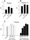
Neither wild-type β1 nor C121Wβ1 increases sodium current amplitude. A–C, Mean amplitudes of currents elicited by depolarizations to 0 mV from a holding voltage of −90 mV, for SNaIIA cells (A), CNahIII-12 cells (B), and Xenopus oocytes expressing rat Nav1.2a (C).
Fig. 11.
Fig. 11.
The C121W mutation disrupts β1–β1 homophilic interactions. A, Western blot analysis of β1 subunit expression in transfected S2 cells. Wild-type β1- or C121Wβ1-transfected S2 cells were solubilized in 5% SDS and boiled in SDS-PAGE sample buffer containing 5% β-mercaptoethanol. Samples were separated by 10% acrylamide SDS-PAGE and transferred to nitrocellulose. The Western blot was probed with anti-β1EX antibody (1:500 dilution) and then with horseradish peroxidase-conjugated goat anti-rabbit antibody (1:100,000 dilution). Immunoreactive bands were visualized with Westdura chemiluminescent substrate (Pierce). B, C121Wβ1 subunit expression does not promote S2 cell aggregation. Transfected S2 cells were induced in the presence of 0.7 mmCuSO4. An aliquot of each cell line was removed and stained for β1 or C121Wβ1 expression at the cell surface (left panel). Cells were viewed with a confocal microscope. Scale bar, 10 μm. In the remaining cells, aggregation was induced by rotary shaking. Cells were viewed with a phase-contrast microscope. Scale bar, 100 μm. Aggregation was not observed in cells transfected with C121Wβ1 in any field of view.
Fig. 12.
Fig. 12.
C121Wβ1 does not exert a dominant-negative effect on cell adhesion in Drosophila S2 cells. Stable S2β1 cell lines (Malhotra et al., 2000) were transfected with increasing amounts of C121Wβ1 plasmid, as indicated above, using Fugene reagent. Selection (250 μg/ml hygromycin) was started 48 hr after transfection and continued for 30 d. Transfected or untransfected S2 cells were induced in the presence of 0.7 mm CuSO4, and then aggregation was induced by rotary shaking. Cells were viewed with a phase-contrast microscope. Representative fields of view are presented for 0 (S2β1), 1, and 5 μg of transfected C121Wβ1 plasmid, respectively. Untransfected S2 cells are presented for comparison. Scale bar, 100 μm.

Similar articles

Cited by

References

    1. Abou-Khalil B, Ge Q, Desai R, Ryther R, Bazyk A, Baily R, Haines JL, Sutcliff JS, George AL., Jr Partial and generalized epilepsy with febrile seizures plus and a novel SCN1A mutation. Neurology. 2001;57:2265–2272. - PubMed
    1. Alekov AK, Rahman M, Mitrovic N, Lehmann-Horn F, Lerche H. A sodium channel mutation causing epilepsy in man exhibits subtle defects in fast inactivation and activation in vitro. J Physiol (Lond) 2000;529:533–539. - PMC - PubMed
    1. Alekov AK, Rahman M, Mitrovic N, Lehmann-Horn F, Lerche H. Enhanced inactivation and acceleration of activation of the sodium channel associated with epilepsy in man. Eur J Physiol. 2001;13:2171–2176. - PubMed
    1. Armstrong CM, Bezanilla F. Inactivation of the sodium channel. II. Gating current experiments. J Gen Physiol. 1977;70:567–590. - PMC - PubMed
    1. Barek S. Site-directed mutagenesis by double polymerase chain reaction: megaprimer method. In: White BA, editor. Methods in molecular biology, Vol 15: PCR protocols, current methods and applications. Humana; Totowa, NJ: 1993. pp. 277–286. - PubMed

Publication types

MeSH terms

LinkOut - more resources