. 2002 Dec 15;22(24):10751-60.
doi: 10.1523/JNEUROSCI.22-24-10751.2002.

Identification of an axotomy-induced glycosylated protein, AIGP1, possibly involved in cell death triggered by endoplasmic reticulum-Golgi stress

Affiliations

Identification of an axotomy-induced glycosylated protein, AIGP1, possibly involved in cell death triggered by endoplasmic reticulum-Golgi stress

Shunsuke Aoki et al. J Neurosci. .

Abstract

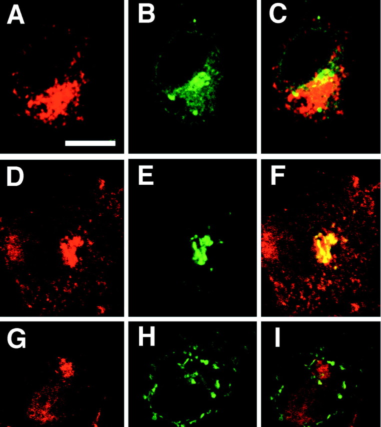
We developed a new method, designated N-linked glycosylation signal (NGS) differential display (DD)-PCR, that enables the identification of genes encoding N-linked glycosylated molecules that exhibit varying patterns of expression. Using this innovative technique, we identified an N-linked glycosylated 11-transmembrane domain protein that is upregulated in response to axotomy. Expression levels increased 3 d after axotomy, reached maximal levels at approximately postoperative days 5-7, and then gradually decreased through day 20. The protein was termed axotomy-induced glycosylated/Golgi-complex protein 1 (AIGP1). AIGP1 immunoreactivity is specifically localized in neurons, with subcellular localization within the Golgi, indicating that AIGP1 is a resident Golgi protein. Moreover, AIGP1 gene expression in cultured neurons is specifically induced by the endoplasmic reticulum (ER)-Golgi stressors tunicamycin and brefeldin A. We observed that the frequency of cell death is increased by AIGP1 overexpression and that the corresponding region of the protein implicated in the activity involves the large eighth and ninth transmembrane loops. Our results suggest that AIGP1 gene activation and protein accumulation in the Golgi complex in response to axotomy-induced ER-Golgi stress may contribute to signaling during programmed cell death in damaged neurons.


Figures

Fig. 1.
Fig. 1.
Isolation of a gene encoding an N-linked glycosylated protein from axotomized hypoglossal nuclei by NGS DD-PCR.A, The primer sequence used in NGS DD-PCR (N = random nucleotide). B,35S-labeled PCR products (from NGS DD-PCR) in hypoglossal nuclei from normal (cont) and operated (op.) sides (7 d after injury). Thearrowhead indicates the band in the op.lane corresponding to a differentially expressed gene.C, Expression of the isolated cDNA fragment was further examined by in situ display. A section was obtained from a mouse 7 d after it had undergone unilateral hypoglossal nerve transection. The arrow in the top panelindicates increased expression of the candidate gene on the injured/right side only. A bright-field micrograph of Nissl stain shows localization of mRNA on neurons in the hypoglossal nucleus (bottom). Scale bar: top, 2 mm;bottom, 50 μm. D, mRNA expression profile after hypoglossal nerve transection. mRNA signal intensity in control (○) and operated sides (●) was measured and presented as mean ± SD obtained from at least eight sections from five mice; *p < 0.01 (ANOVA). E, AIGP1 is an N-linked glycosylated protein. COS-7 cells were transfected with a negative control mock construct (lanes 1,2) or N-terminal FLAG-tagged AIGP1 expression construct (lanes 3, 4, 5). Cell lysates were treated without (lanes 1, 3,4) or with (lanes 2,5) 5 U/ml N-glycosidase F and subjected to SDS-PAGE (10 μg protein per lane). Lysates from PC12 cells were treated without (lanes 6, 7) or with (lane 8) 5 U/ml N-glycosidase F and subjected to SDS-PAGE (30 μg protein per lane). Blots were probed with an antibody to AIGP1 (ABEP56). Cell lysates not subjected to 0.5% Nonidet P-40 treatment were analyzed by Western blot (lanes 3, 6). The results are representative of three separate experiments that yielded similar results.
Fig. 2.
Fig. 2.
Localization of AIGP1 immunoreactivity in the mouse brain. A, Expression of AIGP1 (green) in the mouse brain examined by fluorescent immunohistochemistry with polyclonal antibody ABEP56. A section (25 μm) was obtained from a unilateral hypoglossal nerve-transected mouse 7 d after surgery. B, Staining with ABEP56 and propidium iodide (red) revealed AIGP1 localization (green) as a dot-like area adjacent to the nucleus. CE, Tissue localization of AIGP1 in the cortex (C), cerebellum (D), and hippocampus (E). Coronal sections were stained with ABEP56 and Alexa Flour 488-labeled secondary antibody (green). Confocal images are presented.F, Mouse cortical neurons were lysed, and total protein was subjected to Western blotting with preimmune (pre) or ABEP56, respectively. 4v, Fourth ventricular; cont, control side;op, operated side; DG, dentate gyrus;gl, granule cell layer; pl, Purkinje cell layer; ml, molecular cell layer; py, pyramidal cell layer. Scale bars: A, C,D, E, 300 μm; B, 15 μm.
Fig. 3.
Fig. 3.
Localization of AIGP1 immunostaining in mouse cultured cortical neurons and PC12 cells. Primary cultured cortical neurons were fixed at 8 hr (A, B), 24 hr (C), and 48 hr (D) after plating and stained with polyclonal antibody ABEP56 and Alexa Flour 488-labeled secondary antibody (green). Specificity of staining was verified by preabsorption of ABEP56 with 100-fold excess peptide EP56 (A) or negative control peptide (B). E, Primary cultured cortical neurons were fixed 13 d after plating and coimmunostained with ABEP56 and Alexa Flour 594 (red) and monoclonal anti-synapsin-I and Alexa Flour 488 (green). F, Primary cultured cortical neurons were fixed 48 hr after plating and immunostained with polyclonal anti-membrin and Alexa Flour 594 (red).G–I, PC12 cells were coimmunostained with ABEP56 (red) (G) and monoclonal anti-TGN46 (green) (H). I, Merged image ofG and H. Both Alexa Flour 488-labeled anti-rabbit IgG and Alexa Flour 594-labeled anti-mouse IgG were used as secondary antibodies for double-staining (G–I).AD andEI are confocal and cooled CCD images, respectively. Scale bars: A–D, 10 μm;E, F, 15 μm;GI, 10 μm.
Fig. 4.
Fig. 4.
AIGP1 immunostaining is specifically localized to the Golgi in transfected COS-7 cells. COS-7 cells were cotransfected with N-terminal FLAG-tagged AIGP1 and pEYFP constructs: pEYFP–ER (A–C), pEYFP–Golgi (D–F), or pEYFP–mito (G–I). Cells were fixed 24 hr after transfection and stained with anti-FLAG M2. Confocal images of FLAG immunostaining with Alexa Fluor 594-labeled anti-mouse IgG (A,D, G) and EYFP fluorescence (B, E, H).C, F, and Irepresent merged images. Similar results were obtained in four independent experiments. Scale bar, 10 μm.
Fig. 5.
Fig. 5.
ER–Golgi stress specifically induced AIGP1 expression in cultured cortical neurons. A, After 40 cycles of PCR with primers that recognize either β-actin or AIGP1 cDNA sequences, reaction products (β-actin = 118 bp; AIGP1 = 93 bp) were resolved by 2% agarose gel electrophoresis. B, Three days after plating, cortical neurons were cultured for 12 hr under the following stress conditions: B27 supplement deprivation [B27()], 10 μg/ml tunicamycin (TN), 10 μg/ml brefeldin A (BrA), 0.2 μm staurosporin (STS), 250 μm H2O2(H202), or 1 μm cytochalasin D (CyD). Each cDNA was prepared from random hexamer primers and total RNA from stress-treated cells. Real-time RT-PCR assays of AIGP1 mRNA expression were performed with SYBR green and ABI7700 sequence detection system. β-actin was used as an internal control. Similar results were obtained in three separate experiments using independent cortical neuron cultures. Error bars represent the SD calculated from quadruplicate samples at a minimum (from culture wells). **p < 0.01.
Fig. 6.
Fig. 6.
AIGP1 expression increases the frequency of cell death. A, COS-7 cells were transfected with constructs encoding β-galactosidase (b-gal), TPO1-myc, AIGP1-myc, N-FLAG–presenilin-1 (PS1), or N-FLAG-AIGP1. At 48 hr after transfection, cells were fixed and stained with anti-β gal (A, bottom;b-gal), anti-myc 9E10 (A,bottom; TPO myc and AIGP1 myc), or anti-FLAG M2 (A, bottom;FLAG PS1 and FLAG AIGP1). Cell death was determined by fluorescein TUNEL assay (A,bottom; TUNEL). Confocal images of immunostaining and the fluorescein TUNEL stains are presented (A, bottom). The frequency of cell death (the number of fluorescein TUNEL-positive cells per β-gal, 9E10-, or FLAG-positive cells) was counted and presented as mean percentages (A, top). Error bars represent SD calculated from quadruplicate samples. *p < 0.05; **p < 0.01. Scale bar, 20 μm. B, Nuclear crumpling of a Neuro2a cell transfected with an AIGP1 expression construct. Neuro2a cells were transfected with constructs encoding TPO1-myc or AIGP1-myc. At 72 hr after transfection, cells were fixed and stained with CYTO green (nuclear stain; top) and anti-myc 9E10 (bottom). Scale bar, 10 μm.
Fig. 7.
Fig. 7.
C-terminal deletion mutants of AIGP1 and induction of cell death. A, Schematic representation of AIGP1 structure and C-terminal deletion mutants. Locations of amino acid residues are indicated by numbers, and transmembrane domains are shown as gray boxes (Grossman et al., 2000). B, Loss of AIGP1 activity after deletion of the region containing the eighth and ninth helices and loops. COS-7 cells were transfected with a series of deletion mutants, fixed 48 hr after transfection, and stained with anti-FLAG M2 antibody (B, right, top). Cell death was analyzed by the fluorescein TUNEL method (B,right, middle). Confocal images of FLAG immunostaining with Alexa Flour 594-labeled anti-mouse IgG and fluorescein TUNEL stains are presented (B,right, top and middle). The frequency of cell death (TUNEL-positive cells per FLAG-positive cells) was counted and is presented as mean percentages (B, left). Expression levels of mutant constructs were analyzed by Western blots with anti-FLAG M2 (8 μg protein per lane) (B, right,bottom). Error bars represent SD calculated from at least four different samples. **p < 0.01. Scale bar, 20 μm.

Similar articles

Cited by

References

    1. Abeijon C, Hirschberg CB. Topography of glycosylation reactions in the endoplasmic reticulum. Trends Biochem Sci. 1992;17:32–36. - PubMed
    1. Aoki K, Sun YJ, Aoki S, Wada K, Wada E. Cloning, expression and mapping of a gene that is upregulated in adipose tissue of mice deficient in bombesin receptor subtype-3. Biochem Biophys Res Commun. 2002;290:1282–1288. - PubMed
    1. Baba N, Koji T, Itoh M, Mizuno A. Reciprocal changes in the expression of Bcl-2 and Bax in hypoglossal nucleus after axotomy in adult rats: possible involvement in the induction of neuronal cell death. Brain Res. 1999;827:122–129. - PubMed
    1. Cameron AA, Cliffer KD, Dougherty PM, Willis WD, Carlton SM. Changes in lectin, GAP-43 and neuropeptide staining in the rat superficial dorsal horn following experimental peripheral neuropathy. Neurosci Lett. 1991;131:249–252. - PubMed
    1. Cockcroft S. Mammalian phosphatidylinositol transfer proteins: emerging roles in signal transduction and vesicular traffic. Chem Phys Lipids. 1999;98:23–33. - PubMed

Publication types

MeSH terms