. 2002 Dec 15;22(24):10781-9.
doi: 10.1523/JNEUROSCI.22-24-10781.2002.

Involvement of tissue plasminogen activator in onset and effector phases of experimental allergic encephalomyelitis

Affiliations

Involvement of tissue plasminogen activator in onset and effector phases of experimental allergic encephalomyelitis

Weiquan Lu et al. J Neurosci. .

Abstract

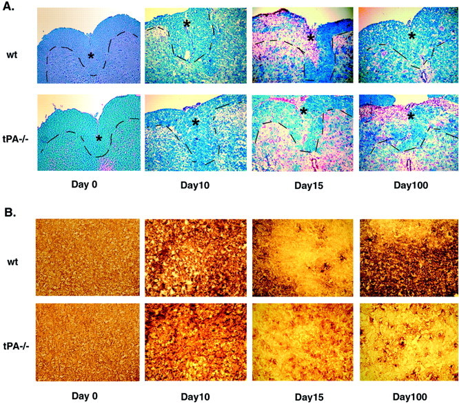
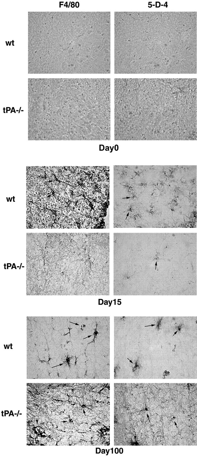
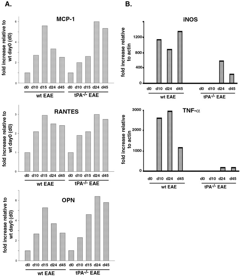
Inflammation, demyelination, and neurodegeneration are pathological features of multiple sclerosis (MS). In the brains of MS patients, tissue plasminogen activator (tPA) mRNA and protein are upregulated, and changes in the levels of tPA correlate with progression of the disease. However, the role of tPA in MS is as yet unknown. tPA functions in the CNS in neuronal plasticity and cell death. tPA also mediates the activation of microglia, the CNS "immune cells." In this study, we establish that tPA activity increases during major oligodendrocyte glycoprotein-induced experimental allergic encephalomyelitis (EAE) in normal mice. To explore the role of tPA in this disease as a model for MS, we have examined the EAE course and expression of histopathological markers in mice lacking tPA (tPA(-/-)). We find that tPA(-/-) mice have a delayed onset of EAE but then exhibit increased severity and delayed recovery from the neurological dysfunction. Demyelination and axon degeneration are delayed, microglial activation is attenuated, and the production of chemokines is decreased. Our results suggest that tPA and activated microglia have complex roles in MS/EAE, and that these roles are harmful during the onset of the disease but beneficial in the recovery phase. A temporally restricted attenuation of tPA activity could have therapeutic potential in the management of MS.


Figures

Fig. 1.
Fig. 1.
Levels of tPA activity increase during the clinical course of MOG-induced EAE in wt mice. Lumbar spinal cord lysates were prepared from wt EAE mice at different time points after MOG injection. Uninjected adult female mice were used as controls.Top, To quantitatively determine tPA activity, amidolytic assays were performed as described in Materials and Methods. Total protein content in aliquots of each sample was determined using the Bradford assay. Note that tPA activity significantly increased during the disease but returned to near normal levels after recovery. Although this assay does not discriminate between uPA and tPA activity, uPA is unlikely to have contributed to the increased activity observed for two reasons: (1) previous reports (Akenami et al., 1996; Cuzner et al., 1996) using tPA-specific assays have demonstrated that it is the activity of tPA rather than uPA that becomes upregulated, and (2) we performed in situ zymographic assays on spinal cord sections in the presence or absence of amiloride, a specific uPA inhibitor, and observed no differences in activity (data not shown). This result is consistent with our previous report that uPA mRNA and protein are not detected in the mouse CNS (Tsirka et al., 1997).Bottom, PAI-1 expression was determined by Western blotting. FluorImager was used for the quantification of the bands. Note that the level of PAI-1 expression also increased during this period, although less quickly or dramatically than tPA, and returned to baseline levels after recovery. The data are presented as mean ± SEM (n = 3 mice). *p < 0.05; Student's t test.
Fig. 2.
Fig. 2.
Altered progression of EAE in tPA−/− mice. The wt and tPA−/− mice were injected with MOG35–55 peptide in CFA and pertussis toxin (PT) to induce EAE. The disease severity was scored on a clinical scale from 0 to 5 as described in Materials and Methods. The average score for each day was calculated by averaging the clinical score for that day for each mouse in the group (n = 12 mice for each group). tPA−/− mice showed a significant delay in the onset of EAE, followed by a delay in recovery. Table 2 presents the statistical significance in the day of onset, average maximum clinical score, and score at day 50.
Fig. 3.
Fig. 3.
Delayed demyelination in tPA−/− mice during EAE. Frozen cross sections of spinal cords from wt and tPA−/− mice at different time points during the EAE course were stained with LFB and Nuclear Red (A). Note the extensive demyelination and infiltration of inflammatory cells evident in sections of wt mice at day 15 and in sections of tPA−/− mice until day 100. The dashed line demarcates the border of the dorsal horn (as denoted by asterisks), where demyelination is observed. B, Immunohistochemistry using an antibody to visualize MBP revealed similar patterns of demyelination (600× magnification).
Fig. 4.
Fig. 4.
Delayed axonal damage in tPA−/− mice during EAE. Using an antibody directed against APP, neuronal degeneration was detected in the white matter of spinal cord sections from wt mice as early as day 10 and more strongly at day 15. For the tPA−/− mice, in contrast, only minimal APP was detected, and even that was not observed until day 15 (600× magnification).
Fig. 5.
Fig. 5.
Attenuated microglial activation in tPA−/− mice during the course of EAE. Frozen cross sections of spinal cords from wt and tPA−/− mice at different time points of EAE were probed using antibodies directed against either the mature macrophage/microglia-specific antigen F4/80 or the microglial-specific cell surface antigen 5-D-4. Highly activated microglial cells were found in wt mice at day 15. In contrast, only attenuated microglial activation was noted in tPA−/− mice, and even that was only seen at day 100. Arrows point to individual activated macrophage/microglial cells (600× magnification).
Fig. 6.
Fig. 6.
Altered expression of chemokines and cytokines in tPA−/− mice during the course of EAE.A, Quantitative Western blotting analysis for chemokines was performed using spinal cord lysates from wt and tPA−/− mice at days 0, 10, 15, 24, and 45 of EAE, using either anti-MCP-1 antibody, anti-RANTES antibody, or anti-OPN. Note the delayed expression of all three chemokines and persistent expression of MCP-1 and OPN in the tPA−/− mice during the course of EAE. B, Quantitative RT-PCR analysis for TNF-α and iNOS. RNA from lumbar spinal cords of wt and tPA−/− mice at days 0, 10, 24, and 45 after MOG injection was used to perform RT-PCR in a real-time LightCycler system. Specific primer sets were designed to detect the cytokines iNOS and TNF-α (described in Table 1). The level of specific mRNAs was quantified in the exponential phase of PCR product accumulation and normalized by comparison with standard curves obtained from serial dilutions of plasmids encoding cDNAs for each gene.
Fig. 7.
Fig. 7.
Alteration of a T cell proliferation/activation marker but not a B cell marker in tPA−/− mice during EAE. A, Quantitative Western blotting analysis was performed using spinal cord lysates from wt and tPA−/− mice at days 0, 10, 15, 24, and 45 of EAE, using either anti-T cell/CD3 or anti-B cell/CD22. Note that the expression of the T cell marker (but not that of the B cell marker) showed a delayed rise in tPA−/− mice during EAE. No differences in marker expression were observed at day 0.B, Quantitative RT-PCR analysis of CD8α, a marker for cytotoxic T cells. RNA from lumbar spinal cords of wt and tPA−/− mice at days 0, 10, 24, and 45 after MOG injection was used to perform RT-PCR in a real-time LightCycler system. Specific primer sets were designed as described in Table 1. The level of specific mRNAs was quantified in the exponential phase of PCR product accumulation and normalized by comparison with standard curves obtained from serial dilutions of a plasmid containing the CD8α cDNA.

Similar articles

Cited by

References

    1. Akassoglou K, Kombrinck K, Degen J, Strickland S. Tissue plasminogen activator-mediated fibrinolysis protects against axonal degeneration and demyelination after sciatic nerve injury. J Cell Biol. 2000;149:1157–1166. - PMC - PubMed
    1. Akenami F, Sirén V, Koskiniemi M, Siimes M, Teräväinen H, Vaheri A. Cerebrospinal fluid activity of tissue plasminogen activator in patients with neurological diseases. J Clin Pathol. 1996;49:577–580. - PMC - PubMed
    1. Akenami F, Koskiniemi M, Mustjoki S, Siren V, Farkkila M, Vaheri A. Plasma and cerebrospinal fluid activities of tissue plasminogen activator and urokinase in multiple sclerosis. Fibrinolysis Proteolysis. 1997;11:109–113.
    1. Akenami F, Siren V, Wessman M, Koskiniemi M, Vaheri A. Tissue plasminogen activator gene expression in multiple sclerosis brain tissue. J Neurol Sci. 1999;165:71–76. - PubMed
    1. Andrade-Gordon P, Strickland S. Interaction of heparin with plasminogen activators and plasminogen: effects on the activation of plasminogen. Biochemistry. 1986;25:4033–4040. - PubMed

Publication types

MeSH terms

Substances