. 2002 Dec 15;22(24):10847-55.
doi: 10.1523/JNEUROSCI.22-24-10847.2002.

Rostral ventromedial medulla neurons that project to the spinal cord express multiple opioid receptor phenotypes

Affiliations

Rostral ventromedial medulla neurons that project to the spinal cord express multiple opioid receptor phenotypes

Silvia Marinelli et al. J Neurosci. .

Abstract

The rostral ventromedial medulla (RVM) forms part of a descending pathway that modulates nociceptive neurotransmission at the level of the spinal cord dorsal horn. However, the involvement of descending RVM systems in opioid analgesia are a matter of some debate. In the present study, patch-clamp recordings of RVM neurons were made from rats that had received retrograde tracer injections into the spinal cord. More than 90% of identified spinally projecting RVM neurons responded to opioid agonists. Of these neurons, 53% responded only to the mu-opioid agonist D-Ala2, N-Me-Phe4, Gly-ol5 enkephalin, 14% responded only to the kappa-opioid agonist U-69593, and another group responded to both mu and kappa opioids (23%). In unidentified RVM neurons, a larger proportion of neurons responded only to mu opioids (75%), with smaller proportions of kappa- (4%) and mu/kappa-opioid (13%) responders. These RVM slices were then immunostained for tryptophan hydroxylase (TPH), a marker of serotonergic neurons. Forty-percent of spinally projecting neurons and 11% of unidentified neurons were TPH positive. Of the TPH-positive spinally projecting neurons, there were similar proportions of mu- (33%), kappa- (25%), and mu/kappa-opioid (33%) responders. Most of the TPH-negative spinally projecting neurons were mu-opioid responders (67%). These findings indicate that functional opioid receptor subtypes exist on spinally projecting serotonergic and nonserotonergic RVM neurons. The proportions of mu- and kappa-opioid receptors expressed differ between serotonergic and nonserotonergic neurons and between retrogradely labeled and unlabeled RVM neurons. We conclude that important roles exist for both serotonergic and nonserotonergic RVM neurons in the mediation of opioid effects.


Figures

Fig. 1.
Fig. 1.
Electrophysiologically characterized serotonergic RVM cell that was retrogradely labeled from the dorsal spinal cord. This cell responded to both μ- and κ-opioid receptors.A, Low-magnification image of the cell on the midline of the brainstem, ∼600 μm dorsal to the pyramidal tract.B, High-magnification image of the biocytin-filled cell.C, High-magnification image of the fluorescent microspheres present within the cell. Inset, Extent in the coronal plane of the spinal injection site of the fluorescent microspheres. D, High-magnification image of TPH-ir in the biocytin-filled cell. The high-magnification images were all collected at the same optical plane using confocal microscopy. Scale bars: A, 100 μm; BD, 10 μm.
Fig. 2.
Fig. 2.
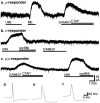
Three distinct categories of opioid-responding bulbospinal cells in RVM. Typical current traces of μ- (a), κ- (b), and μ/κ- (c) opioid-responding RVM neurons. The membrane current was recorded in RVM neurons during the superfusion of the opioid agonists U69593 (U69) (1 μm), DAMGO (3 μm), and met-enkephalin (ME) (10 μm). The effects of DAMGO and U69593 were reversed by the μ- and κ-opioid-selective antagonists CTAP (1 μm) and nor-BNI (300 nm). Current traces ac are from different neurons that were voltage clamped at −60 mV. The action potential tracesfor df are from different neurons in current-clamp mode that are typical of the opioid-responding types in ac, respectively.
Fig. 3.
Fig. 3.
The proportion of opioid-responding subtypes differs between spinally projecting and randomly selected RVM neurons. Bar charts display the percentage of μ-, κ-, μ/κ-opioid-responding neurons in recordings from retrogradely labeled neurons using fluorescence illumination (Retro-labelled) and from randomly sampled neurons with the fluorescence illumination off (Random). Both groups of neurons are from animals that had received previous spinal injections of fluorescent microspheres. *p < 0.05.
Fig. 4.
Fig. 4.
TPH immunoreactivity of electrophysiologically characterized RVM neurons. AC, RVM neuron that expressed TPH-ir. This cell responded to μ- and κ-opioid receptor activation. A, Merged confocal images showing both the biocytin-filled cell (red) and TPH-ir (green). B,C, Separate images showing TPH-ir (B) and biocytin (C) labeling of the filled cell. DF, RVM neuron that did not express TPH-ir. This cell responded to μ-opioid receptor activation and had an action potential shape typical of μ responders. D, Merged confocal images showing both the biocytin-filled cell (red) and TPH-ir (green). Note the lack of double labeling.E, F, Separate images showing TPH-ir (E) and biocytin (F) labeling of the filled cell. Both neurons were identified as containing fluorescent microspheres before recording. Scale bars: 25 μm (bar inD applies to A and D; bar in C applies to B, C,E, and F).
Fig. 5.
Fig. 5.
The proportion of TPH-immunoreactive neurons differs between spinally projecting and randomly selected RVM neurons. Bar charts display the percentage of TPH-positive neurons (TPH+ve, filled bar) and TPH-negative neurons (TPHve, open bar) in recordings from retrogradely labeled neurons using fluorescence illumination (Retrolabeled) and from randomly sampled neurons with the fluorescence illumination off (Random). Both retrolabeled and random neurons are from animals that had received previous spinal injections of fluorescent microspheres. The proportion of TPH-positive neurons was higher for the retrogradely labeled neurons than for the randomly selected neurons (p < 0.005; see Results).
Fig. 6.
Fig. 6.
The proportion of opioid-responding subtypes differs between spinally projecting TPH-positive and TPH-negative RVM neurons. The bar chart displays the percentage of μ-, κ-, μ/κ-opioid-responding neurons in recordings of cells retrogradely labeled from the spinal cord that were identified as TPH positive (TPH+ve) or TPH negative (TPHve). **p < 0.0001; *p < 0.005.
Fig. 7.
Fig. 7.
Distribution of characterized RVM cells. Biocytin-filled cells were categorized by their responses to selective μ- and κ-opioid agonists and by the presence of TPH-ir (serotonergic). These cells were mapped onto drawings of caudal, intermediate, and rostral RVM (based on the presence of the seventh cranial nerve, the inferior olive, or the superior olive).A, Retrogradely labeled neurons that were first identified as containing fluorescent beads after spinal injections of fluorescent microspheres (Retrolabeled).B, Randomly selected cells in animals that had been injected spinally with fluorescent microspheres [Random (injected)]. C, Randomly selected cells in animals that had not received spinal tracer injections [Random (non-injected)]. A, C,Filled circles indicate TPH positive; open circles indicate TPH negative. B, Filled symbols indicate TPH positive; open symbolsindicate TPH negative; stars indicate cells found post hoc to be retrogradely labeled from the spinal cord; circles indicate cells that were not retrogradely labeled. Scale bar, 1 mm. 6, Abducens nucleus; 7, facial nucleus; 7n, facial nerve; 8vn, vestibular root vestibulocochlear nerve;CG, central gray; DR, dorsal raphe nucleus; DT, dorsal tegmental nucleus;g7, genu of the facial nerve; Gi, gigantocellular reticular nucleus; LC, locus coeruleus;Lve, lateral vestibular nucleus; mcp, middle cerebellar peduncle; ml, medial lemniscus;Mo5, motor trigeminal nucleus; Mve, medial vestibular nucleus; PR5VL, principal sensory trigeminal nucleus, ventrolateral part; Pr5, principal sensory trigeminal nucleus; py, pyramidal tract;RMg, raphe magnus nucleus; s5, sensory root of the trigeminal nerve; scp, superior cerebellar peduncle; SO, Superior olive; Sp5O, spinal trigeminal nucleus, oral part; SPO, superior paraolivary nucleus; tz, trapezoid body;Tz, nucleus of the trapezoid body; VCA, ventral cochlear nucleus, anterior part; VCP, ventral cochlear nucleus, posterior part.
Fig. 8.
Fig. 8.
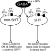
Proposed model of RVM opioid actions.Schematic diagram illustrates the opioid receptor subtypes on the different populations of RVM neurons. Of the RVM neurons that project to the spinal cord dorsal horn, 40% are serotonergic. These serotonergic projection neurons (5HT) have μ-, κ- and μ/κ-opioid-responding subtypes. The functional role of each of these neuronal subtypes is unknown, and they might have distinct spinal targets. The nonserotonergic projection neurons (non-5HT) are composed of μ- and μ/κ-opioid-responding subtypes and may code for ON- versus OFF-neurons, respectively. RVM neurons that were not retrogradely labeled were nonserotonergic and mostly μ-opioid responsive. Our model predicts that some of these nonretrogradely labeled neurons are GABAergic interneurons (GABA) that might also correspond to a population of ON-cells. In addition, a small population of projection neurons (∼10%) did not respond to opioids. These could correspond to either NEUTRAL-cells or OFF-cells.

Similar articles

Cited by

References

    1. Ackley MA, Hurley RW, Virnich DE, Hammond DL. A cellular mechanism for the antinociceptive effect of a kappa opioid receptor agonist. Pain. 2001;91:377–388. - PubMed
    1. Arvidsson U, Dado RJ, Riedl M, Lee JH, Law PY, Loh HH, Elde R, Wessendorf MW. δ-Opioid receptor immunoreactivity: distribution in brainstem and spinal cord, and relationship to biogenic amines and enkephalin. J Neurosci. 1995;15:1215–1235. - PMC - PubMed
    1. Azami J, Llewelyn MB, Roberts MH. The contribution of nucleus reticularis paragigantocellularis and nucleus raphe magnus to the analgesia produced by systemically administered morphine, investigated with the microinjection technique. Pain. 1982;12:229–246. - PubMed
    1. Bederson JB, Fields HL, Barbaro NM. Hyperalgesia during naloxone-precipitated withdrawal from morphine is associated with increased on-cell activity in the rostral ventromedial medulla. Somatosens Mot Res. 1990;7:185–203. - PubMed
    1. Christie MJ. Do medullary serotonergic neurons tonically modulate nociceptive transmission. Pain Forum. 1998;7:155–158.

Publication types

MeSH terms