. 2002 Dec 15;22(24):10914-23.
doi: 10.1523/JNEUROSCI.22-24-10914.2002.

Induction of hippocampal long-term potentiation during waking leads to increased extrahippocampal zif-268 expression during ensuing rapid-eye-movement sleep

Affiliations

Induction of hippocampal long-term potentiation during waking leads to increased extrahippocampal zif-268 expression during ensuing rapid-eye-movement sleep

Sidarta Ribeiro et al. J Neurosci. .

Abstract

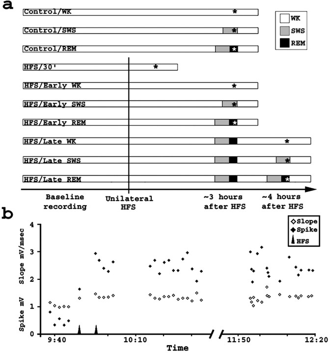
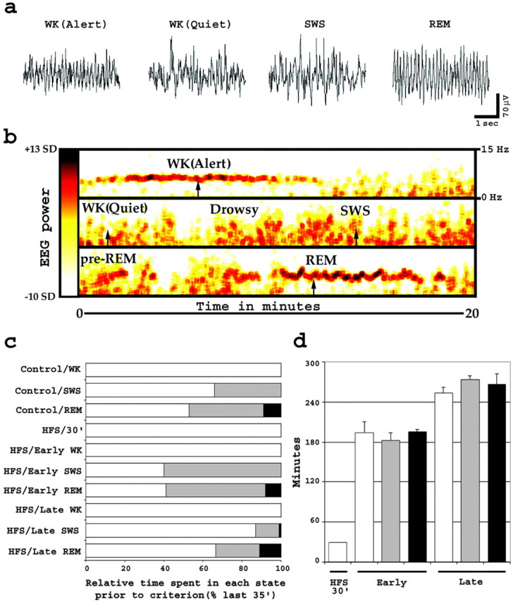
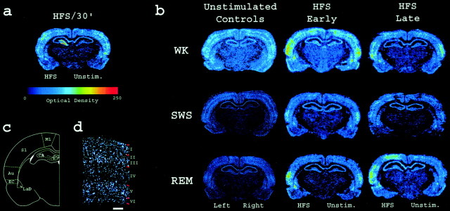
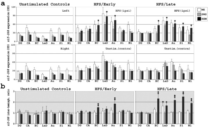
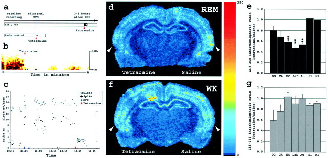
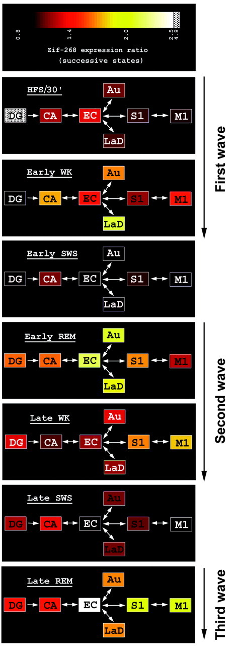
Rapid-eye-movement (REM) sleep plays a key role in the consolidation of memories acquired during waking (WK). The search for mechanisms underlying that role has revealed significant correlations in the patterns of neuronal firing, regional blood flow, and expression of the activity-dependent gene zif-268 between WK and subsequent REM sleep. Zif-268 integrates a major calcium signal transduction pathway and is implicated by several lines of evidence in activity-dependent synaptic plasticity. Here we report that the induction of hippocampal long-term potentiation (LTP) during WK in rats leads to an upregulation of zif-268 gene expression in extrahippocampal regions during subsequent REM sleep episodes. This upregulation occurs predominantly in the amygdala, entorhinal, and auditory cerebral cortices during the first REM sleep episodes after LTP induction and reaches somatosensory and motor cerebral cortices as REM sleep recurs. We also show that hippocampal inactivation during REM sleep blocks extrahippocampal zif-268 upregulation, indicating that cortical and amygdalar zif-268 expression during REM sleep is under hippocampal control. Thus, expression of an activity-dependent gene involved in synaptic plasticity propagates gradually from the hippocampus to extrahippocampal regions as REM sleep recurs. These findings suggest that a progressive disengagement of the hippocampus and engagement of the cerebral cortex and amygdala occurs during REM sleep. They are also consistent with the view that REM sleep constitutes a privileged window for hippocampus-driven cortical activation, which may play an instructive role in the communication of memory traces from the hippocampus to the cerebral cortex.


Figures

Fig. 1.
Fig. 1.
Experimental design and LTP induction.a, Seven HFS and three unstimulated control groups were studied. Acquisition of baseline field potentials was followed by HFS of one hemisphere (vertical line) to produce unilateral LTP. Group criteria are indicated by asterisks. Notice that all animals were kept awake during the last 30 min of the experiment. b, HFS potentiated mPP–DG synapses. A representative experiment is shown. Plotted are EPSP slope and population spike amplitudes from evoked field potentials recorded in the DG after single-pulse stimulation of the mPP. HFS (arrows) was followed by sustained potentiation of evoked responses.
Fig. 2.
Fig. 2.
Assessment of wake and sleep in rats. a, Characteristic wake–sleep EEG traces recorded in the DG. Notice the presence of theta rhythm (∼7 Hz) during both alert WK and REM sleep states. b, EEG spectral analysis used for quantification of wake–sleep states. Plotted is a spectrogram (frequency and power over time) of a representative 60-min-long EEG segment. Power is coded according to thecolor bar to the left, which runs linearly between −10 and +13 SDs of the logarithm of the power between 6 and 9 Hz. Frequencies are depicted in a linear scale according to the references on the top right. Arrows, Time points used to sample the EEG traces depicted in a.c, Wake–sleep state composition of experimental and control groups. Shown are the mean relative times spent in WK, SWS, and REM sleep, in percentage of the last 35 min before criterion. Histograms are colored according to the key on Figure1a. d, Survival times after HFS. Plotted are the average elapsed times between HFS and kill (mean ± SEM). Histograms are colored according to the key on Figure1a.
Fig. 3.
Fig. 3.
Post-LTP zif-268 expression profile across wake–sleep cycles. a, Phosphorimager autoradiogram of a representative zif-268-hybridized section. The ipsilateral (but not contralateral) DG showed markedzif-268 upregulation 30 min after unilateral HFS.b, Zif-268 expression levels in the brain during early and late WK, SWS, and REM sleep depend on previous WK stimulation. Both HFS-stimulated and unstimulated controls showed a generalized decrease in brain zif-268 expression in association with SWS, whereas HFS-stimulated hemispheres in both HFS early and late groups showed a selective increase inzif-268 expression in the cortex and amygdala in association with REM sleep. c, Brain regions in whichzif-268 expression was quantified. d,Zif-268 cortical reinduction during REM sleep occurred predominantly in layers II, III, and V. Shown is a dark-field view of the auditory cortex in a representative section from the HFS/early REM group, hybridized to zif-268 and exposed to autoradiographic emulsion. White–silver grains denote gene expression; red marks on the rightindicate cell layer boundaries, as determined by cresyl violet counterstaining. Scale bar, 250 μm.
Fig. 4.
Fig. 4.
Quantification of zif-268expression across the wake–sleep cycle. a, Absolute levels of zif-268 expression. Both hemispheres of unstimulated controls showed decreased zif-268expression levels during SWS and REM sleep compared with WK. The same general pattern was observed in the unstimulated (contralateral) hemispheres of HFS animals. In contrast, several brain structures in the HFS (ipsilateral) hemispheres of both HFS/early and HFS/late groups showed high zif-268 expression during WK and REM sleep and low zif-268 expression during SWS.Zif-268 reinduction occurred in proximal extrahippocampal regions (EC, Au, and LaD) during Early REM sleep and reached distal extrahippocampal areas (S1 and M1) during Late REM sleep (Bonferroni post hoc tests; *p < 0.05). OD, Optical density. b, Interhemisphericzif-268 expression ratios. Histograms are according to the key in a. Whereas unstimulated controls did not show differences between left and right hemispheres (two-way ANOVA; F(12,63) = 0.33;p = 0.98), significant interactions were detected in HFS groups (two-way ANOVA; F(30,126)= 3.72; p < 0.0001), because of higherzif-268 interhemispheric ratios (HFS/unstimulated) in the EC, LaD, Au, S1, and M1 during REM sleep than during preceding SWS (Bonferroni post hoc tests; *p < 0.05; **p < 0.01). Notice that the interhemispheric zif-268 expression ratio was also significantly higher for LaD in the HFS/late WK group than in the HFS/late SWS group.
Fig. 5.
Fig. 5.
Effect on extrahippocampal zif-268expression of hippocampal inactivation during REM sleep and WK.a, Experimental design of the hippocampal inactivation experiment (see Materials and Methods). b, Tetracaine infusion strongly reduced EEG power in the DG immediately after the beginning of injection. Amplitude is color coded as in Figure2b. c, Tetracaine effects on evoked potentials. A representative experiment is shown; notice that potentials almost recovered to previous potentiated levels right before kill, at 14:15. d, Zif-268 brain expression levels during early REM sleep, after bilateral HFS and unilateral tetracaine infusion. Shown is a phosphorimager autoradiogram of a representative brain section hybridized forzif-268. There was a marked reduction ofzif-268 expression in the left EC, Au, and LaD (tetracaine side) compared with the right hemisphere (saline side).Arrowheads, Auditory cortex (see Fig. 3cfor anatomical localization). e, Zif-268expression interhemispheric ratios (tetracaine/saline) in bilateral HFS and early REM animals (*p < 0.05 and **p < 0.01). f,Zif-268 brain expression levels during WK, after unilateral tetracaine infusion. Zif-268 expression levels were comparable between hemispheres. Arrowheads, Auditory cortex. g, Zif-268 expression interhemispheric ratios (tetracaine/saline) in WK control animals.
Fig. 6.
Fig. 6.
Hippocampal LTP is followed by a series ofzif-268 induction waves that progressively disengage the hippocampus and engage the cerebral cortex and the amygdala as the wake–sleep cycle recurs. Shown is a diagram of zif-268expression across states, organized according to the intrinsic and extrinsic connectivity of the hippocampus with brain areas showingzif-268 reinduction. The mean ratios ofzif-268 expression across successive states (HFS/30′ over control/WK; HFS/early WK over HFS/ 30′; HFS/early SWS over HFS/early WK, etc.) are plotted in color according to the key at the top.Panels are labeled with the name of the later state.Boxes, Brain regions; arrows, connections between them. Three distinct waves of experience-dependentzif-268 upregulation generated during REM sleep or WK and separated by periods of SWS-associated gene downregulation were observed.

Similar articles

Cited by

References

    1. Aston-Jones G, Bloom FE. Activity of norepinephrine-containing locus ceruleus neurons in behaving rats anticipates fluctuations in the sleep-waking cycle. J Neurosci. 1981;1:876–886. - PMC - PubMed
    1. Basheer R, Sherin JE, Saper CB, Morgan JI, McCarley RW, Shiromani PJ. Effects of sleep on wake-induced c-fos expression. J Neurosci. 1997;17:9746–9750. - PMC - PubMed
    1. Ben-Ari Y, Le Gal la Salle G. Plasticity at unitary level. II. Modifications during sensory-sensory association procedures. Electroencephalogr Clin Neurophysiol. 1972;32:667–679. - PubMed
    1. Bliss TV, Collingridge GL. A synaptic model of memory: long-term potentiation in the hippocampus. Nature. 1993;361:31–39. - PubMed
    1. Bliss TV, Lomo T. Long-lasting potentiation of synaptic transmission in the dentate area of the anaesthetized rabbit following stimulation of the perforant path. J Physiol (Lond) 1973;232:331–356. - PMC - PubMed

Publication types

MeSH terms

LinkOut - more resources