

Modulation by central and basolateral amygdalar nuclei of dopaminergic correlates of feeding to satiety in the rat nucleus accumbens and medial prefrontal cortex
- PMID: 12486191
- PMCID: PMC6758436
- DOI: 10.1523/JNEUROSCI.22-24-10958.2002
Modulation by central and basolateral amygdalar nuclei of dopaminergic correlates of feeding to satiety in the rat nucleus accumbens and medial prefrontal cortex
Abstract
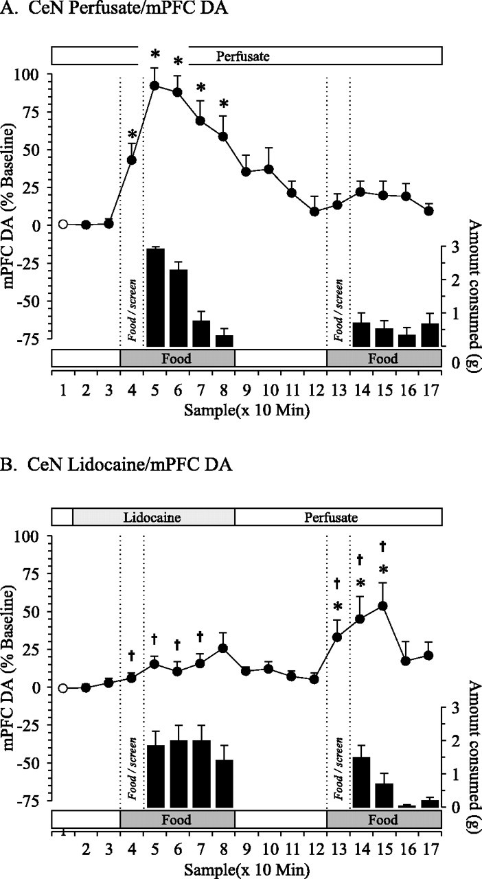
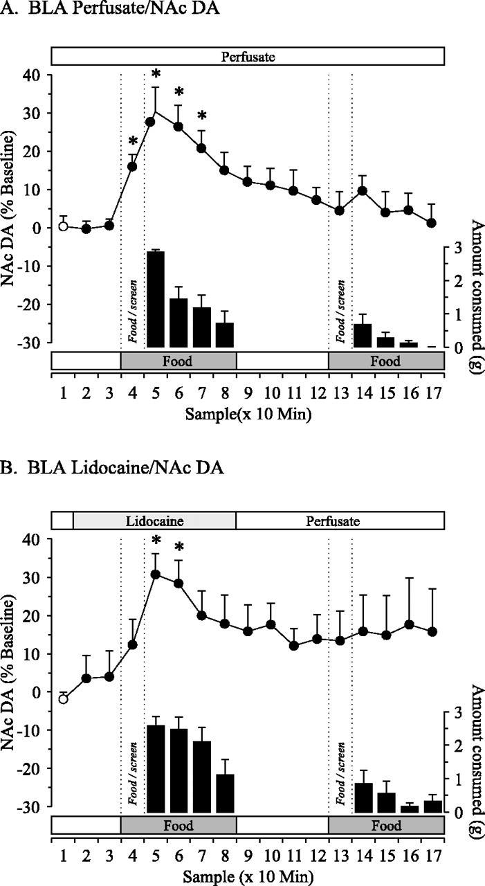
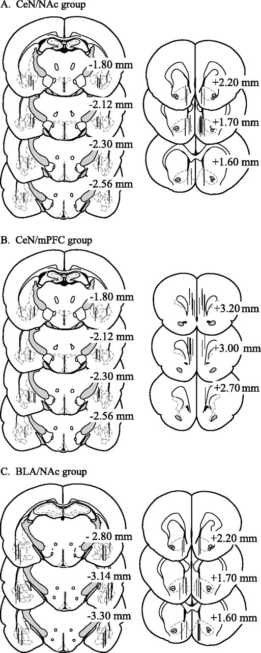
Current studies raise the possibility that subregions within the amygdala may interact with the mesocorticolimbic dopamine (DA) system to subserve specific psychological processes underlying food reward. The present study compared the effect of reversible inactivation of the central nucleus (CeN) versus the basolateral amygdala (BLA) on DA efflux in the nucleus accumbens (NAc) and medial prefrontal cortex (mPFC) in hungry rats that were tested in a food-devaluation procedure. During DA microdialysis experiments, lidocaine, a sodium channel blocker, was delivered via reverse dialysis into the CeN or BLA while rats were given two consecutive meals of Froot Loops. Loss of CeN function impaired the development of satiety during an initial meal and, consequently, diminished the effect of devaluation by satiety on intake of the same food during a second meal. Inactivation of the CeN was also associated with decreased basal levels of DA efflux in the NAc before food intake and attenuated increases in DA efflux related to anticipatory and consummatory aspects of feeding in both the NAc and mPFC. In contrast, inactivation of the BLA did not affect feeding behavior or DA efflux. Overall, these findings indicate that the CeN and BLA independently modulate DA transmission in both terminal regions. It is proposed that interaction between the CeN and mesocorticolimbic DA activity may be a mechanism by which hunger and satiety signals influence the value of food reward, or alternatively, a mechanism by which memory for a recently consumed food regulates food intake.
Figures

References
-
- Aggleton JP, Passingham RE. An assessment of the reinforcing properties of foods after amygdaloid lesions in rhesus monkeys. J Comp Phyiol Psychol. 1982;96:71–77. - PubMed
-
- Ahn S, Phillips AG (2002) Independent modulation of basal and feeding-evoked dopamine efflux in the nucleus accumbens and medial prefrontal cortex by the central and basolateral amygdalar nuclei in the rat. Neuroscience, in press. - PubMed
-
- Bakshi VP, Kelley AE. Sensitization and conditioning of feeding following multiple morphine microinjections into the nucleus accumbens. Brain Res. 1994;648:342–346. - PubMed
Publication types
MeSH terms
Substances
LinkOut - more resources
Full Text Sources
Other Literature Sources