. 2002 Dec 15;22(24):10966-75.
doi: 10.1523/JNEUROSCI.22-24-10966.2002.

Feedforward mechanisms of excitatory and inhibitory cortical receptive fields

Affiliations

Feedforward mechanisms of excitatory and inhibitory cortical receptive fields

Randy M Bruno et al. J Neurosci. .

Abstract

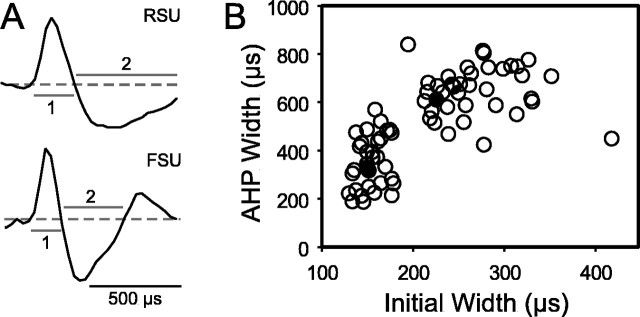
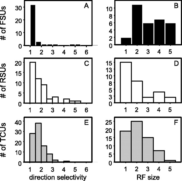
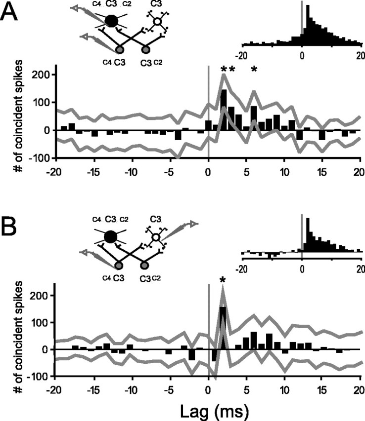
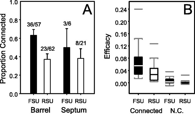
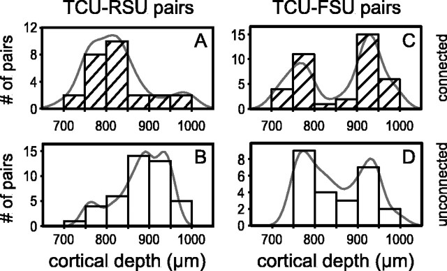
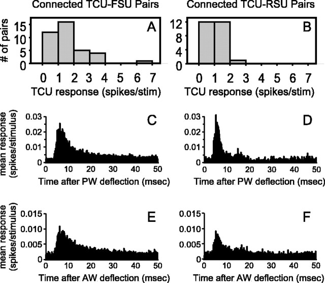
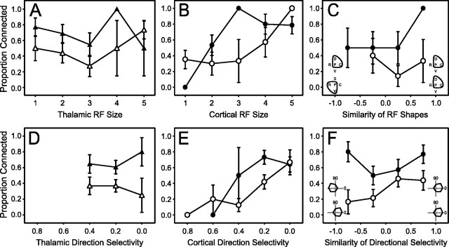
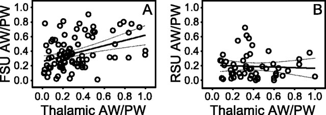
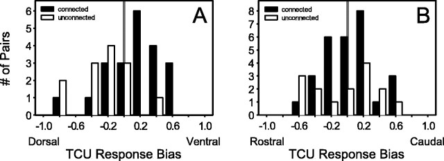
Excitatory and inhibitory cortical layer IV neurons have distinctive response properties. Thalamocortical connectivity that may underlie differences was examined using cross-correlation analyses of pairs of thalamic and cortical neurons in the rat whisker/barrel system. Cortical layer IV cells discharging fast spikes, presumed inhibitory neurons, were distinguished from regular-spike units, presumed excitatory neurons, by the extracellular waveform shape. Regular-spike neurons fired less robustly and had smaller receptive fields (RFs) and greater directional tuning than fast-spike cells. Presumed excitatory neurons were less likely to receive thalamocortical connections, and their connections were, on average, weaker. RF properties of thalamic inputs to both cell types were equivalent, except that the most highly responsive thalamic cells contacted only fast-spike neurons. In contrast, the size and directional tuning of cortical RFs were related to the number of detectable thalamocortical inputs. Connected thalamocortical pairs were likely to have matching RF characteristics. The smaller, more directionally selective RFs of excitatory neurons may be a consequence of their weaker net thalamic drive, their more nonlinear firing characteristics and pervasive feedforward inhibition provided by strongly driven, broadly tuned inhibitory neurons.


Figures

Fig. 1.
Fig. 1.
Cortical cell type can be inferred from extracellular waveform shape. A, Example waveforms of an RSU and an FSU. Dashed line indicates baseline;1 and 2 denote initial and secondary phases. B, Scatterplot of width measurements of initial and secondary phases. Filled circles, examples shown inA. AHP, Afterhyperpolarization.
Fig. 2.
Fig. 2.
RF properties of FSUs (A,B), RSUs (C,D), and TCUs (E,F). A, C, E, Histograms of the direction selectivity index (response to whisker deflection at angle evoking the maximum number of spikes per average response over all eight angles). B, D, F, Histograms of number of whiskers evoking a response significantly greater than spontaneous activity.
Fig. 3.
Fig. 3.
Representative and average cross-correlograms of thalamocortical pairs. A, Representative stimulus-corrected cross-correlogram for a connected TCU–FSU pair.x-axis, Milliseconds by which cortical spikes follow (positive lags) or precede (negative lags) reference thalamic spikes.y-axis, Number of coincident spikes relative to baseline (see Materials and Methods). Gray lines indicate 99% confidence limits for individual bins. In this example, the +2, +3, and +6 bins (asterisks) are significantly >0 (baseline).Npre = 2427 spikes;Npost = 9412 spikes. Left inset, Experimental design and hypothetical circuit showing multi-whisker thalamic neurons (gray), an inhibitory cortical neuron with a larger RF (black), and an excitatory neuron with a smaller RF (white).Right inset, Average of peak-normalized cross-correlograms of all putatively connected TCU–FSU pairs.B, Same as A, but for a connected TCU–RSU pair (Npre = 11,090 spikes;Npost = 8036 spikes) and the average of all TCU–RSU pairs.
Fig. 4.
Fig. 4.
RSUs receive both less numerous and weaker thalamocortical connections than do FSUs. A, Summary of the proportions of connected pairs observed for each anatomical category studied (see Results). Error bars indicate SEs of the proportion. B, Standard box plots of the efficacy of pairs of thalamic and barrel FSUs and RSUs found to be connected and not connected (N.C.). Boxes indicate 25–75 percentile ranges of the distributions; lines through boxes indicate medians; vertical lines indicate tails of distributions; isolated horizontal linesindicates outliers.
Fig. 5.
Fig. 5.
Connected TCU–RSU pairs tend to involve RSUs located superficially in layer IV. Shown are histograms of microdrive depth readings for connected (A, C) and unconnected (B,D) pairs involving RSUs (A,B) and FSUs (C,D).
Fig. 6.
Fig. 6.
RSUs but not FSUs receive inputs exclusively from thalamic neurons with low evoked firing rates. A,B, Distributions of responses to PW deflections for thalamic neurons found to be connected to FSUs (A) and RSUs (B).C–F, Average PSTHs of thalamic responses to PW (C,D) and AW (E,F) deflections for FSUs (C,E) and RSUs (D,F). Bin width is 100 μsec.
Fig. 7.
Fig. 7.
RF size and direction selectivity of cortical layer IV neurons reflect the amount of nonspecific convergence of thalamocortical input. A–F, open symbols, TCU–RSU pairs; filled symbols, TCU–FSU pairs. Error bars indicate SEs of the proportion. Connected proportion of pairs as a function of A, the RF size (number of responsive whiskers) of the thalamic neuron.B, The RF size of the cortical neuron. C, The similarity of thalamic and cortical RF shapes withinsets schematically depicting examples of perfectly nonmatching (left) and matching (right) thalamic and cortical RF shapes. P, Principal whisker;C, D, R, and V, caudal, dorsal, rostral, and ventral AWs, respectively; lines, more effective whiskers. D, The directional tuning (in log units) of the response of the thalamic neuron to PW deflections.E, The directional tuning (in log units) of the responses of the cortical neuron to PW deflections. F, The similarity (see Results) of the responses of a thalamocortical pair to PW deflections in each of eight directions, withinsets schematically depicting examples of anticorrelated (left) and correlated (right) response profiles. 0, Caudal deflections; 90, dorsal deflections. D, E, Although moderately directionally selective, TCUs have a more limited range of tuning than RSUs. Likewise, tuned FSUs exist, but not in the extreme. Consequently, some datapoints do not exist for TCUs and FSUs.
Fig. 8.
Fig. 8.
RFs of inhibitory but not excitatory barrel neurons have shapes similar to those of their thalamic inputs.A, Relationship of AW/PW response ratios for connected pairs of TCUs and FSUs. The AW/PW ratio is defined as the mean response of a neuron to the deflection of an AW over the mean response to deflection of its PW. B, Same as A, but for connected pairs of TCUs and RSUs. A,B, solid line, Fitted linear regression;dottedlines, 95% confidence limits. Note that both FSUs and RSUs are contacted by TCUs with focused (small ratio) and unfocused (large ratio) RFs.
Fig. 9.
Fig. 9.
Thalamic neurons with spatial response biases are more likely to contact FSUs. A, Histograms of dorsoventral response biases of TCUs classified as connected (filled columns) and unconnected (open columns). Cells responding only to dorsal AW deflections have a bias of −1.0; those responding only to ventral AW deflections have a bias of +1.0. Biases of 0 indicate no difference between dorsal and ventral AW deflections. B, Same as A, but for the rostrocaudal axis.

Similar articles

Cited by

References

    1. Agmon A, Connors BW. Thalamocortical responses of mouse somatosensory (barrel) cortex in vitro. Neuroscience. 1991;41:365–379. - PubMed
    1. Agmon A, Connors BW. Correlation between intrinsic firing patterns and thalamocortical synaptic responses of neurons in mouse barrel cortex. J Neurosci. 1992;12:319–329. - PMC - PubMed
    1. Agmon A, Yang LT, Jones EG, O'Dowd DK. Topological precision in the thalamic projection to neonatal mouse barrel cortex. J Neurosci. 1995;15:549–561. - PMC - PubMed
    1. Alonso J-M, Martinez LM. Functional connectivity between simple cells and complex cells in cat striate cortex. Nat Neurosci. 1998;1:395–403. - PubMed
    1. Alonso J-M, Usrey WM, Reid RC. Rules of connectivity between geniculate cells and simple cells in cat primary visual cortex. J Neurosci. 2001;21:4002–4015. - PMC - PubMed

Publication types

LinkOut - more resources