. 2002 Dec 15;22(24):11065-79.
doi: 10.1523/JNEUROSCI.22-24-11065.2002.

Electroconvulsive seizure behavior in Drosophila: analysis of the physiological repertoire underlying a stereotyped action pattern in bang-sensitive mutants

Affiliations

Electroconvulsive seizure behavior in Drosophila: analysis of the physiological repertoire underlying a stereotyped action pattern in bang-sensitive mutants

Jisue Lee et al. J Neurosci. .

Abstract

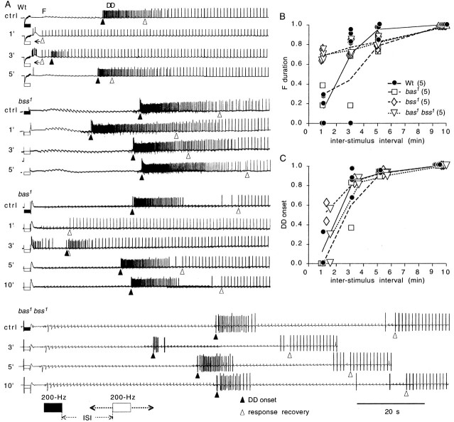
Drosophila bang-sensitive mutants display a remarkable stereotyped behavioral sequence during mechanical disturbances. This seizure repertoire consists of initial and delayed bouts of spasm interposed with paralysis and followed by recovery of activity and a period of refractoriness to further stimulation. Electroconvulsive stimuli across the brain induced a similar seizure behavior in tethered flies, in which corresponding electrophysiological events could be readily recorded in indirect flight muscles [dorsal longitudinal muscles (DLMs)] of the giant fiber (GF) pathway. The DLM physiological repertoire consisted of initial and delayed discharges (IDs and DDs), a response failure and recovery, followed by a refractory period. Interestingly, wild-type flies also displayed the same electroconvulsive repertoire, albeit inducible only at higher stimulus intensities and with briefer expression. The DLM repertoire presumably originated from activities of distinct neural circuits subserving normal function and reflected the general sequence of excitation and depression of the nervous system as a whole, as shown by simultaneous recordings along the different body axes. The well characterized GF pathway facilitated localization of circuits responsible for response failure and ID and DD motor patterns by surgical manipulations, recording-stimulating site analysis, and genetic mosaic studies. A flight pattern generator is most likely the major contributor to shaping the DD pattern, with modifications by active integration of individual motor neurons and associated interneurons. The robust electroconvulsive repertoire of DLMs provides a convenient window for further genetic analysis of the interacting neural mechanisms underlying a stereotyped action pattern in Drosophila, which shows striking parallels with aspects of seizure in mammalian species.


Figures

Fig. 1.
Fig. 1.
The giant fiber (GF) pathway and stimulation protocol for electroconvulsive seizure.A, Electrical stimuli were delivered across the brain or thorax, and physiological responses were recorded at the various outputs of the GF pathway. One of the bilaterally symmetrical GF pathway pairs, which is responsible for jump-and-flight escape reflex, is schematized. The GF neuron in the brain activates a TTMn (jump) directly but recruits DLMns (flight) via the PSI interneuron in the thorax. Three different types of identified synapses (glutaminergic, cholinergic,electrical) are indicated. GF, Giant fiber; PSI, peripherally synapsing interneuron,DLMaf, a stack of six dorsal longitudinal muscle fibers; DLMn, DLM motor neuron;TTM, tergotrochanteral muscle; TTMn, TTM motor neuron. B, Electroconvulsive stimulation protocol. Electroconvulsive stimuli (200 Hz of 0.1 msec pulses) were followed by test pulses (1 Hz of 0.1 msec pulses) to detect transmission failure in the GF pathway during seizure. Stimulus strength of the 200 Hz stimulus train could vary in voltage (V) or duration (t).
Fig. 2.
Fig. 2.
Mechanically induced seizure in the bang-sensitivebas, bss, and bas bssflies. A, Seizure repertoire in bssmutants. Mechanical shock (10 sec vortexing; see Materials and Methods) induced a stereotyped behavioral sequence of initial spasm, paralysis, delayed spasm, and recovery of normal posture in both male (top panels) and female (bottom panels) mutant flies. Seizing females often lay eggs (arrowheads). The initial spasm, which occurred during the first few seconds after vortexing, was not captured in videotaping. B, Duration of seizure induced by vortexing vials containing five or fewer flies. Onset time of delayed spasm (hatched bar) and time to recovery (open bar) are quantified on the basis of thenumbers of flies indicated. C, Seizure susceptibility in different genotypes as indicated by percentage of flies displaying the seizure repertoire after vortexing. In this and the following figures, error bars indicate SD. **p< 0.01; ***p < 0.001.
Fig. 3.
Fig. 3.
Electroconvulsively induced seizure behavior and its physiological correlates in the DLM flight muscle of abss1 fly. A, Electroconvulsive stimulation applied to a tethered fly induced a behavioral repertoire of initial spasm, paralysis, delayed spasm, and recovery to normal posture. B, The same electroconvulsive stimulation in A induced a physiological repertoire of initial discharge (ID), which occurred during initial spasm, response failure (F), occurring during paralysis, delayed discharge (DD), during delayed spasm, and response recovery (R), concurring with recovery of normal posture in a DLM. In this and the following figures, the filled triangle indicates onset of DD, and the open triangle indicates R (recovery of consecutive responses) in traces of physiological recording.
Fig. 4.
Fig. 4.
The DLM physiological repertoire and induction threshold in wild-type and bang-sensitive mutants.A, Wild type (Wt),bss1,bas1, andbas1bss1 all showed the same stereotyped sequence of initial discharge (ID), response failure (F), delayed discharge (DD), and recovery (R) in the DLM. Note response failures (indicated by stimulus artifacts of 1 Hz test pulses) before recovery (open triangles) of the DLM action potentials (see Results). B, Profiles of the physiological repertoires in the bang-sensitive mutants. ID, DD, and F for each genotype were characterized based on the number of flies indicated. Time periods of DLM response failure (F) and recovery of GF pathway transmission (R) are indicated. For this figure and Figure 6, statistics are pooled from different alleles for single mutants (bas:bas1,bas2; bss:bss1,bss2), and the double-mutant data are based on the bas1bss1 combination. C, Duration-voltage relationship of stimulus trains for electroconvulsive seizure induction. Increasing the 200 Hz pulse train duration lowered the adequate voltage for induction of the DLM repertoire. Data points represent threshold levels of 50% success. One fly contributed only one test result at each intensity. The ranges of sample sizes for data points of each genotype are indicated. At stimulus duration of ≤0.5 sec, seizure induction in wild-type flies was not attainable (C), and the voltage required for seizure induction correlated with the sensitivity to mechanical stimulation (compare with Fig. 2C).
Fig. 5.
Fig. 5.
General nerve activities evoked by electroconvulsive brain stimulation in wild-type andbss1 flies. A,B, Correlation of DLM activities with differential recording between the electrodes positioned in the head and abdomen in wild-type (A) andbss1 (B) flies. These paired recordings demonstrated activity suppression and bursting events in the CNS that corresponded to DLM response failure (F) and discharges (ID,DD). However, individual spikes in the DLM were not coincidental with those in head–abdomen recordings (segmentsad are expanded 20× for temporal comparison; a, b, for wild-type;c, d, forbss1 flies). C, Signal trafficking between the head and thorax detected in the cervical connectives correlated with simultaneous DLM recording after an electroconvulsive stimulus in a wild-type fly (e, 20×).DLM, Muscle recording;h-a, differential recording between head and abdomen; cer, cervical connective recording.
Fig. 6.
Fig. 6.
Electroconvulsion stimulus intensity-response relationship in wild-type and bang-sensitive mutant flies.A, Stimulus intensity-dependent expression of the DLM physiological repertoire in a wild-type fly. ID evoked by 200 Hz, 2 sec stimulus trains showed a nonlinear dependence on stimulus intensity, reaching a maximum ∼70 V but disappearing at 100 V. In contrast, F and DD were lengthened at increasing stimulus intensity, reaching a plateau above 70 V. B–D, Induction probabilities of ID, F, and DD at 50 V with varying stimulus train durations. Probabilities of induction were determined by the fraction of flies displaying ID, F, and DD. The range of the sample size is indicated, with each fly subjected to only a single trial at each stimulus duration. Maximal ID induction was shifted toward lower stimulus intensities in the mutant flies (B). Single-mutant bss and double-mutant bas1bss1 flies exhibited greatly enhanced sensitivity for F induction (C) but only mild alterations in DD induction probability (D). Note that induction curves for ID, F, and DD displayed different profiles within each genotype, suggesting involvement of distinct neural circuits.
Fig. 7.
Fig. 7.
Stereotyped action patterns in the DLM physiological repertoire of wild-type,bas1,bss1, andbas1bss1 flies. A DLM repertoire once initiated completed the sequence even when interrupted by a second stimulus. Two identical suprathreshold stimuli (200 Hz, 2 sec, except for bas1bss1, which was 1 sec; note that ID was suppressed by the suprathreshold stimuli) were delivered at different ISIs. A second stimulus (open rectangle) delivered before the onset of DD produced no change in the DLM repertoire initiated by the first (filled rectangle), whereas deliveries after the onset of DD introduced a brief suppression of DD and a slight delay in response recovery (compare second and third traces with control in the top trace in each panel). Traces shown are sequential recordings from the same fly with at least 10 min rests between trials.
Fig. 8.
Fig. 8.
Refractoriness of the DLM repertoire in wild-type and bang-sensitive mutant flies. A, Refractoriness indicated by reduced effectiveness of the second electroconvulsive stimulus. F and DD in the responses to the second stimuli were either modified because of relative refractoriness or missing because of absolute refractoriness. In each paired-stimulation trial, the first stimulus (200 Hz, 2 sec; filled rectangle) was delivered to a fly rested for at least 10 min, and the second stimulus (open rectangle) followed at different ISIs. In the sequential pair-stimulus trials (at 1, 3, 5, or 10 min ISI, executed in a reversal order in the same fly), the top trace for each genotype (Wt, bas1,bss1, andbas1bss1) represents control (ctrl) taken from one of the first responses in the pairs. Note the reappearance of ID in Wt andbas1 traces during the relative refractory period, which was suppressed by the suprathreshold stimuli in control traces. B, C, Restoration of response during refractory periods as indicated by recovery of F duration (B) and DD onset time (C), determined in the number of flies indicated. At each ISI, recovery is normalized to control values with line segments connecting the average values for each genotype.
Fig. 9.
Fig. 9.
Effects of GABAergic blockade on spontaneous activity and electroconvulsive repertoire of DLMs. Wild-type flies were fed with 1 mm picrotoxin (ptx-fed) in food dye-colored medium. A, DLMs of control flies (fed with medium without drug) could express three distinct activities: sporadic spontaneous bursts (S) during the rest period, sustained flight activity (Fl), and the electroconvulsive repertoire (E).B, Picrotoxin-fed flies exhibited periodic discharges (S) in the DLM and failure of transmission in the GF pathway before experiencing an electroconvulsive shock (data not shown). The periodic bursts appeared to occlude electroconvulsive induction of ID and DD (compare traces E in A andB), suggesting that GABAergic blockade induces another seizure-like state. Suprathreshold stimuli (100 V, 200 Hz for 2 sec) were applied immediately before traces E in bothA and B. An arrow next to the open triangle indicates that no recovery from GF pathway failure (F) was observed throughout the recording.
Fig. 10.
Fig. 10.
Localization of DD pattern generation to the thorax and F induction along the GF pathway. A,B, Intact and decapitated (decap) preparations were examined for the effect of direct thorax (thx) electroconvulsion (second andthird traces) bypassing the brain, as compared with the standard brain (br) electroconvulsion (top traces) in wild-type (A) andbss1 mutant (B) flies. Thorax electroconvulsive stimulation (200 Hz, 2 sec, suprathreshold) could induce characteristic DD discharges of the DLM repertoire with or without the head, suggesting the thoracic localization of DD pattern generator(s). Test pulses (1 Hz) applied to the thorax after thorax electroconvulsion detected no occurrence of F in DLMs, indicating direct activation of motor neurons by test pulses.C, Both brain and thorax electroconvulsion induced failure in DLM response to test pulses delivered to the brain (first and third traces), but not to the thorax (second trace), suggesting multiple sites along the GF–PSI pathway, but excluding the motor neurons, as the sites of transmission failure. Open triangles withbackward-directed arrows indicate the lapse of manual switching time before the onset of test pulses.
Fig. 11.
Fig. 11.
Electroconvulsive repertoires in different motor outputs of the GF pathway in wild-type andbas1bss1 double-mutant flies. Theinset shows a schematic diagram of one half of the bilaterally symmetrical GF pathway (see Materials and Methods). Cells with somata on the left (l) side are filled, and those on the right (r) are open. Isomorphic representations of the GF pathway shown next to individual traces indicate the left- or right-side locations of the neuronal somata and muscle fibers involved. A, Discharges in DLM pairs driven by the same versus different motor neurons. A pair of DLM fibers (lDLMa and lDLMb) that share input from the same motor neuron showed synchronized spike firing (left panel). In contrast, firing in lDLMa and lDLMc, which are driven by separate motor neurons, lacked synchronicity despite their similar temporal characteristics of ID, DD, and R (right panel). This indicates that motor neuron activity rather than muscle excitability determines the electroconvulsive spike discharge patterns. B,C, Electroconvulsive responses of bilateral muscle pairs in wild-type (B) and bas bssdouble-mutant (C) flies. Both ID and DD were missing in TTM responses, indicating specificity of the ID and DD pattern generators for DLMs. TTM recovery was faster than that of DLMs, demonstrating a delay imposed by the mutant PSI (comparelDLMarTTM in C). Note also the temporal coupling between TTM recovery and DLM DD onset (lDLMarTTM pairs), coinciding with the resumption of general CNS activities (compare Fig. 5). Left and right pairs of DLM fibers (lDLM andrDLM), although receiving input from two bilateral GF–PSI chains, showed similar temporal sequence of ID, DD, and R in both wild-type (B) and mutant (C) flies (simultaneous recordings, each pair from a different fly).
Fig. 12.
Fig. 12.
Mutational effects on the identified neurons and muscles of the GF pathway in bas1bss1 bilateral mosaic flies.A, The inset shows one GF pathway of the bilaterally symmetrical pair (compare Fig. 10, inset) with “control” nerve or muscle elements (filled) on the left(ipsi) side and “mutant” elements (open) on the right(contra) side. Also shown is a schematic bilateral mosaic with the phenotypically control (bas1bss1/+ +) and mutant (bas1bss1/O) tissue on theleft and right, respectively. The predicted genotypes of GF and PSI, and DLM, TTM, and their motor neurons (mn) are determined by whether the soma is located on the ipsilateral (i) control or contralateral (c) mutant side of the body, as shown in the relevant portion of the GF pathway for DLM and TTM inputs next to individual traces. Suprathreshold electroconvulsive stimuli (filled bars) were applied to compare modifications in the DLM physiological repertoire for DLMs on the mutant side [DLMa (trace 3,cDLMa: no mutant neuron in the pathway) vsDLMc (trace 4, cDLMc: one mutant neuron)] and on the control side [DLMc(trace 5, iDLMc: two mutant neurons) vsDLMa (trace 6, iDLMa: three mutant neurons)]. Note that the number of mutant neurons along the GF pathway correlates with DLM recovery time (open triangle). In contrast, bilateral TTMs showed similar recovery regardless of the number of mutant neurons (trace 1,iTTM: all normal cells; trace 2,cTTM: all mutant cells), suggesting bilateral coupling for the GF pathways. Note also the similar timing of DLM DD onset and TTM response recovery on both sides regardless of the number of mutant elements along the GF pathway (traces 1–6; compare Fig.10). B, C, Consistent results were obtained from four bas1bss1 bilateral mosaics. Separate symbols were used to distinguish data from different mosaic flies. F duration of DLM recovery lengthened with increased numbers of predicted mutant neurons in the GF pathway (B). In contrast, DD onset time was independent of the number of mutant neurons in the GF–PSI–mn chain (C).

Similar articles

Cited by

References

    1. Baird DH, Koto M, Wyman RJ. Dendritic reduction in Passover, a Drosophila mutant with a defective giant fiber neuronal pathway. J Neurobiol. 1993;24:971–984. - PubMed
    1. Benzer S. From the gene to behavior. JAMA. 1971;218:1015–1022. - PubMed
    1. Blagburn JM, Alexopoulos H, Davies JA, Bacon JP. Null mutation in shaking-B eliminates electrical, but not chemical, synapses in the Drosophila giant fiber system: a structural study. J Comp Neurol. 1999;404:449–458. - PubMed
    1. Burg MG. PhD thesis. University of Iowa; 1987. Genetic and mosaic analysis of mutations which alter nerve and muscle excitability in Drosophila melanogaster: effects on development and behavior.
    1. Burg MG, Wu C-F. A class of Drosophila behavioral mutants that are sensitive to both mechanical vibrations and temperature conditions. Soc Neurosci Abstr. 1987;13:619.

Publication types

MeSH terms

Substances

LinkOut - more resources