. 2002 Dec 24;99(26):16543-50.
doi: 10.1073/pnas.012669599. Epub 2002 Dec 16.

Effects of strand and directional asymmetry on base-base coupling and charge transfer in double-helical DNA

Affiliations

Effects of strand and directional asymmetry on base-base coupling and charge transfer in double-helical DNA

Melanie A O'Neill et al. Proc Natl Acad Sci U S A. .

Abstract

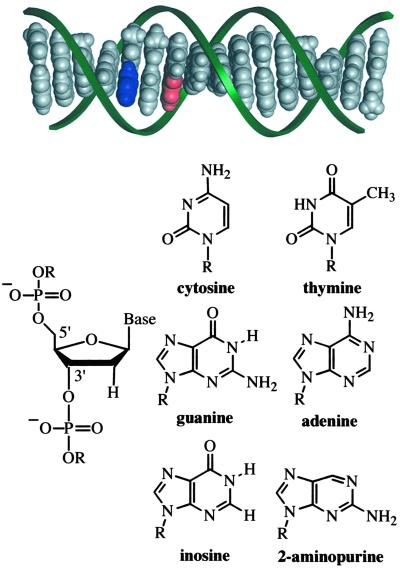
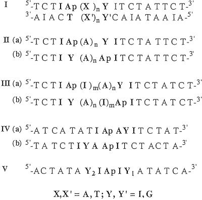
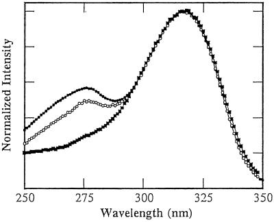
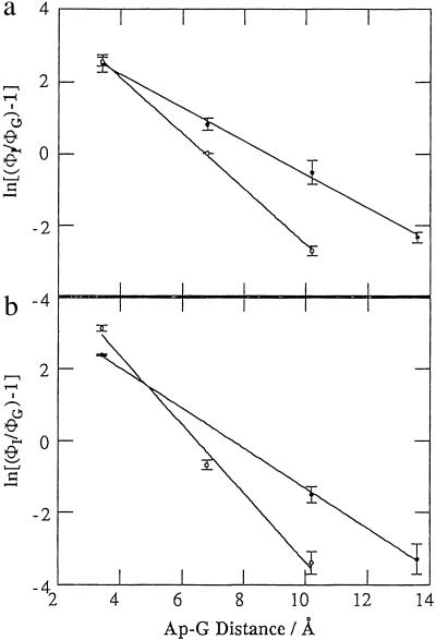
Mechanistic models of charge transfer (CT) in macromolecules often focus on CT energetics and distance as the chief parameters governing CT rates and efficiencies. However, in DNA, features unique to the DNA molecule, in particular, the structure and dynamics of the DNA base stack, also have a dramatic impact on CT. Here we probe the influence of subtle structural variations on base-base CT within a DNA duplex by examining photoinduced quenching of 2-aminopurine (Ap) as a result of hole transfer (HT) to guanine (G). Photoexcited Ap is used as a dual reporter of variations in base stacking and CT efficiency. Significantly, the unique features of DNA, including the strandedness and directional asymmetry of the double helix, play a defining role in CT efficiency. For an (AT)n bridge, the orientation of the base pairs is critical; the yield of intrastrand HT is markedly higher through (A)n compared with (T)n bridges, whereas HT via intrastrand pathways is more efficient than through interstrand pathways. Remarkably, for reactions through the same DNA bridge, over the same distance, and with the same driving force, HT from photoexcited Ap to G in the 5' to 3' direction is more efficient and less dependent on distance than HT from 3' to 5'. We attribute these differences in HT efficiency to variations in base-base coupling within the DNA assemblies. Thus base-base coupling is a critical parameter in DNA CT and strongly depends on subtle structural nuances of duplex DNA.


Figures

Fig 1.
Fig 1.
Idealized model (insight ii) of a B-DNA duplex (5′-TCTIApAGITCTATTCT-3′ and complement) and structures of the molecular constituents. The aromatic stack of DNA bases, shown in gray, blue (Ap), and red (guanine, G), is distinctly visible within the sugar phosphate backbone (green ribbons). The connection of deoxyribose sugar units via phosphate groups at distinct 5′ and 3′ positions imparts the DNA strand with directional asymmetry.
Fig 2.
Fig 2.
Sequences of DNA duplexes designed to examine the influence of base–base coupling on DNA CT. Intrastrand and interstrand coupling involving adenine and thymine bridges is investigated by using series I duplexes (X,X′ = A, T; Y,Y′ = I, G, C). The duplexes of series II—V (complements not shown) are designed to probe the influence of base-step direction on base–base coupling and CT in DNA (Y = I, G). The adenine analog, 2-Ap, is paired with thymine, whereas the guanine analog, inosine, is paired with cytosine.
Fig 3.
Fig 3.
Excitation spectra of Ap in DNA duplexes demonstrating the influence of sequence and base-step direction on base–base energy transfer and stacking interactions. The following duplexes (5 μM duplex in 100 mM sodium phosphate buffer, pH 7, at 5°C) are shown: series IIa, 5′-TCTIApAAAIITCTTCT-3′ (•); series IIb, 5′-TCTIIAAAApTCTTCT-3′ (○); and series IIIa, 5′-TCTCIApIAAIITCTTCT (▪).
Fig 4.
Fig 4.
Variation in the yield of intrastrand CT between Ap* and G as a function of distance determined from steady-state fluorescence measurements of redox-active (ΦG) and redox-inactive (ΦI) duplexes (ln[(ΦIg − 1)] = −γr): (a) series II duplexes, IIa (•), γ = 0.46(2) and IIb (○), γ = 0.77(1); (b) series III duplexes, IIIa (•), γ = 0.56(1) and IIIb (○), γ = 0.95(9).
Fig 5.
Fig 5.
Qualitative representation of HOMOs of 5′ApG-3′ versus 5′-GAp-3′ (a) and 5′ApA-3′ versus 5′-AAp-3′ (b) steps. The extent of orbital overlap and delocalization is notably greater for the 5′ApG-3′ and 5′-ApA-3′ steps. The HOMOs were calculated by using HYPERCHEM 5.1 (restricted Hartree–Fock level with 3-21G* basis set). The atomic coordinates of the bases were obtained from insight ii based on standard B-DNA geometry and the sugar-phosphate backbones were replaced with a methyl group.

Similar articles

Cited by

References

    1. Dekker C. & Ratner, M. A. (2001) Phys. World 14, 29-33.
    1. Boon E. M. & Barton, J. K. (2002) Curr. Opin. Struct. Biol. 12, 320-329. - PubMed
    1. Giese B. (2002) Annu. Rev. Biochem. 71, 51-70. - PubMed
    1. Schuster G. B. (2000) Acc. Chem. Res. 33, 253-260. - PubMed
    1. Lewis F. D., Letsinger, R. L. & Wasielewski, M. R. (2001) Acc. Chem. Res. 34, 159-170. - PubMed

Publication types

LinkOut - more resources