. 2002 Dec 24;99(26):17055-9.
doi: 10.1073/pnas.232588899. Epub 2002 Dec 16.

Sensitization of regulated exocytosis by protein kinase C

Affiliations

Sensitization of regulated exocytosis by protein kinase C

Hongliang Zhu et al. Proc Natl Acad Sci U S A. .

Abstract

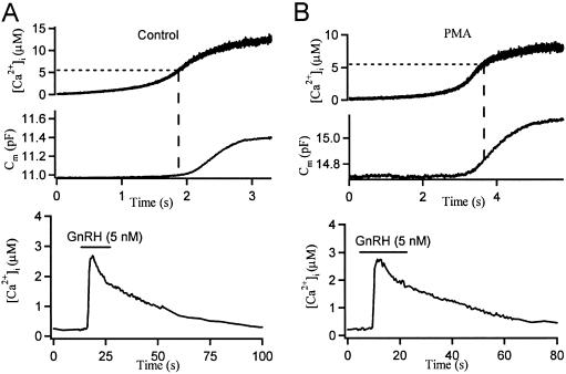
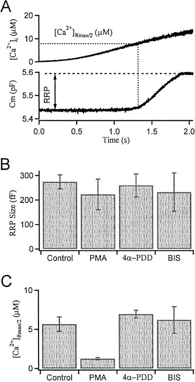
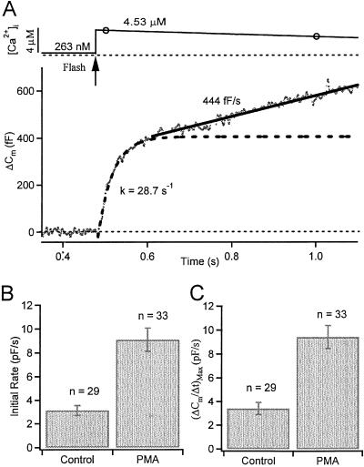
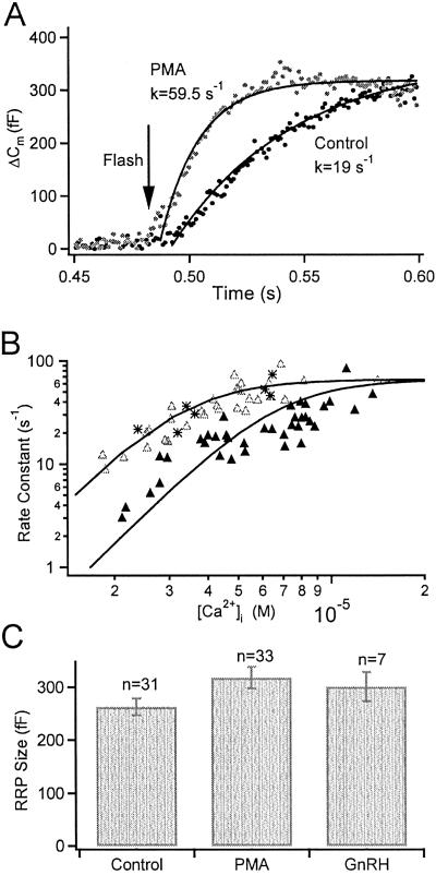
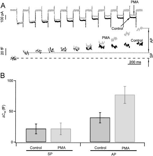
Activation of protein kinase C (PKC) increases vesicular secretion in many cell types. We determined the calcium dependence of secretion and the size of the readily releasable pool of secretory granules in pituitary gonadotropes by photorelease of caged-calcium. The calcium affinity for exocytosis was roughly doubled by activation of PKC by a phorbol ester, whereas the size of the readily releasable pool was not greatly increased. The effect was due to activation of PKC, because it was blocked by a PKC inhibitor and was not mimicked by an inactive phorbol ester analogue. A similar increase in calcium sensitivity was induced by preincubation with gonadotropin-releasing hormone, the physiological releasing hormone. These findings provide direct evidence for physiological regulation of secretion by enhancement of Ca2+-sensing steps. Because exocytosis depends on the third- to fourth-power of intracellular free Ca2+ concentration, this mechanism ensures a powerful up-regulation of hormone release and may explain how PKC can stimulate exocytosis without an increase of Ca2+ above the resting level.


Figures

Fig 1.
Fig 1.
PMA lowers the calcium threshold for secretion in gonadotropes. Steady UV illumination generates ramp [Ca2+]i increases with intracellular caged Ca2+ and measures [Ca2+]i by using a Ca2+ indicator, fura-6F. Simultaneous time courses of [Ca2+]i and Cm for a single control gonadotrope (A) and for a PMA-treated (for 2–3 min) gonadotrope (B). Note at the same [Ca2+]i level, Cm only begins to increase in the control cell, whereas it already takes off in the PMA-treated cell. At the end of the experiments, the cells were challenged by 5 nM GnRH to verify them as gonadotropes (Lower).
Fig 2.
Fig 2.
Summary of PMA actions on exocytosis in gonadotropes. (A) Simultaneous time courses of [Ca2+]i and Cm for a single control gonadotrope illustrating how the RRP size and the [Ca2+]i threshold are determined by ramp [Ca2+]i experiments. (B) Pretreatments with PMA (100 nM), 4α-PDD (1 μM), and BIS (500 nM) have no significant effect on the RRP size (defined in text as ΔCm). (C) The calcium threshold (measured as [Ca2+]Rmax/2) is lowered by PMA, but not by 4α-PDD or by PMA in combination with BIS.
Fig 3.
Fig 3.
PMA increases the initial rate of exocytosis in gonadotropes. (A) Simultaneous measurements of [Ca2+]i level and membrane area Cm after a UV flash (arrow) uncages Ca2+ within 2 ms. Open circles are measured calcium points (continuously measured every 0.5 s). Cm increases in an early burst (fitted with single exponential with the rate constant, k = 1/τ indicated) followed by a sustained component (fitted with a straight line of the indicated slope). (B and C) PMA increases the initial rate of secretion (measured 20 ms after the flash) and the maximum rate.
Fig 4.
Fig 4.
PMA facilitates exocytosis in gonadotropes by increasing the calcium sensitivity of release without significantly altering RRP size. (A) Averaged Cm responses for control (filled symbols) and PMA (100 nM, open symbols) treated gonadotropes, summarizing experiments that had postflash [Ca2+]i values between 4 and 6 μM. Superimposed curves are single exponential fits with the rate constants indicated. (B) PMA increases the Ca2+ sensitivity of release (all experiments plotted individually). Rate constants for exponential fits of the exocytotic burst are plotted against calcium levels for control (filled triangles), PMA-treated (open triangles, 100 nM for 2–3 min), and GnRH-treated (asterisks, 5 nM for 2 min) gonadotropes, respectively. The continuous curves represent the equation: Rate = R/(1 + (Kd/[Ca2+]i)3). (C) RRP size is unchanged by PMA or GnRH preincubation.
Fig 5.
Fig 5.
PMA augments the size of the asynchronous pool (AP) but not the synchronous pool (SP) of secretory granules. (A) Calcium currents and Cm response to a train of ten 100-ms depolarizing pulses from −70 to 0 mV. Traces displayed are from the same cell, before and 3 min after PMA treatment. The change of Cm in response to the first depolarization defines the SP size and the subsequent increase in Cm defines the AP. PMA increased AP by a factor of 2 (despite a reduction on calcium currents), but left SP unaltered. (B) Summary of the effect of PMA on SP and AP from six cells.

Comment in

References

    1. Chen Y. A., Duvvuri, V., Schulman, H. & Scheller, R. H. (1999) J. Biol. Chem. 274, 26469-26476. - PubMed
    1. Evans G. J. O., Wilkinson, M. C., Graham, M. E., Turner, K. M., Chamberlain, L. H., Burgoyne, R. D. & Morgan, A. (2001) J. Biol. Chem. 276, 47877-47885. - PubMed
    1. Gillis K. D., Mossner, R. & Neher, E. (1996) Neuron 16, 1209-1220. - PubMed
    1. Smith C., Moser, T., Xu, T. & Neher, E. (1998) Neuron 20, 1243-1253. - PubMed
    1. Ashery U., Varoqueaux, F., Voets, T., Betz, A., Thakur, P., Koch, H., Neher, E., Brose, N. & Rettig, J. (2000) EMBO J. 19, 3586-3596. - PMC - PubMed

Publication types

LinkOut - more resources