

doi: 10.1002/prot.10311.
Deep trefoil knot implicated in RNA binding found in an archaebacterial protein
Affiliations
- PMID: 12486711
- PMCID: PMC2792022
- DOI: 10.1002/prot.10311
Item in Clipboard
Deep trefoil knot implicated in RNA binding found in an archaebacterial protein
Proteins.
.
No abstract available
Figures
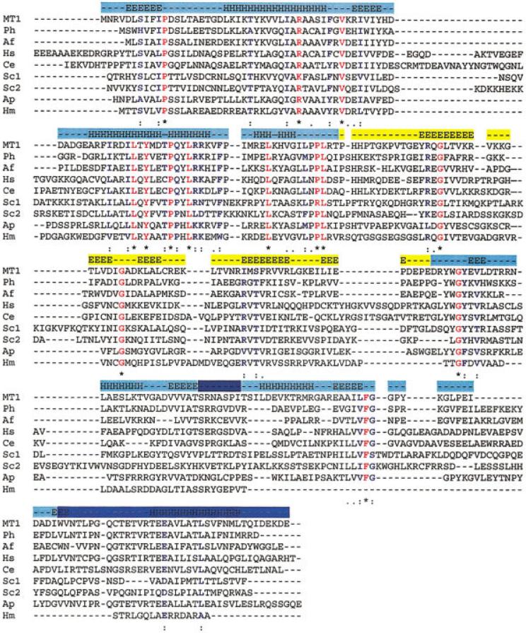
Sequence alignment of MT1 with proteins from a representative set of organisms using the program CLUSTALW. Completely conserved residues are highlighted in red; other conserved residues are highlighted in blue. Secondary structural elements are based on the crystal structure of MT1 and are indicated above the sequences. The half-TIM barrel domain is marked in shades of blue with a knot loop in dark blue and threaded C-terminal sequence in light-blue. The MT1-CSD domain is marked with yellow. The sequences are represented by the initials of the genus and species of its host organism: Ph, Pyrococcus horikoshii; Af, Archaeoglobus fulgidus; Hs, Homo sapiens; Ce, Caenorhabditis elegans; Sc, Saccharomyces cerevisiae; Ap, Aeropyrum pernix; Hm, Haloarcula marismortii. The NCBI Genbank ID numbers (gi) of the included sequences are as follows: MT1, gi|15678032|; Ph, gi|14591536|; Af, gi|11499510|; Hs, gi|5817110|; Ce, gi|1730629|; Sc1, gi|6323970|; Sc2, gi|6321722|; Ap, gi|5103617|; Hm, gi|140984|.

Overall structure of MT1 dimer. Stereoview is along non-crystallographic two-fold axis. Each monomer is separately colored and the knot region is marked in both monomers. The loop is dark blue and the C-terminal sequence threaded through the loop is red. The N- and C-termini are labeled “N” and “C,” respectively. Figure was prepared with WebLabPro.

Secondary structure and topology map of MT1 showing connectivity β-strands and α-helices. All secondary structural elements that are part of the TIM barrel are numbered according to the corresponding elements of MTHFR (see text). The remaining elements (part of the MT1-CSD and linker) are designated with a prime.

Cα trace and stereo view of MT1-DD and MT1-CSD structural overlap. (a) The MT1-DD (red) is shown overlapped with the corresponding domain of MTHFR (green). Corresponding α-helices and β-strands are labeled as α and β, respectively, according to numbering convention for MTHFR. (b) The small domain of MT1 (red) is shown overlapped with 1SRO (green), 1MJC (blue), and 1FJF_Q (orange). Figures were prepared with the program MOLSCRIPT.33 Residues were deleted from proteins where appropriate to simplify the diagram. Refer to text for PDB identifiers.

The MT1 trefoil knot. Residues 1–190 and 199–229 are shown in solvent accessible surface representation (1.4 Å radius). The knot loop (residues 191–198) is in blue and the polypeptide chain threaded through the loop (residues 230–264) is labeled red. Crossover residues (Ser194 and Asn234) as well as Arg191 on the loop, Trp232 and Glu239 on threaded through chain are marked as a reference points. Carboxy terminus is labeled “C”. Figure was prepared with WebLabPro.
References
-
- Wierenga RK. The TIM-barrel fold: a versatile framework for efficient enzymes. FEBS Lett. 2001;492:193–198. - PubMed
-
- Hegyi H, Gerstein M. The relationship between protein structure and function: a comprehensive survey with application to the yeast genome. J Mol Biol. 1999;288:147–164. - PubMed
-
- Hennig M, Schlesier B, Dauter Z, Pfeffer S, Betzel C, Höhne WE, Wilson KS. A TIM barrel protein without enzymatic activity? Crystal-structure of narbonin at 1.8 Å resolution. FEBS Lett. 1992;306:80–84. - PubMed
-
- Farber GK, Petsko GA. The evolution of α/β enzymes. Trends Biochem Sci. 1990;15:228–234. - PubMed
Publication types
MeSH terms
Substances
Associated data
- Actions
- Actions
- Actions
- Actions
Grants and funding
LinkOut - more resources
Full Text Sources
