Comment
. 2002 Dec;110(12):1849-57.
doi: 10.1172/JCI14218.

Increased bone mass is an unexpected phenotype associated with deletion of the calcitonin gene

Affiliations
Comment

Increased bone mass is an unexpected phenotype associated with deletion of the calcitonin gene

Ana O Hoff et al. J Clin Invest. 2002 Dec.

Abstract

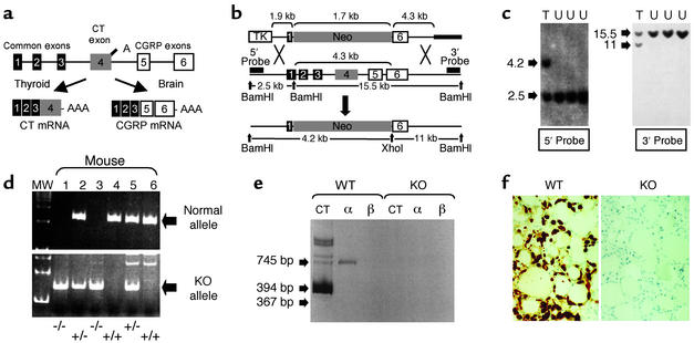
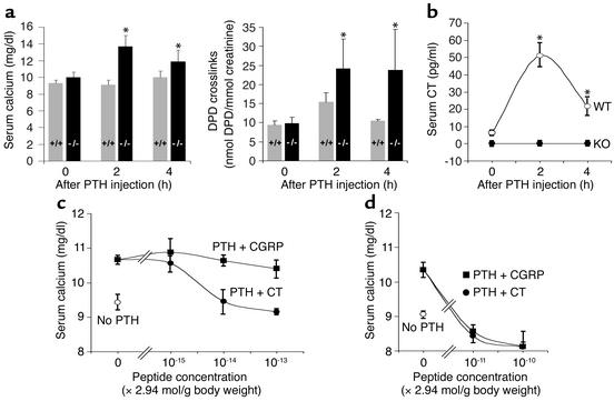
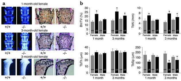
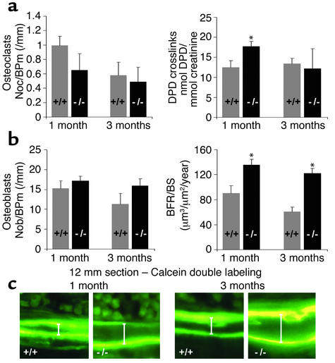
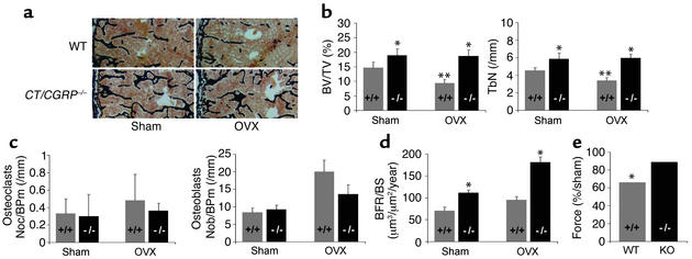
Calcitonin (CT) is a known inhibitor of bone resorption. Calcitonin gene-related peptide-alpha (CGRPalpha), produced by alternative RNA processing of the CT/CGRP gene, has no clearly defined role in bone. To better understand the physiologic role of the CT/CGRP gene we created a mouse in which the coding sequences for both CT and CGRPalpha were deleted by homologous recombination. The CT/CGRP(-/-) knockout (KO) mice procreated normally, there were no identifiable developmental defects at birth, and they had normal baseline calcium-related chemistry values. However, KO animals were more responsive to exogenous human parathyroid hormone as evidenced by a greater increase of the serum calcium concentration and urine deoxypyridinoline crosslinks, an effect reversed by CT and mediated by a greater increase in bone resorption than in controls. Surprisingly, KO mice have significantly greater trabecular bone volume and a 1.5- to 2-fold increase in bone formation at 1 and 3 months of age. This effect appears to be mediated by increased bone formation. In addition, KO mice maintain bone mass following ovariectomy, whereas wild-type mice lose approximately one-third of their bone mass over 2 months. These findings argue for dual roles for CT/CGRP gene products: prevention of bone resorption in hypercalcemic states and a regulatory role in bone formation.


Figures

Figure 1
Figure 1
Creation of mice null for the CT/CGRP gene. (a) Schematic representation of CT/CGRP gene tissue-specific alternative RNA processing and precursor peptide processing. (b) Schematic of recombination of targeting vector with CT/CGRP gene. Black bars indicate general location of DNA probes used for Southern screening. (c) Representative Southern analysis of BamHI/XhoI-digested DNA isolated from doubly resistant embryonic stem cells showing targeted (T) and untargeted (U) clones. A restriction map outlining the origin of individual bands is provided in b. (d) Identification of mouse genotype by PCR analysis of tail-derived DNA (see Methods). PCR product in the upper gel shows the presence of normal WT allele (+/+); the lower gel shows rearranged KO allele (–/–). Both alleles are detected in heterozygous animals (+/–). MW, 100-bp ladder molecular weight marker. (e) RT-PCR analysis of mRNA isolated from the thyroid glands of WT or KO animals (see Methods). Primer pairs used detect mRNAs for CT, CGRPα (α) and CGRPβ (β), which have predicted band sizes of 394 bp, 745 bp, and 367 bp, respectively. (f) Calcitonin immunohistochemical staining of thyroid from WT and KO animals (see Methods).
Figure 2
Figure 2
PTH-stimulated bone resorption in KO mice causes hypercalcemia and is blocked by CT administration. Groups of five WT (gray bars) or KO (black bars) male mice were injected intraperitoneally with human PTH or vehicle (PBS with 0.1 mM HCl and 0.01% BSA) and sacrificed at the indicated times with collection of serum and urine. (a) In KO animals, human PTH stimulates a 4 mg% rise in serum calcium concentration and a doubling of urine DPD during the first 2 hours of the experiment. No significant change of serum calcium or urine DPD was seen in WT animals treated with PTH or vehicle at any timepoint. (b) Measurement of serum CT demonstrated a significant increase in WT animals (open circles) and no detectable CT in KO animals (filled circles). (c) Groups of male mice were injected intramuscularly in the thigh with vehicle, rat CT (10, 100, or 1,000 pg/g mouse body weight) (filled circles), or rat CGRP (11.2, 112, or 1,120 pg/g mouse body weight) (filled squares), followed 3 minutes later by an intraperitoneal injection with human PTH (or vehicle, in the case of the control). Blood was collected 1 hour after the initial intramuscular injection. (d) Experiments were performed as in c but at higher dosage. All experiments included five animals per group with bars representing mean ± SD. *P < 0.05.
Figure 3
Figure 3
KO of the CT/CGRP gene results in increased bone in mice. (a) Radiographic and histologic analysis of vertebrae from 1-month-old and 3-month-old WT and KO mice, and of the proximal tibiae from 3-month-old WT and KO mice. (b) Histomorphometric analysis of trabecular bone volume in vertebrae of WT (gray bars) and KO (black bars) mice. Graphs provide data for trabecular bone volume as a ratio of total bone volume (BV/TV), trabecular number (TbN), trabecular thickness (TbTh), and trabecular spacing (TbSp). Bars represent mean ± SD (n = 5). *Statistically significant difference between WT and KO animals (P < 0.05) as determined by Student t test.
Figure 4
Figure 4
Comparison of bone resorption and formation in female WT and KO animals. (a) Parameters of bone resorption. Histomorphometric analysis of osteoclast number per mm bone perimeter (Noc/BPm) and urinary DPD crosslinks (a marker of bone resorption) was performed in 1- and 3-month-old WT (gray bars) and KO (black bars) mice. (b) Parameters of bone formation. Histomorphometric analysis of osteoblast number per mm bone perimeter (Nob/BPm) (left graph) and in vivo analysis of bone formation (right graph) in 1- and 3-month-old WT and KO mice. The rate of bone formation was determined by in vivo dual calcein labeling; the graphs provide quantification of the bone formation rate per bone surface (BFR/BS). (c) Representative fluorescent micrographs demonstrate the two labeled mineralization fronts, with the arrows indicating growth between labelings. Note the greater distance between the two labeled areas in the KO mice than in the WT animals. All experiments included five animals per group; bars represent mean ± SD. *Statistically significant difference between WT and KO mice (P < 0.05).
Figure 5
Figure 5
The absence of the CT/CGRP gene is protective against estrogen deficiency–mediated bone loss. At 3 months, WT and KO female mice received ovariectomy or a sham procedure. Animals were sacrificed at age 5 months following dual calcein labeling. (a) Histologic analysis of the vertebrae of WT and KO animals after ovariectomy or a sham procedure (see Methods). (b) Static histomorphometric analysis of vertebral bodies from WT (gray bars) and KO (black bars) mice. Graphs provide data for trabecular bone volume as a ratio of total bone volume (BV/TV) and trabecular number (TbN). Significant differences were observed between WT sham and WT OVX animals (**P < 0.01), and between WT and KO animals in both groups (*P < 0.05). (c) Histomorphometric analysis of osteoclast number and osteoblast number per mm bone perimeter. (d) Dynamic histomorphometric analysis of bone formation rate per bone surface in WT and KO mice after ovariectomy or a sham procedure. (e) Biomechanical properties of femora after ovariectomy or a sham procedure. Ovariectomy resulted in decreased force needed to break bone as determined by a three-point bending test (*P < 0.05). All experiments included five animals per group; bars represent mean ± SD.

Comment on

References

    1. Copp DH, Davidson AGF, Cheney BA. Evidence for a new parathyroid hormone which lowers blood calcium. Proc. Canad. Fed. Biol. Soc. 1961;4:17.
    1. Copp DH, Cameron EC, Cheney BA, Davidson AGF, Henze KG. Evidence for calcitonin—a new hormone from the parathyroid that lowers blood calcium. Endocrinology. 1962;70:638–649. - PubMed
    1. Hirsch PF, Gauthier GF, Munson PL. Thyroid hypocalcemic principle and recurrent laryngeal nerve injury as factors affecting response to parathyroidectomy in rats. Endocrinology. 1963;73:244–251. - PubMed
    1. Foster GV, MacIntyre I, Pearse AGE. Calcitonin production and the mitochondrion-rich cells of the dog thyroid. Nature. 1965;203:1029–1031. - PubMed
    1. Foster GV, et al. Thyroid origin of calcitonin. Nature. 1964;202:1303–1305. - PubMed

Publication types

MeSH terms