. 2003 Jan;72(1):48-61.
doi: 10.1086/345379. Epub 2002 Dec 13.

Characterization of genetic miscoding lesions caused by postmortem damage

Affiliations

Characterization of genetic miscoding lesions caused by postmortem damage

M Thomas P Gilbert et al. Am J Hum Genet. 2003 Jan.

Abstract

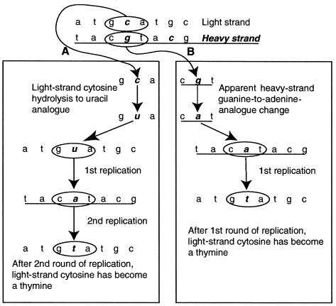
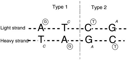
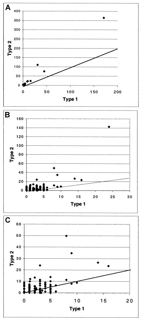
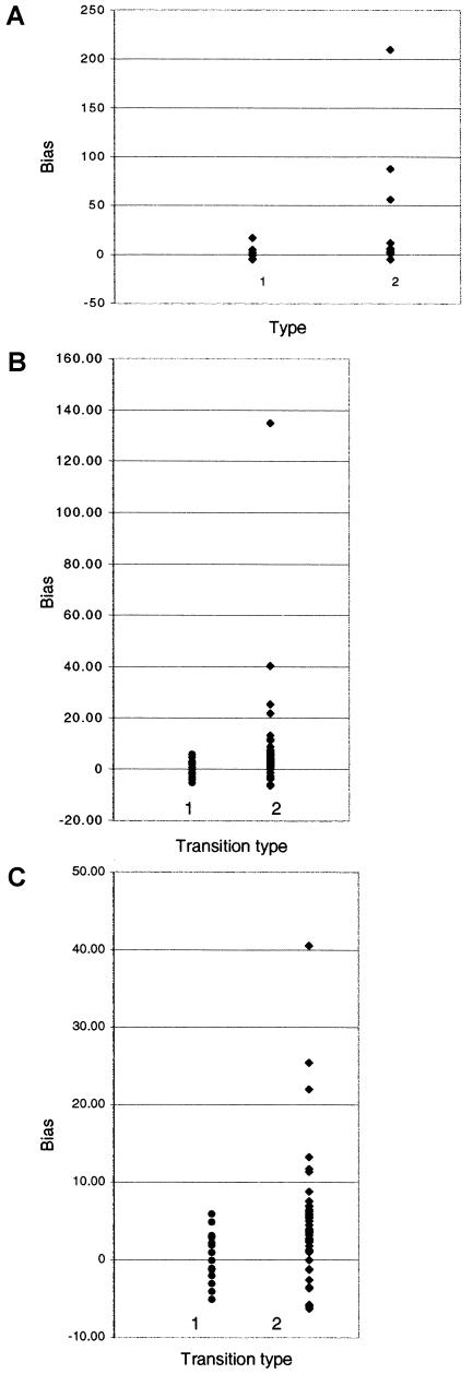
The spectrum of postmortem damage in mitochondrial DNA was analyzed in a large data set of cloned sequences from ancient human specimens. The most common forms of damage observed are two complementary groups of transitions, termed "type 1" (adenine-->guanine/thymine-->cytosine) and "type 2" (cytosine-->thymine/guanine-->adenine). Single-primer extension PCR and enzymatic digestion with uracil-N-glycosylase confirm that each of these groups of transitions result from a single event, the deamination of adenine to hypoxanthine, and cytosine to uracil, respectively. The predominant form of transition-manifested damage varies by sample, though a marked bias toward type 2 is observed with increasing amounts of damage. The two transition types can be used to identify the original strand, light (L) or heavy (H), on which the initial damage event occurred, and this can increase the number of detected jumping-PCR artifacts by up to 80%. No bias toward H-strand-specific damage events is noted within the hypervariable 1 region of human mitochondria, suggesting the rapid postmortem degradation of the secondary displacement (D-loop) H strand. The data also indicate that, as damage increases within a sample, fewer H strands retain the ability to act as templates for enzymatic amplification. Last, a significant correlation between archaeological site and sample-specific level of DNA damage was detected.


Figures

Figure  1
Figure 1
Determination of a strand of origin for postmortem-DNA-damage events by using type 2 (C→T/G→A) transitions as an example. A, L-strand C→T transitions after two cycles of amplifications, resulting in a permanent L-strand change. B, A theoretical H-strand G→A change, producing the L-strand phenotype of C→T change following one cycle of amplification. However, since a direct G→A postmortem modification is chemically impossible, the example depicted in this panel is not possible. Thus, all C→T changes observed on the L strand must have occurred as L-strand C→T postmortem damage, and all G→A changes on the L strand must have occurred as H-strand C→T postmortem damage.
Figure  2
Figure 2
Type 1 and type 2 damage–induced transitions. Circled letters represent the principle modifications observed in cloned sequences (e.g., deamination of C→U [read as T] or A→HX [read as G]). Changes introduced on the complementary strand when the damaged bases are subsequently copied are shown in italics. By convention, sequences are referred to in the L-strand orientation. Therefore, if an amplified sequence was initiated from an original H-strand template, then the type 1 and type 2 errors observed are expected to be T→C and G→A, respectively.
Figure  3
Figure 3
Jumping PCR (Pääbo et al. 1990). Strands i–v represent five sequences obtained from the cloned product of an individual PCR based on one extraction, using a low-error-rate enzyme such as Platinum Taq Hifidelity (Invitrogen). Positions 1–9 represent nucleotide positions that differ between strands, with the altered nucleotide marked above the strand. The shared adenine (a) base on strands i–iv at position 1 helps determine that they derive from one source (though not template molecule) of DNA, with other differences arising due to hydrolytic damage and jumping PCR. Positions 2, 4, and 7 on strands i–iv are base changes resulting from DNA damage. Differences in strand v at positions 1, 3, 5, 6, 8, and 9 identify it as a contaminant. Under the assumption that transitions at identical positions are rare, the shared thymine (t) at position 2 indicates that strands i and ii derived from one template molecule with damage at position 2. The shared adenine (a) base at position 7 on strands ii and iv, in contrast to differences at position 2, indicates jumping PCR between the two strands. Finally, position 9 on strands iii and v represents apparent damage to strand C, arising from jumping with the contaminant strand v.
Figure  4
Figure 4
Type 1 versus type 2 damage. A, Damage per study. All studies demonstrate a type 2 bias. B, Damage per clone region. Although the type 2 bias is significant, many samples demonstrate a type 1 bias. This is seen clearly in panel C, in which the outlier is removed, to give greater resolution.
Figure  5
Figure 5
Damage bias within transition types. Positive Y-axis values represent a bias toward type 1 A→G transitions and type 2 C→T transitions. Negative values demonstrate a bias toward type 1 G→C transitions and type 2 T→C transitions. A, Data sets from whole studies. B, Data from individual cloned regions. C, Same as panel B but with high values removed to increase overall resolution.

Similar articles

Cited by

References

    1. Anderson S, Bankier A, Arrell B, de Bruijn M, Coulson A, Drouin J, Eperon I, Nierlich D, Roe B, Sanger F, Schreier P, Smith A, Staden R, Young I (1981) Sequence and organisation of the human mitochondrial genome. Nature 290:457–465 - PubMed
    1. Ånensen H, Provan F, Lian A, Reinertsen S-H, Ueno Y, Matsuda A, Seeberg E, Bjelland S (2001) Mutations induced by 5-formyl-2′-deoxyuridine in Escherichia coli include base substitutions that can arise from mispairs of 5-formyluracil with guanine, cytosine and thymine. Mutat Res 476:99–107 - PubMed
    1. Bada J, Wang X, Hamilton H (1999) Preservation of key biomolecules in the fossil record: current knowledge and future challenges. Philos Trans R Soc Lond B Biol Sci 354:77–87 - PMC - PubMed
    1. Barnes I, Matheus P, Shapiro B, Jensen D, Cooper A (2002) Dynamics of Pleistocene population extinctions in Beringian brown bears. Science 295:2267–2270 - PubMed
    1. Burger J, Hummel S, Herrmann B, Henke W (1999) DNA preservation: a microsatellite-DNA study on ancient skeletal remains. Electophoresis 20:1722–1728 - PubMed

Publication types

Substances

LinkOut - more resources