. 2003 Jan 7;100(1):205-10.
doi: 10.1073/pnas.262655099. Epub 2002 Dec 18.

Characterization of mouse clonogenic megakaryocyte progenitors

Affiliations

Characterization of mouse clonogenic megakaryocyte progenitors

Thanyaphong Na Nakorn et al. Proc Natl Acad Sci U S A. .

Abstract

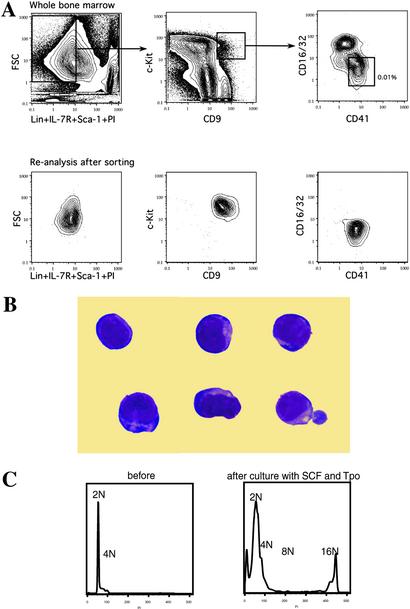
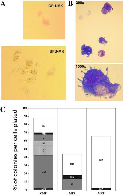
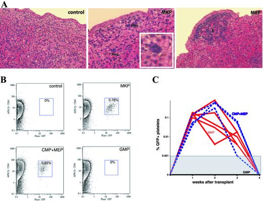
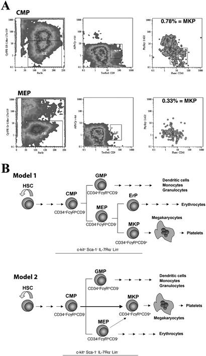
Although it has been shown that unfractionated bone marrow, hematopoietic stem cells, common myeloid progenitors, and bipotent megakaryocyteerythrocyte progenitors can give rise to megakaryocyte colonies in culture, monopotent megakaryocyte-committed progenitors (MKP) have never been prospectively isolated from the bone marrow of adult mice. Here, we use a monoclonal antibody to the megakaryocyte-associated surface protein, CD9, to purify MKPs from the c-kit(+)Sca-1(-)IL7Ralpha(-)Thy1.1(-)Lin(-) fraction of adult C57BLKa-Thy1.1 bone marrow. The CD9(+) fraction contained a subset of CD41(+)FcgammaR(lo)CD34(+)CD38(+) cells that represent approximately 0.01% of the total nucleated bone marrow cells. They give rise mainly to colony-forming unit-megakaryocytes and occasionally burst-forming unit-megakaryocytes, with a plating efficiency >60% at the single-cell level. In vivo, MKPs do not have spleen colony-forming activity nor do they contribute to long-term multilineage hematopoiesis; they give rise only to platelets for approximately 3 weeks. Common myeloid progenitors and megakaryocyteerythrocyte progenitors can differentiate into MKPs after 72 h in stromal cultures, indicating that MKPs are downstream of these two progenitors. These isolatable MKPs will be very useful for further studies of megakaryopoiesis as well as the elucidation of their gene expression patterns.


Figures

Figure 1
Figure 1
Identification of MKPs in mouse bone marrow. (A) Flow cytometric analysis of bone marrow after depleting the lineage-positive cells with magnetic beads. (Upper) The sorting gates for MKPs. The frequency of MKPs is shown as relative to total nucleated bone marrow cells before the negative depletion. Reanalysis after the first round of sorting found MKPs to be cleanly isolatable population (Lower). (B) MKPs were myeloblast-like cells with no characteristic features of megakaryocytes (Giemsa staining, original magnification ×1,000). (C) DNA content analysis showed MKPs to be diploid cells. After a 5-day incubation with SCF and Tpo, polyploid (16N) megakaryocytes were found in the culture.
Figure 2
Figure 2
Clonogenic megakaryocyte colony formation. (A) Morphology of typical CFU-MK and BFU-MK derived from MKP at day 10 of culture (phase contrast; original magnification ×100 for CFU-MK, ×40 for BCU-MK). (B) Mature megakaryocytes from a single colony (Giemsa; original magnification ×200 and ×1,000). (C) Day-10 colony readouts of single progenitors in methylcellulose-containing SCF, Flt3-ligand, IL-3, IL-11, GM-CSF, Epo, and Tpo. A total of 270 wells deposited with single progenitors were scored. CMPs gave rise to all types of myeloid colonies with plating efficiency >80%. MEPs gave rise to both megakaryocyte and erythroid colonies, whereas MKPs formed megakaryocyte colonies exclusively.
Figure 3
Figure 3
In vivo differentiation of MKPs. (A) Histology of the spleen at day 8 (H&E, original magnification: control and MEP, ×200; MKP, ×400; Inset, ×1,000). Mice were lethally irradiated and then injected with no cell (control), 1,000 MKPs, or 500 MEPs. MKPs gave rise to microscopic foci of megakaryocytes (Meg), whereas MEPs formed large colonies composed of only erythroid cells (E). No erythroid or megakaryocyte colonies were observed in the control animals. (B) Representative FACS plots of platelets in the peripheral blood of recipient mice 14 days after receiving the purified progenitors from β-actin GFP transgenic donors. Blood cells were first gated on the forward scatter and side scatter. Percentages of GFP+CD61+ cells in the platelet gate are shown in the boxes. (C) MKPs generated platelets in vivo for 3 weeks. The kinetics of platelet engraftment of 3,000 MKPs (red lines) was similar to 10,000 CMPs and MEPs (blue dashed lines). In contrast, 10,000 GMPs did not generate detectable platelets at any time point of analysis (black line). The threshold for detecting GFP+ platelets in the whole population was 1 in 100,000.
Figure 4
Figure 4
Lineage relationships among myeloid progenitors. (A) Flow cytometric analysis for cells with the MKP phenotype in the culture of CMPs and MEPs. Note more MKPs present in the CMP culture. (B) Two models for the MKP development. The first model suggests that commitment to the megakaryocytic lineage occurs at the level of MEP where the progenitor has to make a choice to become either MKP or erythrocyte progenitor (ErP; yet to be prospectively identified). In the second model, the commitment occurs earlier, at the stage of CMP. Although MEPs are bipotent, they differentiate mainly into the erythroid lineage. The majority of MKPs are the direct descendants of CMPs.

References

    1. Weissman I L, Anderson D J, Gage F. Annu Rev Cell Dev Biol. 2001;17:387–403. - PubMed
    1. Metcalf D. Ann NY Acad Sci. 1999;872:289–303. - PubMed
    1. Bradley T R, Metcalf D. Aust J Exp Biol Med Sci. 1966;44:287–299. - PubMed
    1. Eaves C J. In: Williams Hematology. Beutler E, Lichtman M A, Coller B S, Kipps T J, editors. New York: McGraw–Hill; 1995. pp. L22–L26.
    1. Ichikawa Y, Pluznik D H, Sachs L. Proc Natl Acad Sci USA. 1966;56:488–495. - PMC - PubMed

Publication types